
2023 , Vol. 20 >Issue 04: 462 - 466
DOI: https://doi.org/10.3877/cma.j.issn.1672-6448.2023.04.014
涎腺腺泡细胞癌的超声表现及病理对照分析
Copy editor: 汪荣
收稿日期: 2021-07-28
网络出版日期: 2023-08-07
版权
Ultrasound features of acinic cell carcinoma in the salivary gland
Received date: 2021-07-28
Online published: 2023-08-07
Copyright
探讨涎腺腺泡细胞癌(ACC)的超声声像图表现,对照分析其相应的病理学基础。
回顾性选取2003年7月至2019年11月于上海中医药大学附属市中医医院和上海市奉贤区奉城医院经手术病理证实为ACC的患者39例。所有患者术前均进行涎腺超声检查,对肿瘤边界、内部回声、形态、后方回声、囊实性等超声表现进行总结;同时将超声声像图与病理资料作对照分析。
本组ACC发生于腮腺(含副腮腺)37例(94.9%),2例发生于颌下腺。声像图多数表现为边界清晰(28/39,71.8%),病理对照提示33例(33/39,84.6%)肿瘤有包膜,但包膜多不完整;形态以椭圆形、分叶状较多见(33/39,84.6%);病变以低回声(24/39,61.5%)、混合性回声(14/39,35.9%)为主,多数内部回声不均(25/39,64.1%),82.1%(32/39)的肿块后方回声增强,病理对照示27例(27/39,69.2%)细胞间隙内见大量似胶冻样PAS阳性物质;血流信号不丰富20例(20/39,51.3%),病理对照示肿瘤血管密度稀疏者18例(18/39,46.2%)。
ACC具有一定的超声表现特征及相应的病理学基础,但其恶性特征不具有典型性,因此超声定性诊断存在一定局限性,诊断准确性有待进一步提高。
贾春岩 , 李月河 . 涎腺腺泡细胞癌的超声表现及病理对照分析[J]. 中华医学超声杂志(电子版), 2023 , 20(04) : 462 -466 . DOI: 10.3877/cma.j.issn.1672-6448.2023.04.014
To explore the ultrasonographic features of salivary acinic cell carcinoma (ACC) and analyze the corresponding pathological basis.
Thirty-nine patients with acinic cell carcinoma between July 2003 and November 2019 confirmed by surgery and pathology at Shanghai Municipal Hospital of Traditional Chinese Medicine and Fengcheng Hospital of Fengxian District were retrospectively analyzed. Ultrasonographic features, including the boundary, internal echo, morphology, posterior echo, and cystic and/or solid mixed echo, were summarized. The pathological data and the sonographic images were comparative analyzed.
There were 37 cases (94.9%) occurring in the parotid gland and 2 cases in the submandibular gland. Most of the lesions were showed a clear boundary (28/39, 71.8%). Pathology showed that the tumors had envelopes in 33 cases (33/39 84.6%), the envelopes were incomplete, and oval and lobular morphology was common (33/39, 84.6%). The majority of lesions had hypoechoic (24/39 61.5%) and mixed echo (14/39,35.9%), the internal echo was heterogeneous (25/39, 64.1%), and echo enhancement occurred behind most masses (32/39, 82.1%). Pathological analysis suggested that 27 cases (27/39 69.2%) had a large amount of jelly-like PAS positive substances in the intercellular space. Color Doppler flow imaging (CDFI) signal was not abundant in 20 cases (20/39, 51.3%). Pathology showed that tumor vascular density was sparse in 18 cases (18/39 46.2%).
ACC has some ultrasonographic features and corresponding pathological basis, but its malignant signs are not typical, so the qualitative diagnosis of ACC by ultrasonography has some limitations, and the diagnostic accuracy needs to be further improved.
Key words: Ultrasonography; Salivary glands; Acinic cell carcinoma; Pathology
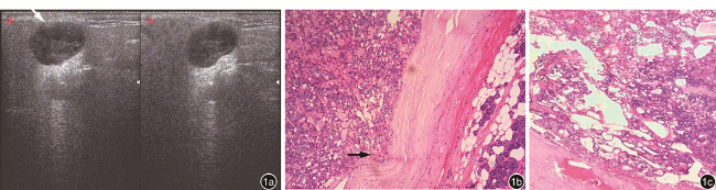
图1 涎腺腺泡细胞癌超声及病理表现。图a为超声声像图(左图为横切面图像,右图为纵切面图像)显示肿瘤形态略有分叶呈蚕豆状;边界基本清晰,箭头处显示局部边界不清(病理提示为局部包膜不完整);回声欠均匀,局部回声极低近似无回声;后方回声增强。图b,c为病理图像,图b可见肿瘤具有完整的包膜,箭头处显示局部包膜内缘受侵呈凹陷改变(HE ×400);图c显示滤泡型结构,内见胶冻样物质淤积(HE ×100) |
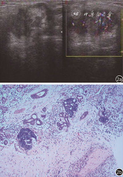
图2 腮腺腺泡细胞癌超声及病理表现。图a为典型涎腺恶性肿瘤声像图表现,形态不规则、边界不清、内部回声不均匀,彩色多普勒示血流信号较丰富;图b为病理图像示肿瘤边界不清,其内可见出血区(HE ×100) |
| 1 |
|
| 2 |
马大权. 涎腺疾病 [M].北京: 人民卫生出版社, 2002: 239-243.
|
| 3 |
吴奇光. 口腔组织病理学 [M]. 3版.北京:人民卫生出版社, 1994: 198-199.
|
| 4 |
|
| 5 |
|
| 6 |
陆林国, 燕山, 徐秋华. 腮腺恶性肿瘤的超声诊断研究 [J]. 中国超声医学杂志, 2001, 17(8): 604-606.
|
| 7 |
|
| 8 |
|
| 9 |
|
| 10 |
|
| 11 |
|
| 12 |
蒋淑婉, 王艳芬, 丁永玲, 等. 涎腺腺泡细胞癌10例临床病理分析 [J]. 医学前沿, 2016, 6(24): 46-48.
|
| 13 |
郭莉, 徐志锋, 刘芳. 涎腺腺泡细胞癌28例临床病理分析 [J]. 肿瘤研究与临床, 2019, 31(2): 124-126.
|
| 14 |
孙妍. 涎腺腺泡细胞癌36例临床病理分析 [J]. 西南军医, 2010, 12(6): 1096-1099.
|
| 15 |
李佳, 熊屏, 龚霞. 腮腺腺泡细胞癌的超声表现 [J]. 中国超声医学杂志, 2014, 30(5): 385-387.
|
| 16 |
董文霞, 江明祥, 邵国良, 等. 腮腺腺泡细胞癌的CT表现分析 [J]. 医学影像学杂志, 2016, 26(4): 596-600.
|
| 17 |
蒯新平, 王胜裕, 范国润, 等. 腮腺腺泡细胞癌的MRI及临床特点 [J]. 临床耳鼻咽喉头颈外科杂志, 2014, 28(24): 1968-1971.
|
| 18 |
朱少明, 陈剑, 赵齐羽, 等. 腮腺多形性腺瘤与Warthin氏瘤的超声图像对比分析 [J]. 中国超声医学杂志, 2013, 29(9): 848-850.
|
/
| 〈 |
|
〉 |
