
2023 , Vol. 20 >Issue 05: 511 - 516
DOI: https://doi.org/10.3877/cma.j.issn.1672-6448.2023.05.008
ACR-TIRADS与C-TIRADS分类分别联合二维剪切波弹性成像对甲状腺结节分类的诊断效能——多中心回顾性研究
Copy editor: 吴春凤
收稿日期: 2022-03-28
网络出版日期: 2023-10-06
基金资助
安徽医科大学校科研基金(2019xkj045)
版权
Comparison of diagnostic efficacy of either ACR-TIRADS or C-TIRADS combined with 2D-SWE imaging technology in thyroid nodules: a multicenter retrospective study
Received date: 2022-03-28
Online published: 2023-10-06
Copyright
探讨美国放射学会(ACR)-甲状腺影像报告和数据系统(TIRADS)、甲状腺结节超声恶性危险分层中国指南(C-TIRADS)分类联合二维剪切波弹性成像(2D-SWE)技术在甲状腺结节良恶性鉴别中的诊断效能。
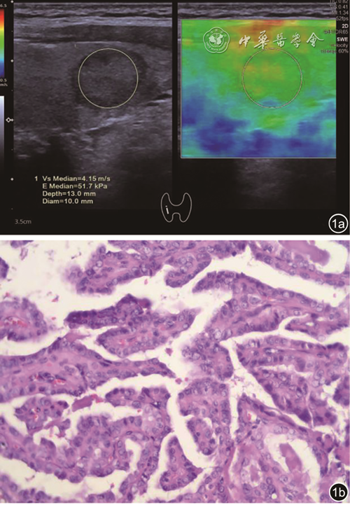
收集省内4家医院(安徽医科大学第二附属医院、安徽省立医院、安徽省立肿瘤医院及安徽医科大学第四附属医院)2021年2月至2022年1月行细针穿刺或手术的甲状腺结节病例,由高年资超声医师经培训后,分别以ACR-TIRADS(2017版)和C-TIRADS对每个结节进行分类。回顾性分析502个甲状腺结节的常规超声及2D-SWE的图像特征,以细针穿刺活检术或手术病理结果为金标准绘制受试者操作特征(ROC)曲线,分析比较ACR-TIRADS、C-TIRADS、2D-SWE以及ACR-TIRADS联合2D-SWE、C-TIRADS联合2D-SWE对甲状腺结节的诊断效能,采用Kappa检验比较2种联合诊断方法的一致性。
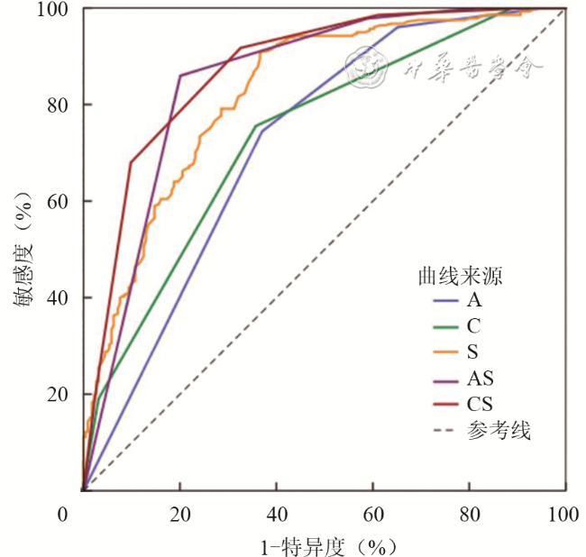
ACR-TIRADS联合2D-SWE、C-TIRADS联合2D-SWE诊断恶性结节的ROC曲线下面积(AUC)依次为0.852、0.875,敏感度为86.00%、91.70%,特异度为79.90%、67.40%,准确性为83.27%、80.79%,其诊断效能优于单独运用ACR-TIRADS分类、C-TIRADS分类和2D-SWE成像技术(P<0.05),C-TIRADS联合2D-SWE诊断的AUC高于ACR-TIRADS联合2D-SWE,差异具有统计学意义(Z=2.090,P=0.037)。ACR-TIRADS联合2D-SWE(以TR5类为截断值)和C-TIRADS联合2D-SWE(以C-TR4B为截断值)的诊断一致性较高(Kappa=0.801,P<0.001)。
ACR-TIRADS和C-TIRADS分类方法分别联合2D-SWE技术均可提高对甲状腺恶性结节的诊断效能,C-TIRADS联合2D-SWE的整体诊断效能优于ACR-TIRADS联合2D-SWE方法。
关键词: 超声检查; 甲状腺结节; 甲状腺影像报告和数据系统; 剪切波弹性成像
郭云云 , 解翔 , 彭梅 , 姜凡 , 毕玉 , 何年安 , 胡蕾 , 杨杨 , 王涛 , 石玉洁 , 陈冬冬 . ACR-TIRADS与C-TIRADS分类分别联合二维剪切波弹性成像对甲状腺结节分类的诊断效能——多中心回顾性研究[J]. 中华医学超声杂志(电子版), 2023 , 20(05) : 511 -516 . DOI: 10.3877/cma.j.issn.1672-6448.2023.05.008
To compare the efficacy of either the 2017 American Society of Radiology thyroid imaging report and data system (ACR-TIRADS) or the Chinese ultrasound thyroid imaging report and data system (C-TIRADS) combined with two-dimensional shear wave elastography (2D-SWE) imaging technology in the differential diagnosis of benign and malignant thyroid nodules.
Cases with thyroid nodules who underwent fine needle aspiration or surgery at four hospitals in Anhui province (the Second Affiliated Hospital of Anhui Medical University, Anhui Provincial Hospital, Anhui Provincial Cancer Hospital, and the Fourth Affiliated Hospital of Anhui Medical University) from February 2021 to January 2022 were collected and classified by trained senior sonographers based on the ACR-TIRADS (2017) and C-TIRADS, respectively. A total of 502 thyroid nodules with the data of conventional ultrasound and 2D-SWE were retrospectively analyzed. Using the fine needle aspiration or surgical pathological results as the gold standard, receiver operating characteristic (ROC) curve analysis was performed to compare the diagnostic efficacy of ACR-TIRADS, C-TIRADS, 2D-SWE technology, ACR-TIRADS combined with 2D-SWE, and C-TIRADS combined with 2D-SWE in thyroid nodules. The consistency of the two combined diagnostic methods was assessed by Kappa test.
The area under the ROC curve (AUC) of ACR-TIRADS combined with 2D-SWE and C-TIRADS combined with 2D-SWE (set cut-off value: C-TR4C) was 0.852 and 0.875, respectively, for the diagnosis of malignant thyroid nodules, with a corresponding sensitivity of 86.00% and 91.70%, specificity of 79.90% and 67.40%, and accuracy of 83.27% and 80.79%; the diagnostic efficacy of these two combinations was significantly better than that of ACR-TIRADS, C-TIRADS, or 2D-SWE technology alone (P<0.05). The AUC of C-TIRADS was higher than that of ACR-TIRADS (Z=2.090, P=0.037). The Kappa value of ACR-TIRADS combined with 2D-SWE and C-TIRADS combined with 2D-SWE was 0.801 (P<0.001).
Either ACR-TIRADS or C-TIRADS combined with 2D-SWE technology can considerably improve the diagnostic efficacy for malignant thyroid nodules, with the latter having better diagnostic efficacy.
图2 不同方法诊断甲状腺结节良恶性的受试者操作特征曲线注:A为ACR-TIRADS分类,C为C-TIRADS分类,S组为2D-SWE技术,AS为ACR-TIRADS+2D-SWE,CS为C-TIRADS+2D-SWE |
表1 不同诊断方法对甲状腺结节良恶性的诊断效能 |
| 组别 | 曲线下面积 | 敏感度(%) | 特异度(%) | 准确性(%) |
|---|---|---|---|---|
| A组 | 0.720ab | 74.50a | 62.90a | 69.32 |
| C组 | 0.735b | 79.90 | 64.30 | 70.52 |
| S组 | 0.825b | 90.60ab | 63.40ab | 78.49 |
| A+S组 | 0.852b | 86.00b | 79.90b | 83.27 |
| C+S组 | 0.875 | 91.70 | 67.40 | 80.79 |
注:A组为美国放射学会(ACR)-甲状腺影像报告和数据系统(TIRADS)分类检验诊断结果,C组为中国(C)-TIRADS分类检验诊断结果,S组为二维剪切波弹性成像(2D-SWE)技术检验诊断结果,A+S组为ACR-TIRADS+2D-SWE检验诊断结果,C+S组为C-TIRADS+2D-SWE检验诊断结果;a与A+S组相比,AUC对比差异具有统计学意义(P<0.05),敏感度、特异度对比,差异具有统计学意义(P′<0.005);b与C+S组相比,AUC对比差异具有统计学意义(P<0.05),敏感度、特异度对比,差异具有统计学意义(P′<0.005) |
| 1 |
梁羽, 岳林先, 陈琴, 等. Kwak与ACR(2017)甲状腺影像报告和数据系统(TI-RADS)分类的诊断效能比较——多中心回顾性研究 [J]. 中华超声影像学杂志, 2019, 28(5): 419-424.
|
| 2 |
中华医学会超声医学分会浅表器官和血管学组, 中国甲状腺与乳腺超声人工智能联盟. 2020甲状腺结节超声恶性危险分层中国指南: C-TIRADS [J]. 中华超声影像学杂志, 2021, 30(3): 185-200.
|
| 3 |
|
| 4 |
陈庆芳, 吴嗣泽. 甲状腺结节恶性风险分层的C-TIRADS与ACR-TIRADS诊断效能比较研究 [J]. 中华超声影像学杂志, 2021, 30(10): 861-867.
|
| 5 |
唐力, 徐辉雄, 李建卫, 等. 新型声触诊组织成像定量剪切波弹性成像技术鉴别甲状腺结节良恶性的价值 [J/CD]. 中华医学超声杂志(电子版), 2015, 12(3): 61-65.
|
| 6 |
|
| 7 |
吕玲, 赵树樊, 牛惠萍. 甲状腺影像报告与数据系统分类和超声弹性成像技术及其联合诊断甲状腺结节研究进展 [J]. 中国医学影像技术, 2021, 37(8): 1238-1241.
|
| 8 |
|
| 9 |
|
| 10 |
|
| 11 |
|
| 12 |
|
| 13 |
|
| 14 |
|
| 15 |
李帅, 樊秀齐, 康春松, 等. 甲状腺结节杨氏模量最大值的影响因素及其对结节性质的鉴别诊断价值 [J]. 中华医学超声杂志(电子版), 2021, 18(12): 1185-1190.
|
| 16 |
|
/
| 〈 |
|
〉 |
