
2023 , Vol. 20 >Issue 08: 844 - 848
DOI: https://doi.org/10.3877/cma.j.issn.1672-6448.2023.08.010
含鳞状细胞癌成分的乳腺化生性癌的超声与病理特征分析
Copy editor: 汪荣
收稿日期: 2022-04-26
网络出版日期: 2023-10-31
版权
Ultrasound and pathological characteristics of breast metaplastic carcinoma containing squamous cell carcinoma components
Received date: 2022-04-26
Online published: 2023-10-31
Copyright
观察含有鳞状细胞癌成分的乳腺化生性癌(MCSC)的超声特征,并与病理学结果对比分析。
回顾性选取湖北省肿瘤医院9例经手术病理检查证实的MCSC患者,采用彩色多普勒超声扫查双侧乳腺及腋窝,观察肿瘤的超声特征,并对乳房肿瘤进行乳腺影像报告与数据系统(BI-RADS)分类及Adler血流分级。所有MCSC患者术后均进行大体、镜下病理和免疫组化检查,将肿瘤超声特征与病理学结果进行对比分析。
9例MCSC病理分型鳞状细胞癌3例,腺鳞癌1例,伴间叶分化的化生性癌1例,混合型化生性癌4例。肿瘤最大径1.07~15.00 cm,中位径线为4.48 cm。5例内部回声呈混合性回声,4例呈低回声;9例后方回声均可见不同程度增强;7例边界欠清晰或模糊不清;8例形态不规则;3例伴钙化;超声检查有4例腋下淋巴结肿大。Adler血流分级:8例为Ⅲ级,1例为I级。BI-RADS分级:1例4b类,其余4c~5类。术后病理大体标本8例可见大小不等的囊腔,仅1例未见囊腔。免疫组化显示雌激素受体(ER)、孕激素受体(PR)、人表皮生长因子受体-2(Her-2)三阴者即三阴性乳腺癌4例(4/9)。
MCSC中以混合型化生性癌最为常见,超声声像图呈恶性肿瘤的超声表现,肿瘤体积较大、内部为混合性回声伴后方回声增强、肿瘤实质部分血供丰富的超声表现对诊断MCSC有意义。
郏亚平 , 曾书娥 . 含鳞状细胞癌成分的乳腺化生性癌的超声与病理特征分析[J]. 中华医学超声杂志(电子版), 2023 , 20(08) : 844 -848 . DOI: 10.3877/cma.j.issn.1672-6448.2023.08.010
To explore the ultrasound characteristics of breast metaplastic carcinoma containing squamous cell carcinoma components (MCSC) and compare them with pathological results.
Nine patients with MCSC confirmed by surgical and pathological examination at Hubei Cancer Hospital were retrospectively selected. Color Doppler ultrasound was used to scan both the breasts and armpits to observe the ultrasound characteristics of the tumors. BI-RADS classification and Adler blood flow grading were performed on the breast tumors. All MCSC patients underwent gross, microscopic, and immunohistochemical examinations after surgery, and the ultrasound features of the tumors were compared with pathological results.
There were 3 cases of squamous cell carcinoma, 1 case of adenosquamous carcinoma, 1 case of metaplastic carcinoma with mesenchymal differentiation, and 4 cases of mixed metaplastic carcinoma. The maximum diameter of the tumors ranged from 1.07-15.00 cm, with a median diameter of 4.48 cm. Five cases showed mixed internal echoes, and 4 showed hypoechogenicity; 9 showed varying degrees of enhancement in posterior echoes; 7 had unclear or unclear boundaries; 8 had an irregular morphology; 3 were accompanied by calcification. Ultrasound examination showed 4 cases of axillary lymph node enlargement. According to Adler blood flow grading, 8 cases were classified as Grade III, and 1 was classified as Grade I. BI-RADS grading suggested category 4b in 1 case and categories 4c-5 in the remaining. Postoperative pathological examination of gross specimens showed cysts of varying sizes in 8 cases, with only 1 case showing no cysts. Immunohistochemistry showed that there were 4 cases (4/9) of triple negative breast cancer.
Mixed metaplastic carcinoma is the most common type in MCSC, and ultrasound imaging shows the appearance of a malignant tumor. The ultrasound features of a larger tumor volume, mixed echoes with enhanced posterior echoes, and abundant blood supply in the tumor parenchyma are meaningful for the diagnosis of MCSC.
表1 9例MCSC的超声表现与病理结果 |
| 病例序号 | 年龄(岁) | 肿瘤位置 | 肿瘤最大径线(cm) | 内部回声 | Adler血流分级 | 后方回声增强 | 钙化 | BI-RADS分级 | 腋下淋巴结肿大 | 病理分型 |
|---|---|---|---|---|---|---|---|---|---|---|
| 1 | 51 | 右乳、外上 | 4.12 | 实质性 | Ⅲ | 有 | 有 | 5 | 有 | 混合型化生性癌 |
| 2 | 57 | 左乳、外上 | 4.04 | 囊实性 | Ⅲ | 有 | 无 | 4c | 有 | SCC |
| 3 | 47 | 右乳、外上 | 2.78 | 实质性 | I | 有 | 有 | 5 | 无 | ASC |
| 4 | 47 | 右乳内侧 | 1.07 | 实质性 | Ⅲ | 有 | 无 | 4b | 无 | SCC |
| 5 | 44 | 右乳 | 15.00 | 囊实性 | Ⅲ | 有 | 无 | 5 | 有 | 混合型化生性癌 |
| 6 | 45 | 左乳、外上 | 2.74 | 实质性 | Ⅲ | 有 | 无 | 4c | 有 | 混合型化生性癌 |
| 7 | 79 | 右乳、外上 | 3.54 | 囊实性 | Ⅲ | 有 | 有 | 5 | 无 | 混合型化生性癌 |
| 8 | 46 | 右乳、外上 | 2.95 | 实质性 | Ⅲ | 有 | 无 | 5 | 无 | SCC |
| 9 | 51 | 左乳、外上 | 4.09 | 囊实性 | Ⅲ | 有 | 无 | 4c | 无 | 伴间叶分化的化生性癌 |
注:MCSC为含有鳞状细胞癌成分的乳腺化生性癌;SCC为单纯性鳞状细胞癌;ASC为腺鳞癌;BI-RADS为乳腺影像报告与数据系统 |
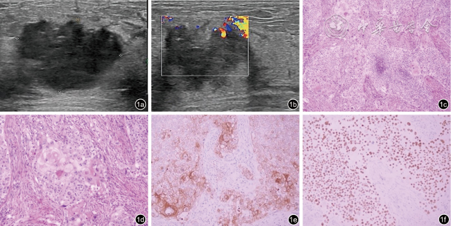
图1 含鳞状细胞癌成分的乳腺化生性癌的超声及病理图像(病例8)。图a为超声图像示肿块呈低回声,边界模糊形态不规则,未显示明显钙化;图b为彩色多普勒图像示肿块血流信号增多;图c为病理图像示肿瘤呈实性片状伴局部坏死,无腺管形成,可见角化珠和细胞间桥(HE ×100);图d为病理图像示肿瘤细胞核深染,核浆比增大,胞浆嗜酸,核分裂象可见(HE ×200);图e为免疫组织化学图像示肿瘤细胞CK5/6胞质阳性(EnVision法×200);图f为免疫组织化学图像示肿瘤细胞P40胞核弥漫强阳性(EnVision法 ×200) |
表2 9例MCSC的病理分型与腋窝淋巴结转移情况 |
| 病例序号 | 年龄(岁) | 病理分型 | 腋窝淋巴结转移 | 免疫组化 |
|---|---|---|---|---|
| 1 | 51 | 混合型化生性癌 | 腺癌(4/19枚) | ER(-)PR(-)Her-2(浸润性导管癌部分3+,鳞癌部分1+) |
| 2 | 57 | SCC | 无 | ER(10%,2+)PR(2%,2+)Her-2(-) |
| 3 | 47 | ASC | 无 | ER(1+,1%)PR(-)Her-2(-) |
| 4 | 47 | SCC | 无 | ER(-)PR(-)Her-2(2+) |
| 5 | 44 | 混合型化生性癌 | 鳞状细胞癌(1/14枚),腺癌(10/14枚) | ER(5%,2+)PR(2%,1+)Her-2(3+) |
| 6 | 45 | 混合型化生性癌 | 腺癌(2/21枚) | ER(-)PR(-)Her-2(-) |
| 7 | 79 | 混合型化生性癌 | 无 | ER(-)PR(-)Her-2(-) |
| 8 | 46 | SCC | 无 | ER(-)PR(-)Her-2(-) |
| 9 | 51 | 伴间叶分化的化生性癌 | 无 | ER(-)PR(-)Her-2(1+) |
注:MCSC为含有鳞状细胞癌成分的乳腺化生性癌;SCC为单纯性鳞状细胞癌;ASC为腺鳞癌;ER为雌激素受体;PR为孕激素受体;Her-2为人表皮生长因子受体-2 |
| 1 |
|
| 2 |
|
| 3 |
|
| 4 |
|
| 5 |
|
| 6 |
|
| 7 |
|
| 8 |
|
| 9 |
|
| 10 |
|
| 11 |
|
| 12 |
|
| 13 |
|
| 14 |
|
| 15 |
|
/
| 〈 |
|
〉 |