
2023 , Vol. 20 >Issue 08: 860 - 865
DOI: https://doi.org/10.3877/cma.j.issn.1672-6448.2023.08.013
胎儿超声心动图测量McGoon指数在评价胎儿肺血管发育中的应用
Copy editor: 汪荣
收稿日期: 2023-05-15
网络出版日期: 2023-10-31
基金资助
浙江大学科学技术研究院一般横向项目(校合-2021-KYY-518053-0055)
浙江省基础公益研究计划项目(LGF20H180013)
版权
Evaluation of fetal pulmonary development using McGoon index measured by fetal echocardiography
Received date: 2023-05-15
Online published: 2023-10-31
Copyright
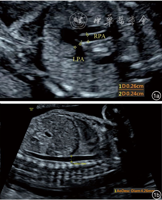
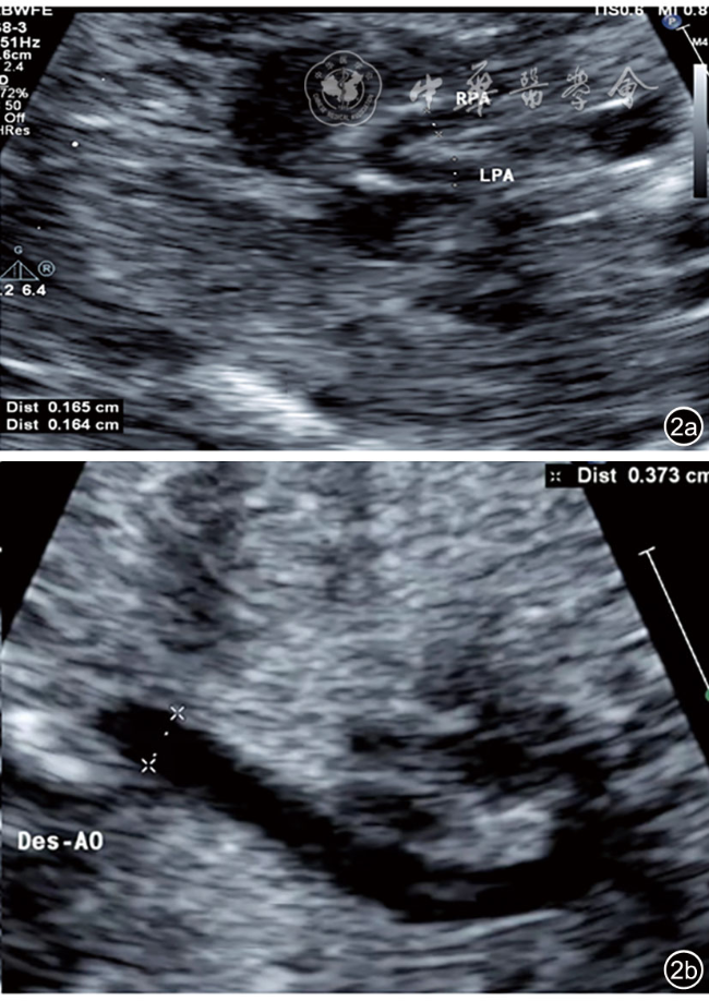
应用胎儿超声心动图测量McGoon指数(MGI),探讨不同孕龄的胎儿及不同肺血流量状态下MGI的变化。
回顾性选取2020年6月至2021年8月在浙江大学医学院附属邵逸夫医院超声科接受超声心动图检查的胎儿。超声心动图检查未见异常的218例胎儿为正常组(依据孕龄分为4个亚组);胎儿超声心动图考虑肺动脉血流减少或闭锁的先天性心脏病胎儿66例(病例组1),考虑主动脉血流减少或离断胎儿19例(病例组2)。超声心动图测量胎儿的MGI,比较分析各组之间测量结果的差异。
正常对照组不同孕龄的4个亚组间(18~21+6周、22~25+6周、26~29+6周、30~33+6周)MGI分别为1.39±0.35、1.38±0.20、1.38±0.21、1.40±0.19,差异无统计学意义(P>0.05)。正常对照组MGI为1.38±0.22;肺动脉血流减少或闭锁组MGI为1.16±0.19,低于其他2组,差异有统计学意义(P<0.05);主动脉血流减少或离断组MGI为1.42±0.25,与正常组比较差异无统计学意义(P>0.05)。
孕18~33+6周阶段正常胎儿MGI相对恒定,不随孕龄的增加而改变;超声心动图定量测量胎儿MGI可评价不同先天性心脏病胎儿的肺血流量,一定程度上反映肺血管发育情况。
张璟璟 , 赵博文 , 潘美 , 彭晓慧 , 毛彦恺 , 潘陈可 , 朱玲艳 , 朱琳琳 , 蓝秋晔 . 胎儿超声心动图测量McGoon指数在评价胎儿肺血管发育中的应用[J]. 中华医学超声杂志(电子版), 2023 , 20(08) : 860 -865 . DOI: 10.3877/cma.j.issn.1672-6448.2023.08.013
To investigate the dynamic changes of McGoon index (MGI) in fetuses with different gestational weeks or different pulmonary blood flow patterns by fetal echocardiography.
Fetuses who underwent echocardioimagedata examination at the Department of Diagnostic Ultrasound & Echocardiography, Sir Run Run Shaw Hospital, Zhejiang University School of Medicine from June 2020 to August 2021 were included, of whom 218 with no abnormalities detected by echocardiography were included in a normal group (further subdivided into 4 subgroups based on gestational age), 66 had decreased pulmonary blood flow or pulmonary atresia (case group 1), and 19 had decreased aortic blood flow or interruption of the aortic arch (case group 2). MGI was measured and calculated by echocardiography, and its differences among groups were compared.
MGI in the four subgroups of the normal control group (18-21+6 weeks, 22-25+6 weeks, 26-29+6 weeks, and 30-33+6 weeks) was (1.39±0.35), (1.38±0.20), (1.38±0.21), and (1.40±0.19), respectively, and the difference among the four subgroups was not statistically significant (P>0.05). MGI in the normal control group was (1.38±0.22). MGI in case group 1 was (1.16±0.19), significantly lower than that of the other two groups (P<0.05). MGI in case group 2 was (1.42±0.25), which was comparable to that of the normal control group (P>0.05).
MGI in normal fetuses with a gestational age of 18-33+6 weeks is constant, and it does not change with increasing gestational age. Quantitative measurement of fetal MGI by echocardiography can evaluate the pulmonary blood flow of fetuses with different congenital heart diseases, reflecting, to some extent, the development of pulmonary blood vessels.
 表示,所有资料经正态性检验及方差齐性检验,正常对照组内4个亚组之间的比较及正常对照组与病例组间的比较均采用单因素方差分析,进一步多重比较分析采用LSD-t法。以P<0.05为差异有统计学意义。
表1 正常对照组与病例组各组间超声心动图参数比较( |
| 组别 | 例数 | H/C | PA/AO | MGI |
|---|---|---|---|---|
| 正常对照组 | 218 | 0.25±0.04 | 1.278±0.133 | 1.38±0.22 |
| 肺动脉血流减少或闭锁组 | 66 | 0.28±0.05a | 0.745±0.276ab | 1.16±0.19ab |
| 主动脉血流减少或离断组 | 19 | 0.26±0.03 | 1.302±0.746 | 1.42±0.25 |
| F值 | 7.79 | 117.8 | 27.78 | |
| P值 | <0.01 | <0.01 | <0.01 |
注:H/C为心胸比;PA/AO为肺动脉与主动脉内径比值;MGI为McGoon指数;与正常对照组比较,aP<0.05;与主动脉血流减少或离断组比较,bP<0.05 |
| 1 |
|
| 2 |
|
| 3 |
贺新建, 董凤群, 魏九茹, 等. 超声估算左心室舒张末容积指数、McGoon比值和Nakata指数对法洛四联症术前评估的对比研究[J].中国超声医学杂志, 2010, 26(8): 716-719.
|
| 4 |
|
| 5 |
郭勇, 刘晓伟, 刘文旭, 等. 超声测量肺动脉内径及McGoon指数评估法洛四联症胎儿肺发育的研究[J].中国超声医学杂志, 2019, 35(3): 256-259.
|
| 6 |
李胜利, 朱军, 李军. 胎儿超声心动图学教程[M]. 北京: 科学出版社, 2018, 5: 63-92.
|
| 7 |
|
| 8 |
李慕子, 李维君, 李健, 等.超声心动图在肺动脉闭锁合并室间隔缺损诊断及外科治疗中的应用价值[J]. 中国循环杂志, 2021, 36(3): 299-304.
|
| 9 |
赵洋, 金岩, 于岩, 等. 多层螺旋CT与超声心动图评估法洛四联症肺动脉发育的对比研究[J].中国心血管病研究, 2021, 19(2): 142-146.
|
| 10 |
|
| 11 |
|
| 12 |
|
| 13 |
|
| 14 |
|
| 15 |
|
| 16 |
|
| 17 |
|
| 18 |
|
| 19 |
|
| 20 |
|
| 21 |
张烨, 何怡华, 孙琳, 等. 胎儿动脉导管血流频谱与右室梗阻性疾病肺动脉发育相关分析[J]. 中国超声医学杂志, 2015, 3(4): 355-357.
|
| 22 |
郑洁怀, 赵博文, 陈欣欣, 等. 卵圆孔瓣膨出指数联合心脏大血管Z-评分在单纯性卵圆孔瓣开放过度胎儿中的应用价值[J].中华超声影像学杂志, 2022, 31(6): 504-510.
|
| 23 |
詹梦娜, 赵博文, 彭晓慧, 等. 胎儿心脏定量分析技术评价左心室流出道梗阻胎儿心脏功能和形态[J].中华超声影像学杂志, 2021, 30(10): 854-860.
|
| 24 |
戚夏近, 赵博文, 郭玉霞, 等. 早期胎儿超声心动图对正常胎儿心室内径及其Z-评分的定量研究[J]. 中华超声影像学杂志, 2020, 29(5): 427-433.
|
| 25 |
朱建菲, 赵博文, 魏秀芝, 等. 定量房室瓣口舒张期彩色血流宽度的Z-评分在胎儿冠状静脉窦扩张中的应用研究[J].中华超声影像学杂志, 2019, 28(1): 42-48.
|
| 26 |
汪琳华, 赵博文, 潘美, 等.实时三维超声Xplane成像技术定量正常胎儿心房容积的Z-评分研究[J]. 中华超声影像学杂志, 2018, 27(10): 841-845.
|
/
| 〈 |
|
〉 |
 )
)