
2023 , Vol. 20 >Issue 10: 1061 - 1067
DOI: https://doi.org/10.3877/cma.j.issn.1672-6448.2023.10.010
胎儿孤立性完全型肺静脉异位引流的超声心动图特征及高分辨率血流联合时间-空间相关成像的应用
Copy editor: 汪荣
收稿日期: 2022-09-01
网络出版日期: 2024-01-08
基金资助
广西科技计划项目(桂科AB22080074)
广西医疗卫生适宜技术开发与推广应用项目(S2019032)
广西医药卫生自筹经费计划课题(Z20200142)
版权
Echocardioimagedata characteristics of fetal isolated total anomalous pulmonary venous connection and application of high definition flow imaging and spatio-temporal image correlation
Received date: 2022-09-01
Online published: 2024-01-08
Copyright
分析胎儿孤立性完全型肺静脉异位引流(TAPVC)的二维灰阶及彩色多普勒超声心动图特征,探讨四维超声时间-空间相关成像(STIC)及高分辨率血流成像(HD-flow)在产前诊断孤立性TAPVC中的应用。
本研究回顾性纳入广西壮族自治区妇幼保健院产前诊断的孤立性TAPVC 16例。其中2016年1月至2018年1月应用二维灰阶及彩色多普勒超声诊断9例;2018年2月至2021年3月,在二维超声心动图基础上应用四维STIC HD-flow诊断7例。均通过超声直接与间接征象对孤立性胎儿TAPVC进行筛查诊断,并对其预后结局进行随访。
16例胎儿中,心上型12例(12/16),心内型2例(2/16),心下型2例(2/16)。8例(8/16)首先由直接征象发现,表现为四腔心切面未显示肺静脉与左心房相连,而汇合成共同腔。8例(8/16)首先由间接征象发现,其中冠状静脉窦扩张2例,三血管气管切面出现垂直静脉或上腔静脉增宽5例,肝门区域显示异常血管1例。7例四维STIC HD-flow重建图像均显示了4条肺静脉与共同腔相连,通过垂直静脉或冠状静脉窦汇入体静脉(右心房)。
产前二维灰阶及彩色多普勒超声心动图直接征象与间接征象联合更有助于诊断孤立性胎儿TAPVC,四维STIC HD-flow可为胎儿TAPVC的诊断提供更多有价值的信息。
杨水华 , 何桂丹 , 覃桂灿 , 梁蒙凤 , 罗艳合 , 李雪芹 , 唐娟松 . 胎儿孤立性完全型肺静脉异位引流的超声心动图特征及高分辨率血流联合时间-空间相关成像的应用[J]. 中华医学超声杂志(电子版), 2023 , 20(10) : 1061 -1067 . DOI: 10.3877/cma.j.issn.1672-6448.2023.10.010
To analyze the two-dimensional gray scale and color Doppler echocardiography characteristics of fetal isolated complete pulmonary venous drainage (TAPVC) , and to explore the application of four-dimensional time-space correlation imaging (STIC) and high definition flow imaging (HD-flow) in the prenatal diagnosis of isolated TAPVC.
A retrospective analysis was performed on 16 cases of fetal isolated TAPVC at Guangxi in Maternity & Child Healthcare Hospital. Nine cases were diagnosed by two-dimensional gray scale and color Doppler echocardiography from January 2016 to January 2018, and seven cases were diagnosed based on two-dimensional echocardiography and four-dimensional STIC and HD-flow from February 2018 to March 2021. All of the above cases were both diagnosed by direct and indirect ultrasonography signs, and all cases were followed up.
The study included supracardiac type TAPVC in 12 (12/16) cases, cardiac type in 2 (2/16), and infracardiac type in 2 (2/16). Eight cases were found with direct ultrasound signs. Eight cases were found with indirect signs, including coronary sinus dilation in 2 cases, vertical vein or superior vena cava widening in 5, and abnormal blood vessels in the hilar area of the liver in 1.The four-dimensional STIC/HD-flow reconstruction images of 7 cases all showed that the 4 pulmonary veins were connected to the common pulmonary vein and joined into the systemic vein through vertical veins or the coronary sinus (right atrium).
The combination of direct and indirect signs of prenatal two-dimensional gray scale and color Doppler echocardiography is more helpful for the diagnosis of isolated fetal TAPVC, and four-dimensional STIC/HD-flow can provide more valuable information.
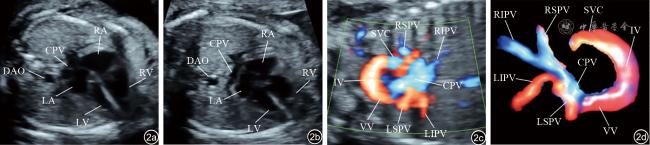
图2 心上型孤立性完全型肺静脉异位引流超声心动图及高分辨率血流联合时间-空间相关成像(STIC HD-flow)。图a为超声心动图检查初次扫查四腔心切面,显示左右心对称,共同腔壁与声束角度小,回声失落,将共同腔误认为左心房的顶部,降主动脉与左心房的距离也误认为正常;图b为再次扫查四腔心切面,调整声束角度,显示共同腔,左心房后壁光滑,肺静脉角消失;图c为HD-flow显示4条肺静脉与共同腔相连,经垂直静脉入无名静脉后汇入上腔静脉,无名静脉血流束明显增宽;图d为STIC HD-flow立体清晰地显示心上型肺静脉异位引流的超声病理解剖线路注:LA为左心房;LV为左心室;RA为右心房;RV为右心室;DAO为降主动脉;LSPV为左上肺静脉;LIPV为左下肺静脉;RSPV为右上肺静脉;RIPV为右下肺静脉;CPA为共同腔;IV为无名静脉;VV为垂直静脉 |
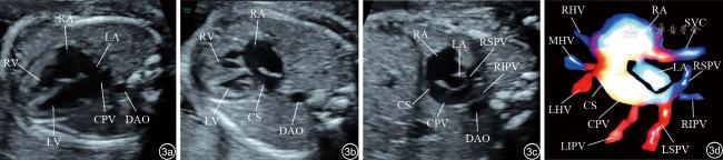
图3 心内型孤立性完全型肺静脉异位引流超声心动图及高分辨率血流联合时间-空间相关成像(STIC HD-flow)。图a为超声心动图扫查四腔心切面,左心房与共同腔相互融合,误认为左右心对称,肺静脉角及降主动脉与左心房距离正常;图b为扫查过程中发现冠状静脉窦扩张;图c显示两房心切面左心房明显小,左心房左后方显示了右肺静脉与共同腔相连汇入冠状静脉窦;图d为STIC HD-flow显示4条肺静脉与共同腔相连,经冠状静脉窦汇入右心房注:LA为左心房;LV为左心室;RA为右心房;RV为右心室;DAO为降主动脉;LSPV为左上肺静脉;LIPV为左下肺静脉;RSPV为右上肺静脉;RIPV为右下肺静脉;CS为冠状静脉窦;LHV为肝左静脉;MHV为肝中静脉;RHV为肝右静脉 |
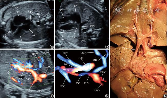
图4 心下型孤立性完全型肺静脉异位引流超声心动图、高分辨率血流联合时间-空间相关成像(STIC HD-flow)及病理解剖图像。图a为超声心动图检查初次扫查四腔心切面,共同腔壁与声束角度小,回声失落,误认为共同腔为左心房的顶部,降主动脉与左心房距离正常;图b为再次扫查四腔心切面,调整声束角度,显示了共同腔;图c为HD-flow显示4条肺静脉与共同腔相连,经垂直静脉入左门静脉;图d为STIC HD-flow清晰地显示心下型肺静脉异位引流的病理解剖线路;图e为病理解剖与产前超声诊断一致注:LA为左心房;LV为左心室;RA为右心房;RV为右心室;DAO为降主动脉;LSPV为左上肺静脉;LIPV为左下肺静脉;RSPV为右上肺静脉;RIPV为右下肺静脉;LPV为左门静脉;DV为静脉导管;IVC为下腔静脉;LPVi为左下门静脉;R为右;L为左;GB为胆囊 |
表1 16例孤立性TAPVC胎儿超声征象及随访结局 |
| 病例 | 年龄(岁) | 孕龄(周) | 类型 | 首要超声切面a | 首要超声线索b | 四维重建 | 垂直静脉梗阻 | 随访结局 |
|---|---|---|---|---|---|---|---|---|
| 1 | 25 | 30 | 心上型 | 四腔心切面 | 肺静脉未与左心房相连 | 否 | 否 | 术后存活 |
| 2 | 27 | 23 | 心上型 | 三血管气管切面 | 上腔静脉增宽、出现垂直静脉 | 否 | 否 | 终止妊娠 |
| 3 | 24 | 23 | 心上型 | 四腔心切面 | 肺静脉未与左心房相连 | 否 | 否 | 术后死亡 |
| 4 | 28 | 21 | 心内型 | 冠状静脉窦切面 | 冠状静脉窦扩张 | 否 | 否 | 终止妊娠 |
| 5 | 33 | 22 | 心上型 | 三血管气管切面 | 上腔静脉增宽、出现垂直静脉 | 否 | 是 | 新生儿期死亡 |
| 6 | 31 | 29 | 心上型 | 四腔心切面 | 肺静脉未与左心房相连 | 否 | 否 | 术后存活 |
| 7 | 35 | 24 | 心上型 | 四腔心切面 | 肺静脉未与左心房相连 | 否 | 否 | 终止妊娠 |
| 8 | 35 | 26 | 心下型 | 四腔心切面 | 肺静脉未与左心房相连 | 否 | 是 | 失访 |
| 9 | 32 | 23 | 心上型 | 四腔心切面 | 肺静脉未与左心房相连 | 否 | 否 | 终止妊娠 |
| 10 | 28 | 24 | 心上型 | 三血管气管切面 | 上腔静脉增宽、出现垂直静脉 | 是 | 否 | 术后存活 |
| 11 | 33 | 28 | 心下型 | 腹部横切面 | 肝门脉区域异常血流 | 是 | 是 | 病理确诊 |
| 12 | 34 | 28 | 心上型 | 四腔心切面 | 肺静脉未与左心房相连 | 是 | 否 | 术后存活 |
| 13 | 31 | 24 | 心上型 | 三血管气管切面 | 上腔静脉增宽、出现垂直静脉 | 是 | 否 | 终止妊娠 |
| 14 | 30 | 24 | 心内型 | 冠状静脉窦切面 | 冠状静脉窦扩张 | 是 | 否 | 术后存活 |
| 15 | 28 | 23 | 心上型 | 三血管气管切面 | 上腔静脉增宽、出现垂直静脉 | 是 | 否 | 新生儿期死亡 |
| 16 | 29 | 25 | 心上型 | 四腔心切面 | 肺静脉未与左心房相连 | 是 | 是 | 术后死亡 |
注:TAPVC为完全型肺静脉异位引流;a表示扫查胎儿心脏可疑TAPVC的第一切面;b表示首要超声切面中观察到的可疑TAPVC的超声表现 |
| 1 |
|
| 2 |
|
| 3 |
|
| 4 |
李博, 孔德璇, 彭芳华, 等.超声在胎儿肺静脉异位引流诊断中的应用价值[J/OL].中华医学超声杂志(电子版), 2023, 20(4): 437-441.
|
| 5 |
张晓花, 王锟, 张平, 等. 区域血流追踪法在超声诊断胎儿肺静脉异位引流中的应用价值[J/OL]. 中华医学超声杂志(电子版), 2021, 18(11): 1073-1077.
|
| 6 |
|
| 7 |
|
| 8 |
许进, 何怡华, 李治安, 等. 时间-空间关联成像联合高分辨率血流显像在产前胎儿静脉系统异常诊断中的应用价值[J]. 中华超声影像学杂志, 2013, 22(4): 300-304.
|
| 9 |
|
| 10 |
International Society of Ultrasound in Obstetrics and Gyne-cology,
|
| 11 |
|
| 12 |
|
| 13 |
|
| 14 |
American Institute of Ultrasound in Medicine. AIUM practice guideline for the performance of fetal echocardiography[J]. J Ultrasound Med, 2013, 32(6): 1067-1082.
|
| 15 |
|
| 16 |
|
| 17 |
吴娟, 刘云, 王铭, 等. 胎儿完全型肺静脉异位引流产前超声诊断要点[J]. 中华围产医学杂志, 2019, 22(5): 296-302.
|
| 18 |
韩建成, 李田静, 王静怡, 等. 正常胎儿左心房后间隙指数及其对胎儿单纯完全性肺静脉异位引流的诊断价值[J]. 中华超声影像学杂志, 2020, 29(9): 743-748.
|
| 19 |
|
| 20 |
|
| 21 |
|
| 22 |
|
| 23 |
|
| 24 |
|
| 25 |
|
| 26 |
|
/
| 〈 |
|
〉 |