
2024 , Vol. 21 >Issue 01: 53 - 56
DOI: https://doi.org/10.3877/cma.j.issn.1672-6448.2024.01.008
上颌前牙牙周生物型的超声影像特征及相关临床参数测量初步探索
Copy editor: 吴春凤
收稿日期: 2023-03-07
网络出版日期: 2024-03-27
基金资助
国家自然科学基金面上项目(82071929)
版权
Ultrasonic imaging characteristics of periodontal biotype of maxillary anterior teeth and measurement of related clinical parameters
Received date: 2023-03-07
Online published: 2024-03-27
Copyright
初步探索利用超声高频线阵探头检查上颌前牙牙周生物型的可行性,并归纳总结其超声影像学特征。
2022年10月至12月通过院内推荐的方式选择25名牙周健康的志愿者,利用发射15 MHz超声波的探头经上唇皮肤,以纵切及横切、平移及扇扫结合的方式检查150颗上颌前牙,然后采集图像,归纳总结其牙周生物型超声影像特征,并在图像上显示相关临床参数测量方法。
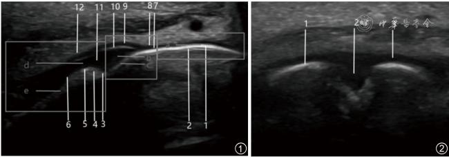
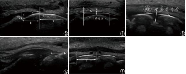
超声图像中牙体及牙槽骨硬组织为强回声,但牙釉质表面光滑,而牙槽骨表面粗糙、呈颗粒感;牙龈软组织显示为低回声,且牙龈表面轮廓光滑;另外,细微结构如游离龈缘、龈沟、牙龈上皮和固有层亦为低回声,而牙周间隙、牙骨质-牙釉质交界部为等回声。牙周生物型相关临床参数可在超声图像上进行标识及测量。
15 MHz的超声波能清晰显示上颌前牙的牙周生物型的特征,并能显示相关临床参数的测量。
徐松城 , 朱曼宁 , 叶瑞忠 , 王立刚 , 侯春杰 , 李建春 , 孙立涛 . 上颌前牙牙周生物型的超声影像特征及相关临床参数测量初步探索[J]. 中华医学超声杂志(电子版), 2024 , 21(01) : 53 -56 . DOI: 10.3877/cma.j.issn.1672-6448.2024.01.008
To investigate the feasibility of using high-frequency linear array ultrasound probe (ML6-15) to detect the periodontal biotype (PB) of maxillary anterior teeth and summarize its ultrasonic imaging characteristics.
A total of 25 volunteers with good periodontal health were selected from October to December 2022, and 150 maxillary anterior teeth were examined through the upper lip skin with a 15 MHz ultrasonic probe by combining longitudinal and transverse cutting, translation, and fan scanning. The images were collected, and the ultrasound imaging characteristics of PB were summarized. The relevant clinical parameter measurement methods were displayed on the images.
The hard tissue of the teeth and alveolar bone showed strong echo in ultrasonic images. The surface of enamel was smooth, while the surface of alveolar bone was rough and granulated. The gingival soft tissue showed low echo, and the gingival surface was smooth. In addition, fine structures such as free gingival margin, gingival groove, gingival epithelium, and lamina propria were hypoechoic, while the periodontal space and cemento-enamel junction were isoechoic. Clinical parameters related to PB can be identified and measured on ultrasonic images.
15 MHz ultrasound can clearly display the characteristics of the PB of maxillary anterior teeth and the measurements of related clinical parameters.
| 1 |
孟焕新. 牙周病学[M]. 4版, 北京: 人民卫生出版社, 2012: 21.
|
| 2 |
|
| 3 |
顾冰霞, 孙江.牙龈生物型对口腔治疗预后影响的研究进展 [J]. 中华口腔医学杂志, 2020, 55(7): 504-508.
|
| 4 |
黄满英, 付云.牙龈生物型的测量方法 [J]. 国际口腔医学杂志, 2019, 46(2): 171-176.
|
| 5 |
|
| 6 |
|
| 7 |
|
| 8 |
张欢, 吴婷婷, 吴川, 等. 超声评估正畸治疗中尖牙牙周膜变化的初步研究 [J]. 中国超声医学杂志, 2022, 38(8): 931-934.
|
| 9 |
|
| 10 |
|
| 11 |
|
| 12 |
|
/
| 〈 |
|
〉 |