
2024 , Vol. 21 >Issue 01: 69 - 74
DOI: https://doi.org/10.3877/cma.j.issn.1672-6448.2024.01.011
双极射频活检针在肝粗针活检止血的研发与初步应用
Copy editor: 吴春凤
收稿日期: 2022-11-22
网络出版日期: 2024-03-27
基金资助
国家重大科研仪器研制项目(82027803)
国家自然科学基金面上项目(81971623)
版权
Development and preliminary application of abipolar radiofrequency biopsy needle in hemostasis after liver coarse needle biopsy in pigs
Received date: 2022-11-22
Online published: 2024-03-27
Copyright
探讨新研发双极射频活检针在肝脏粗针活检后止血的有效性和操作性能,并与明胶海绵填塞方式进行对照研究。
利用16 G活检针对5只家猪的4片肝叶分别行超声引导下粗针活检,首先在无止血措施下进行作为对照组,记录活检后出血量及出血时间。随后,在各穿刺点旁1 cm,再次行穿刺活检,并分别用双极射频活检针和明胶海绵填塞进行止血,其中使用双极射频活检针的射频组分别采用3组参数,输出功率和针头作用区温度分别为40 W,70 ℃;30 W,90 ℃以及40 W,90 ℃,记录止血操作时间、活检后出血量及出血时间。采用Tukey-Kramer事后检验比较4种不同止血方式的实验组和对照组的操作时间、活检后出血量及出血时间的差异。
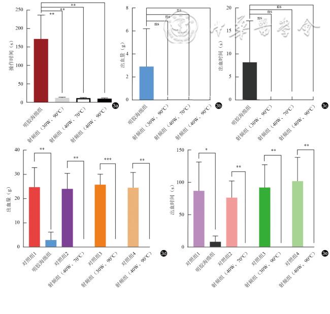
明胶海绵组及3种不同消融参数射频组操作时间为(172.00±28.87)s、(11.40±0.75)s、(12.20±0.80)s、(10.00±0.84)s,射频组操作时间明显短于明胶海绵组(P均=0.005,差值的95%置信区间:81.793~239.407 s、79.085~240.51 s、79.998~244.002 s)。但是,所有止血操作组较其邻近未止血针道的对照组的出血量、出血时间均显著减少,差异均具有统计学意义(出血量:P=0.003、=0.001、<0.001、=0.001,差值的95%置信区间:12.339~31.309 g、15.9~31.964 g、20.331~31.145 g、16.718~32.318 g;出血时间:P=0.018、0.003、0.004、0.003,差值的95%置信区间:22.6~135.0 s、44.4~108.4 s、48.2~135.8 s、56.5~147.4 s),明胶海绵组以及3种射频组彼此间出血量、出血时间比较,差异均无统计学意义(P均>0.05),均能起到较好地针道止血效果。
双极射频活检针能够安全、便捷地对肝脏粗针活检后进行止血,且比明胶海绵填塞操作简便、可靠,具有临床应用价值。
诸佳玮 , 陈强 , 王辉阳 , 蒋天安 . 双极射频活检针在肝粗针活检止血的研发与初步应用[J]. 中华医学超声杂志(电子版), 2024 , 21(01) : 69 -74 . DOI: 10.3877/cma.j.issn.1672-6448.2024.01.011
To investigate the efficacy and serviceability of a novel bipolar radiofrequency biopsy needle for hemostasis after hepatic core needle biopsy by comparing it with gelatin sponge packing.
Ultrasound-guided 16 G biopsy was performed on the four liver lobes of five pigs. First, the bleeding volume and bleeding time after biopsy were recorded without hemostasis. Subsequently, at 1 cm beside each puncture point, puncture biopsy was performed again, and a bipolar radiofrequency biopsy needle and gelatin sponge were used to stop bleeding. The bipolar radiofrequency biopsy needle used three sets of parameters: the output power and temperature of the needle action area were 40 W and 70 ℃, 30 W and 90 ℃, and 40 W and 90 ℃, respectively. Operation time, post-biopsy bleeding volume, and bleeding duration were recorded. The differences in these indicators between the experimental group and control group were compared using the Tukey-Kramer post hoc test.
The average operation time of the gelatin sponge group and the radiofrequency group with different ablation parameters was (172.00±28.87) s, (11.40±0.75) s, (12.20±0.80) s, and (10.00±0.84) s, respectively. The operation time of the radiofrequency group was significantly shorter than that of the gelatin sponge group (P=0.005 for all; 95% confidence interval [CI]: 81.793-239.407, 79.085-240.51, and 79.998-244.002, respectively). However, the bleeding volume and bleeding time of all hemostatic operations were significantly lower than those of the adjacent non-hemostatic needle tracts (bleeding volume: P=0.003, =0.001, <0.001, and =0.001, respectively; 95%CI: 12.339-31.309 g, 15.9-31.964 g, 20.331-31.145 g, and 16.718-32.318 g, respectively; bleeding time: P=0018, 0.003, 0.004, and 0.003, 95% CI: 22.6-135.0 s, 44.4-108.4 s, 482.-135.8 s, and 56.5-147.4 s, respectively). There was no significant difference in bleeding volume or bleeding time between the gelatin sponge group and the radiofrequency group with different ablation parameters (P>0.05).
Porcine hepatic core needle biopsy can be safely performed via the bipolar radiofrequency biopsy needle under ultrasound guidance, which can achieve the same hemostatic effect as gelatin sponge packing, suggesting that it may have potential clinical applications.
 表示,采用Tukey-Kramer事后检验比较4种不同止血方式的实验组和对照组上述资料的差异。P<0.05为差异具有统计学意义。
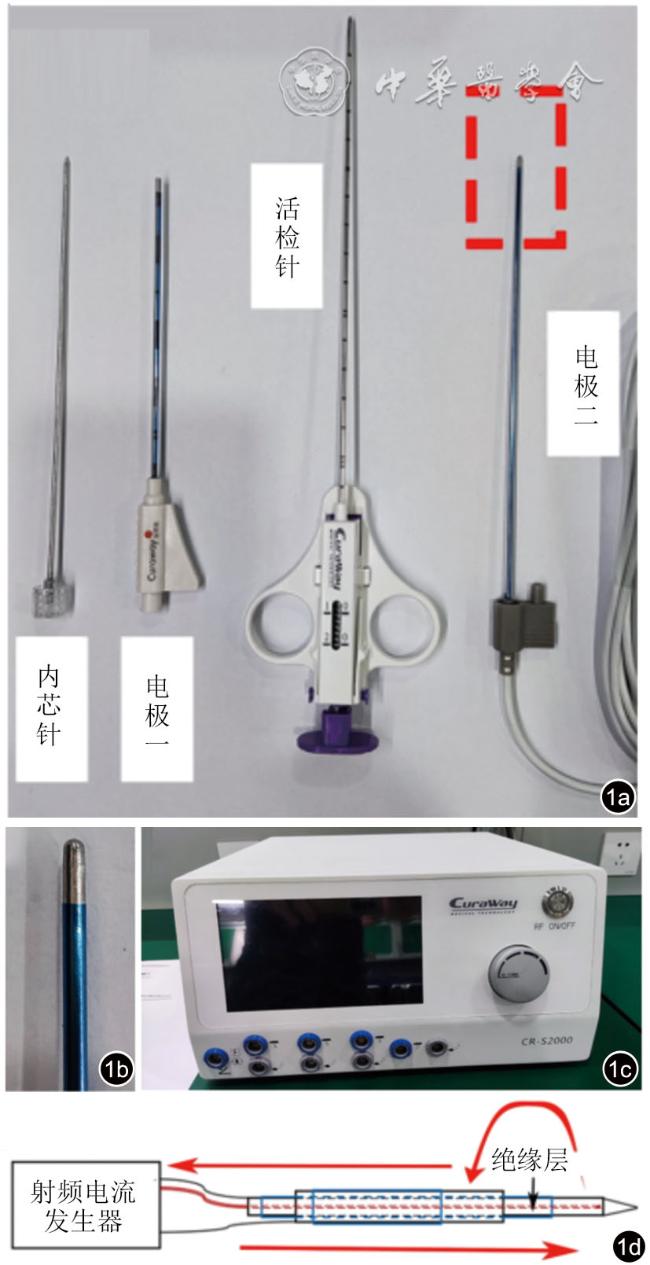
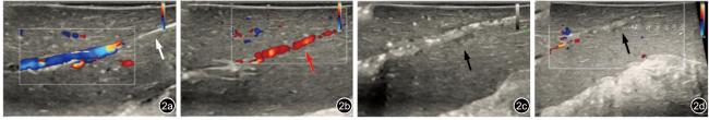
图2 超声引导下双极射频活检针止血过程。图a示避开周围大血管进行肝脏活检(白色箭头代表活检针道);图b:活检完成后,沿针道出现红色反流束,提示存在针道出血(红色箭头);图c和图d:应用双极射频活检针进行针道止血后,灰阶超声显示消融后针道出现气化(图c黑色箭头),彩色多普勒上显示针道血流信号消失(图d黑色箭头) |
图3 4种不同止血措施干预下的操作时间、出血量、出血时间比较。图a、b、c分别示4种不同止血操作的操作时间、出血量和出血时间比较结果,图d、e为4种不同止血操作后的针道出血量、出血时间与对照组比较的结果。射频组操作时间明显短于明胶海绵组,所有止血操作均能起到较好的针道止血效果,均较其邻近未止血针道的出血量、出血时间减少注:ns表示P>0.05,*表示P<0.05,**表示P<0.01,***表示P<0.001 |
表1 猪肝活检术后4种止血措施的操作时间比较结果 |
| 实验分组 | 平均值差值(s) | 标准误差(s) | P值 | 差值的95%置信区间 | |
|---|---|---|---|---|---|
| 下限 | 上限 | ||||
| 明胶海绵组 vs 射频组(40 W、70 ℃) | 160.600 | 28.384 | 0.005 | 81.793 | 239.407 |
| 明胶海绵组 vs 射频组(30 W、90 ℃) | 159.800 | 29.071 | 0.005 | 79.085 | 240.510 |
| 明胶海绵组 vs 射频组(40 W、90 ℃) | 162.000 | 29.535 | 0.005 | 79.998 | 244.002 |
表2 猪肝活检术后4种止血措施干预后的出血量与对照组比较结果 |
| 实验分组 | 平均值差值(g) | 标准误差(g) | P值 | 差值的95%置信区间 | |
|---|---|---|---|---|---|
| 下限 | 上限 | ||||
| 对照组1 vs 明胶海绵组 | 21.824 | 3.416 | 0.003 | 12.339 | 31.309 |
| 对照组2 vs 射频组(40 W、70 ℃) | 23.932 | 2.893 | 0.001 | 15.900 | 31.964 |
| 对照组3 vs 射频组(30 W、90 ℃) | 25.738 | 1.947 | <0.001 | 20.331 | 31.145 |
| 对照组4 vs 射频组(40 W、90 ℃) | 24.518 | 2.809 | 0.001 | 16.718 | 32.318 |
表3 猪肝活检术后4种止血措施干预后的出血时间与对照组比较结果 |
| 实验分组 | 平均值差值(s) | 标准误差(s) | P值 | 差值的95%置信区间 | |
|---|---|---|---|---|---|
| 下限 | 上限 | ||||
| 对照组1 vs 明胶海绵组 | 78.8 | 20.2 | 0.018 | 22.6 | 135.0 |
| 对照组2 vs 射频组(40 W、70 ℃) | 76.4 | 11.5 | 0.003 | 44.4 | 108.4 |
| 对照组3 vs 射频组(30 W、90 ℃) | 92.0 | 15.8 | 0.004 | 48.2 | 135.8 |
| 对照组4 vs 射频组(40 W、90 ℃) | 102.0 | 16.4 | 0.003 | 56.5 | 147.4 |
| 1 |
吕小斌, 罗和生. 肝穿刺活检的临床意义 [J]. 胃肠病学和肝病学杂志, 2020, 29(10): 1192-1196.
|
| 2 |
林振湖, 梁荣喜, 林文金, 等. 肝脏被膜下肿瘤超声引导下穿刺活检的禁忌证 [J]. 中国超声医学杂志, 2021, 37(5): 543-545.
|
| 3 |
钟娴, 谢晓华, 康继辉, 等. 基于超声特点及肿瘤类型的肝恶性肿瘤边缘浸润距离的研究 [J]. 中华超声影像学杂志, 2019, 28(9): 759-765.
|
| 4 |
王利英, 蒋天安, 郑树森. 超声造影引导下穿刺活检在肝占位性病变中的应用价值 [J/CD]. 中华医学超声杂志(电子版), 2018, 15(6): 458-463.
|
| 5 |
|
| 6 |
|
| 7 |
|
| 8 |
饶洁, 阎红琳, 袁静萍,等. 肝脏穿刺病理活检在诊断肝脏恶性肿瘤中的应用及其规范化管理 [J]. 诊断病理学杂志, 2020, 27(12): 909-912.
|
| 9 |
|
| 10 |
|
| 11 |
|
| 12 |
|
| 13 |
|
| 14 |
|
| 15 |
|
| 16 |
|
| 17 |
|
/
| 〈 |
|
〉 |