
2024 , Vol. 21 >Issue 04: 370 - 376
DOI: https://doi.org/10.3877/cma.j.issn.1672-6448.2024.04.004
三维反转成像技术在BI-RADS 4类乳腺肿块应用中的初步研究
Copy editor: 汪荣
收稿日期: 2023-09-19
网络出版日期: 2024-06-13
基金资助
2023年度安徽省高校自然科学研究项目(2023AH040373)
安徽医科大学第二附属医院临床研究培育计划(2021LCZD062021LCYB13)
版权
Application of three-dimensional inversion imaging in diagnosis of nature of Breast Imaging Reporting and Data System category 4 breast masses: a preliminary study
Received date: 2023-09-19
Online published: 2024-06-13
Copyright
探讨三维反转成像技术能否提高BI-RADS 4类乳腺肿块良恶性的鉴别诊断价值,以减少不必要的活检。
收集2021年9月至2023年6月在安徽医科大学第二附属医院经常规超声诊断为BI-RADS 4类并进行反转成像检查的243例患者的247个乳腺肿块。根据反转图像中肿块内深灰色和黑色区域的分布特征,采用半定量5分反转评分法,并以病理结果为金标准,绘制ROC曲线,对比常规超声与联合三维反转成像技术后对BI-RADS 4类乳腺肿块的诊断效能。
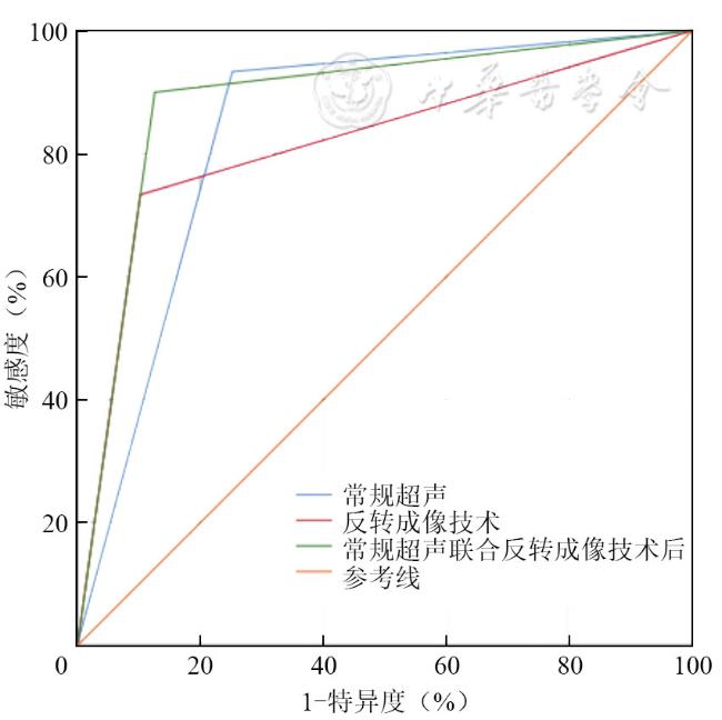
常规超声鉴别BI-RADS 4类乳腺肿块良恶性的ROC曲线下面积(AUC)、准确性、敏感度、特异度、阳性预测值、阴性预测值分别为0.841、83.8%、93.3%、74.8%、77.8%、92.2%;联合诊断后AUC、准确性、敏感度、特异度、阳性预测值、阴性预测值分别为0.887、88.7%、90.0%、87.4%、87.1%、90.2%。联合诊断后有87例BI-RADS 4a类良性肿块降至3类,同时未造成恶性肿块误降为3类而漏诊的情况,可避免84.5%(87/103)的4a类肿块进行不必要的活检。
常规超声联合三维反转成像技术对乳腺肿块良恶性具有良好的诊断效能,尤其特异度和阳性预测值均明显提升,有效减少了4a类肿块不必要的活检。
关键词: 乳腺肿块; 乳腺影像报告和数据系统 4类; 超声检查; 三维反转成像技术
侯中光 , 詹韵韵 , 毕玉 , 王佳佳 , 吴瑕璧 , 彭梅 . 三维反转成像技术在BI-RADS 4类乳腺肿块应用中的初步研究[J]. 中华医学超声杂志(电子版), 2024 , 21(04) : 370 -376 . DOI: 10.3877/cma.j.issn.1672-6448.2024.04.004
To investigate whether three-dimensional inversion imaging can improve the diagnosis of the nature of Breast Imaging Reporting and Data System (BI-RADS) category 4 breast masses to reduce unnecessary biopsies.
A total of 247 breast masses were collected from 243 patients who were diagnosed as having BI-RADS category 4 lesions by conventional ultrasound and underwent three-dimensional inversion imaging from September 2021 to June 2023 at the Second Affiliated Hospital of Anhui Medical University. Based on the distribution characteristics of dark grey and black areas within the mass in the inverted images, the diagnostic efficacy of conventional ultrasound alone and combined with three-dimensional inversion imaging for BI-RADS category 4 breast masses was compared by using a semi-quantitative 5-point inversion scoring method and plotting receiver operating characteristic (ROC) curves with pathological findings as the gold standard.
The area under the ROC curve (AUC), accuracy, sensitivity, specificity, positive predictive value, and negative predictive value of conventional ultrasound for identifying the nature of BI-RADS category 4 breast masses were 0.841, 83.8%, 93.3%, 74.8%, 77.8%, and 92.2%, respectively; the corresponding values of the combined diagnosis were 0.887, 88.7%, 90.0%, 87.4%, 87.1%, and 90.2%. The combined diagnosis resulted in 87 BI-RADS category 4a benign masses being downgraded to category 3, and did not result in malignant masses mistakenly downgraded to category 3, which would have prevented unnecessary biopsies of 84.5% (87/103) of category 4a masses.
Conventional ultrasound combined with three-dimensional inversion imaging has good diagnostic efficacy for benign and malignant breast masses, significantly improving the diagnosic specificity and positive predictive value and thus effectively reducing unnecessary biopsies of category 4a masses.
 表示。计数资料以例(%)表示。良性肿块与恶性肿块的反转评分结果比较采用秩和检验。以最终病理结果作为诊断的金标准,分别构建常规超声及联合三维反转成像技术后判定肿块良恶性的ROC曲线,采用DeLong检验比较曲线下面积(area under the curve,AUC),准确性、敏感度、特异度、阳性预测值、阴性预测值比较采用χ2检验。以P<0.05为差异具有统计学意义。
表1 BI-RADS 4类乳腺良恶性肿块的反转评分结果比较[例(%)] |
| 病理结果 | 肿块数(个) | 反转评分 | ||||
|---|---|---|---|---|---|---|
| 1分 | 2分 | 3分 | 4分 | 5分 | ||
| 良性 | 127 | 30(23.62) | 53(41.73) | 30(23.62) | 13(10.24) | 1(0.79) |
| 恶性 | 120 | 2(1.67) | 9(7.50) | 21(17.50) | 67(55.83) | 21(17.50) |
| Z值 | -10.373 | |||||
| P值 | <0.01 | |||||
表2 BI-RADS 4类乳腺肿块分类反转评分调整前后变化(个) |
| 病理结果 | 肿块数(个) | BI-RADS分类 | ||||
|---|---|---|---|---|---|---|
| 3类 | 4a类 | 4b类 | 4c类 | 5类 | ||
| 分类调整前 | ||||||
| 良性 | 127 | 0 | 95 | 28 | 4 | 0 |
| 恶性 | 120 | 0 | 8 | 37 | 75 | 0 |
| 分类调整后 | ||||||
| 良性 | 127 | 87 | 24 | 11 | 4 | 1 |
| 恶性 | 120 | 0 | 12 | 28 | 25 | 55 |
表3 常规超声及联合三维反转成像技术后对乳腺肿块良恶性的诊断效能比较 |
| 诊断方法 | AUC(95%CI) | 准确性(%) | 敏感度(%) | 特异度(%) | 阳性预测值(%) | 阴性预测值(%) |
|---|---|---|---|---|---|---|
| 常规超声 | 0.841(0.788~0.893) | 83.8 | 93.3 | 74.8 | 77.8 | 92.2 |
| 联合诊断 | 0.887(0.841~0.933) | 88.7 | 90.0 | 87.4 | 87.1 | 90.2 |
| 统计值 | Z=1.622 | χ2=2.456 | χ2=0.800 | χ2=8.000 | χ2=3.935 | χ2=0.275 |
| P值 | 0.105 | 0.117 | 0.371 | 0.005 | 0.047 | 0.600 |
注:AUC为ROC曲线下面积;CI为置信区间 |
| 1 |
国家肿瘤质控中心乳腺癌专家委员会, 中国抗癌协会乳腺癌专业委员会, 中国抗癌协会肿瘤药物临床研究专业委员会, 等. 中国晚期乳腺癌规范诊疗指南(2022版) [J].中华肿瘤杂志, 2022, 44(12): 1262-1287.
|
| 2 |
李宜臻, 郑怡, 邓玉皎, 等. 1990~2019年中国女性乳腺癌疾病负担及危险因素研究 [J]. 中国循证医学杂志, 2021, 21(8): 876-881.
|
| 3 |
|
| 4 |
|
| 5 |
|
| 6 |
张意珍, 黄品同, 洪玉蓉, 等. 超声造影与弹性成像联合评分对乳腺BI-RADS 4类病灶的应用价值[J/CD].中华医学超声杂志(电子版), 2019, 16(2): 120-125.
|
| 7 |
李如冰, 彭梅, 詹韵韵, 等. 多模态超声联合人工智能S-Detect技术校正BI-RADS分类对乳腺肿块的诊断价值 [J/OL].中华医学超声杂志(电子版), 2023, 20(1): 78-83.
|
| 8 |
|
| 9 |
|
| 10 |
|
| 11 |
|
| 12 |
田洁, 刘千琪, 王希, 等. 三维剪切波弹性成像技术在鉴别诊断乳腺良恶性结节中的应用价值 [J/CD]. 中华医学超声杂志(电子版), 2019, 16(8): 586-591.
|
| 13 |
|
| 14 |
|
| 15 |
|
| 16 |
|
| 17 |
代妮娜, 张文君. 乳腺纤维腺瘤的超声诊断及误诊分析 [J/OL].中华医学超声杂志(电子版), 2022, 19(10): 1103-1107.
|
| 18 |
杨嘉嘉, 薛恩生, 林礼务, 等. 乳腺纤维腺瘤的超声诊断及误诊分析 [J].中国医学影像技术, 2017, 33(5): 666-669.
|
| 19 |
张哲元, 袁新春, 张丽丽, 等. 超声弹性应变率比值鉴别乳腺硬化性腺病与浸润性乳腺癌的价值 [J]. 中国超声医学杂志, 2022, 38(8): 847-850.
|
| 20 |
|
| 21 |
耿琛琛, 高晓倩, 杨柳, 等. 乳腺硬化性腺病的超声表现分析 [J]. 中国超声医学杂志, 2018, 34(4): 310-313.
|
| 22 |
中国抗癌协会乳腺癌专业委员会. 中国抗癌协会乳腺癌诊治指南与规范(2021年版) [J]. 中国癌症杂志, 2021, 31(10): 954-1040.
|
| 23 |
金金, 何文, 于腾飞, 等. S-Detect联合超声造影对乳腺BI-RADS4类病灶的应用价值 [J]. 中华超声影像学杂志, 2023, 32(5): 392-398.
|
| 24 |
|
| 25 |
|
| 26 |
陈圆圆, 韦丽艳, 廖新红. 单纯型和混合型乳腺黏液腺癌的超声特征分析 [J]. 中国癌症防治杂志, 2022, 14(3): 310-314.
|
| 27 |
|
| 28 |
詹韵韵, 彭梅, 姜凡. 特殊类型乳腺癌的超声诊断与病理学基础对照分析[J].中国超声医学杂志, 2020, 36(4): 369-373.
|
/
| 〈 |
|
〉 |
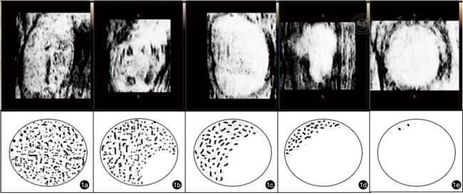
 代表深灰色区域;
 代表反转图像中肿块轮廓