
2024 , Vol. 21 >Issue 04: 391 - 398
DOI: https://doi.org/10.3877/cma.j.issn.1672-6448.2024.04.007
胎儿左心房后间隙指数在胎儿肺动脉瓣缺如综合征中的应用价值
Copy editor: 汪荣
收稿日期: 2024-02-17
网络出版日期: 2024-06-13
基金资助
国家重点研发计划项目(2023YFC2705700)
浙江大学科学技术研究院一般横向项目(校合-2021-KYY-518053-0055)
版权
Application value of post-left atrium space index in fetuses with absent pulmonary valve syndrome
Received date: 2024-02-17
Online published: 2024-06-13
Copyright
定量分析左心房后间隙(PLAS)及左心房后间隙指数(PLASI)在正常胎儿及肺动脉瓣缺如综合征(APVS)胎儿中的变化规律和差异性,探讨PLAS、PLASI在APVS胎儿中的应用价值。
回顾性分析2011年10月至2023年9月在浙江大学医学院附属邵逸夫医院诊断为APVS的胎儿20例作为APVS组,选取 2021年2月至2023年2月连续检查的261例正常胎儿作为对照组。在收缩末期四腔心切面测量PLAS和降主动脉(DA)内径,并计算两者之间的比值,即PLASI。比较对照组与APVS组胎儿的PLAS、DA内径及PLASI,绘制ROC曲线分析PLAS、PLASI诊断APVS的效能。同时,分析对照组及APVS组胎儿的PLAS、DA内径、PLASI与孕龄的关系,以及APVS组胎儿的特征性超声表现及与PLAS、PLASI的相关性。
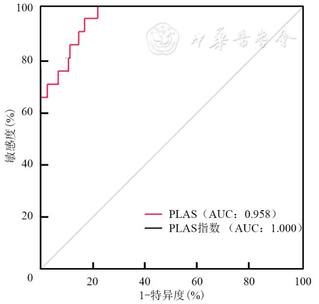
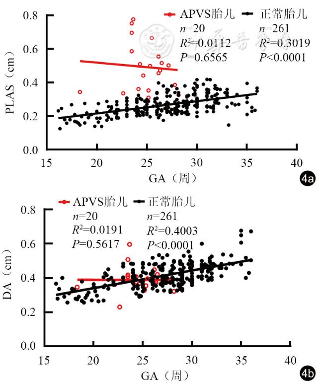
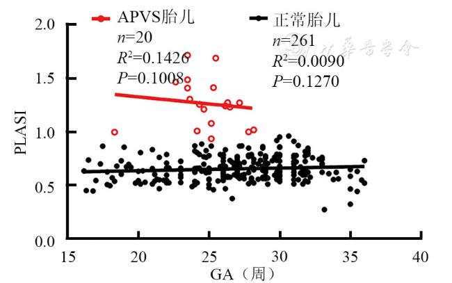
APVS组胎儿的PLAS、PLASI明显高于对照组[(4.74±1.38)mm vs(2.70±0.62)mm、1.225±0.210 vs 0.659±0.111,P均<0.0001]。ROC曲线分析显示,PLAS曲线下面积为0.958,当PLAS>3.3 mm时,PLAS鉴别APVS与正常胎儿的敏感度为90%,特异度为85%;PLASI曲线下面积为1.000,当PLASI>0.93时,PLASI鉴别APVS与正常胎儿的敏感度为100%,特异度为99.2%。在对照组中,PLAS与孕龄呈正相关(r=0.5495,P<0.0001),DA内径与孕龄呈正相关(r=0.6327,P<0.0001);在APVS组中,PLAS、DA内径均与孕龄无显著相关性(P均>0.05)。对照组和APVS组的PLASI与孕龄均无显著相关性(P=0.127、0.1008)。PLAS与肺动脉主干内径无明显相关性(P=0.2255),与心轴呈正相关(r=0.5140,P=0.0204)。PLASI与肺动脉主干内径、心轴均无明显相关性(P=0.2180、0.1299)。
APVS胎儿的PLAS失去正常规律,PLAS与PLASI明显高于正常胎儿。当PLAS>3.3 mm,PLASI介于0.93~1.0之间时,PLAS及PLASI可作为评估APVS的辅助指标。
张盼盼 , 赵博文 , 潘美 , 彭晓慧 , 陈冉 , 田园诗 , 林仙方 , 惠姗姗 , 沈婷婷 . 胎儿左心房后间隙指数在胎儿肺动脉瓣缺如综合征中的应用价值[J]. 中华医学超声杂志(电子版), 2024 , 21(04) : 391 -398 . DOI: 10.3877/cma.j.issn.1672-6448.2024.04.007
To quantitatively analyze the variations of the post-left atrium space (PLAS) and post-left atrium space index (PLASI) in normal fetuses during the mid-to-late gestational period and fetuses with absent pulmonary valve syndrome (APVS), and to explore the application value of PLAS and PLASI in APVS fetuses.
A retrospective analysis was conducted on 20 fetuses diagnosed with APVS at Sir Run Run Shaw Hospital, Zhejiang University College of Medicine from October 2011 to September 2023, as well as 261 normal fetuses consecutively examined between February 2021 and February 2023. Measurements of PLAS and the diameter of the descending aorta (DA) were obtained in the four-chamber view, and PLASI was calculated. PLAS, DA diameter, and PLASI were compared between the control group and the APVS group. Receiver operating characteristic (ROC) curve analysis was performed to assess the diagnostic efficacy of PLAS and PLASI for APVS. Additionally, the relationships between PLAS, DA diameter, PLASI, and gestational age (GA) in both the control and APVS groups were examined. Furthermore, the characteristic ultrasound features of fetuses in the APVS group and their correlations with PLAS and PLASI were analyzed.
Compared with the control group, the APVS group exhibited significantly higher PLAS and PLASI values [(4.74±1.38) mm vs (2.70±0.62) mm and 1.225±0.210 vs 0.659±0.111, respectively, P<0.0001 for both]. ROC curve analysis indicated that the area under the ROC curve of PLAS was 0.958. When PLAS >3.3 mm was used as the cutoff point, the sensitivity and specificity of PLAS in distinguishing APVS from normal fetuses were 90% and 85%, respectively. The area under the ROC curve of PLASI was 1.000, When PLASI >0.93 was used as the cutoff point, the sensitivity and specificity of PLASI in distinguishing APVS from normal fetuses were 100% and 99.2%, respectively. In the control group, PLAS was positively correlated with GA (r=0.5495, P<0.0001), as was the DA diameter (r=0.6327, P<0.0001). In the APVS group, neither PLAS nor DA diameter showed a significant correlation with GA (P>0.05 for both). PLASI was not significantly correlated with GA in either the control group or the APVS group (P=0.127 and 0.1008, respectively). PLAS showed no significant association with the diameter of the main pulmonary artery (MPA) (P=0.2255), but exhibited a positive correlation with the cardiac axis (r=0.5140, P=0.0204). PLASI was not significantly correlated with either MPA diameter or the cardiac axis (P=0.2180 and 0.1299, respectively).
In APVS fetuses, PLAS loses its normal pattern, and both PLAS and PLASI are significantly higher than those in normal fetuses. Considering both sensitivity and specificity, when PLAS>3.3 mm and 1.0>PLASI >0.93 are used as the cutoff points, PLAS and PLASI can serve as effective assistant indicators for predicting APVS.
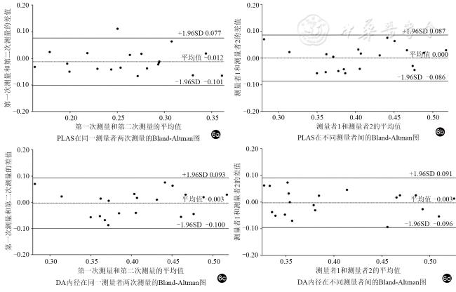
 表示。应用Bland-Altman绘图判断观察者内部和观察者之间的一致性。PLAS、DA内径及PLASI与GA之间的相关性采用Pearson相关分析。应用ROC曲线分析PLASI诊断胎儿APVS的敏感度和特异度。以P<0.05为差异有统计学意义。
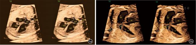
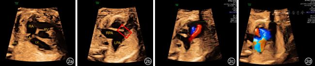
图2 肺动脉瓣缺如综合征胎儿四腔心切面及右心室流出道-肺动脉分支切面超声图像。图a为四腔心切面显示左右心比例不对称,右心明显增大,心轴左偏;图b红框标记为肺动脉瓣环处,未见明显瓣叶结构回声。肺动脉主干及左右分支显著扩张,呈“鲸鱼尾”征;图c,d为彩色多普勒图像,显示肺动脉瓣环处往返于右心室与肺动脉之间的高速“穿梭”样血流信号,其中图c为舒张期,可见由明显扩张的肺动脉主干及分支反流进入右心室的红色血流信号;图d为舒张期,可见右心血流经过肺动脉瓣环冲击肺动脉及其分支的蓝色血流信号注:LA为左心房;LV为左心室;RA为右心房;RV为右心室;LPA为左肺动脉;RPA为右肺动脉;DA为降主动脉 |
表1 对照组中不同孕龄胎儿的PLAS、DA内径测量结果( |
| 孕龄(周) | 例数 | PLAS(mm) | DA内径(mm) |
|---|---|---|---|
| 16~20+6 | 26 | 1.92±0.42 | 3.10±0.40 |
| 21~25+6 | 66 | 2.44±0.37 | 3.90±0.53 |
| 26~30+6 | 114 | 2.76±0.53 | 4.13±0.56 |
| 31~37+1 | 55 | 3.24±0.58 | 4.81±0.75 |
注:PLAS为左心房后间隙;DA为降主动脉 |
表2 对照组与APVS组胎儿的PLAS、DA内径及PLASI比较( |
| 组别 | 例数 | PLAS(mm) | DA内径(mm) | PLASI |
|---|---|---|---|---|
| 对照组 | 261 | 2.70±0.62 | 4.11±0.73 | 0.659±0.111 |
| APVS组 | 20 | 4.74±1.38 | 3.84±0.79 | 1.225±0.210 |
| t值 | -6.56 | 1.48 | -11.93 | |
| P值 | P<0.0001 | P>0.05 | P<0.0001 |
注:APVS为肺动脉瓣缺如综合征;PLAS为左心房后间隙;DA为降主动脉;PLASI为左心房后间隙指数 |
表3 对照组与APVS组胎儿的MPA内径、心轴角度及RA/LA横径比值比较( |
| 组别 | 例数 | MPA内径(mm) | 心轴角度(°) | RAD/LAD |
|---|---|---|---|---|
| 对照组 | 261 | 5.27±0.99 | 47.42±6.12 | 1.11±0.08 |
| APVS组 | 20 | 9.76±1.04 | 74.62±9.04 | 1.17±0.13 |
| t值 | -18.66 | −13.24 | −0.64 | |
| P值 | P<0.0001 | P<0.0001 | P>0.05 |
注:APVS为肺动脉瓣缺如综合征;MPA为肺动脉主干;RAD为右心房横径;LAD为左心房横径 |
| 1 |
|
| 2 |
|
| 3 |
陈建萍, 陈阳, 孙海丽, 等. 三维超声“外科医生观”在胎儿法洛四联症伴肺动脉瓣缺如综合征诊断中的应用1例 [J]. 中华超声影像学杂志, 2022, 7(31): 634-636.
|
| 4 |
何冠南, 罗红, 杨家翔, 等. 胎儿肺动脉瓣缺如综合征产前超声诊断分析[J/CD].中华医学超声杂志(电子版), 2014, 11(10): 3.
|
| 5 |
|
| 6 |
|
| 7 |
|
| 8 |
赵雷生, 张颖, 王彧, 等. 胎儿肺动脉瓣缺如综合征的研究进展 [J]. 中国医学影像学杂志, 2018, 26(12): 957-960.
|
| 9 |
|
| 10 |
|
| 11 |
|
| 12 |
|
| 13 |
|
| 14 |
|
| 15 |
|
| 16 |
|
| 17 |
|
| 18 |
郑凤华, 赵博文, 王蓓, 等. 胎儿四腔心观左心房后壁与降主动脉之间距离的定量研究 [J]. 中华超声影像学杂志, 2016, 25(3): 203-206.
|
| 19 |
张金双, 赵博文, 谭乳燕, 等. 先天性心脏病胎儿心轴变化的定量研究[J/CD].中华医学超声杂志(电子版), 2018, 15(12): 919-924.
|
| 20 |
赵胜, 赵博文, 吴青青, 等. 早孕期胎儿心脏超声专家观点[J/OL].中华医学超声杂志(电子版), 2024, 21(1): 1-9.
|
/
| 〈 |
|
〉 |
 )
) )
)
 )
)