
2024 , Vol. 21 >Issue 07: 657 - 663
DOI: https://doi.org/10.3877/cma.j.issn.1672-6448.2024.07.003
中国经胸超声心动图检查存图及报告质控现状分析
Copy editor: 吴春凤
收稿日期: 2024-05-14
网络出版日期: 2024-07-09
版权
Current status of quality control for image archiving and reporting of transthoracic echocardiography in China
Received date: 2024-05-14
Online published: 2024-07-09
Copyright
通过调查经胸超声心动图检查存图标准及报告模板,旨在获取中国各地区各级医疗机构经胸超声心动图存图及报告质量控制的真实现状,为质量控制工作提供指引。
2023年10月,心脏亚专业超声质量控制工作组在全国范围内开展经胸超声心动图检查存图及报告质量控制问卷调查,分析全国常规经胸超声心动图不同超声模式检查现况。
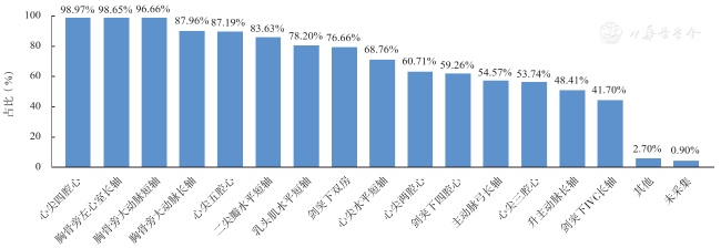
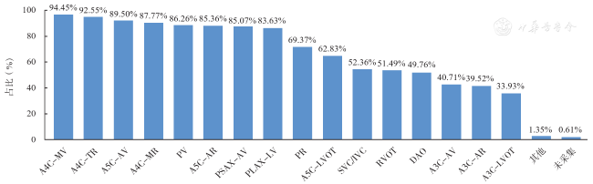
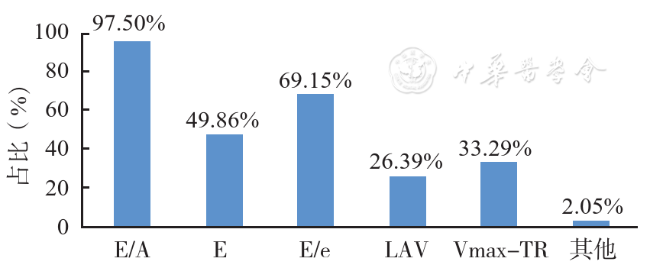
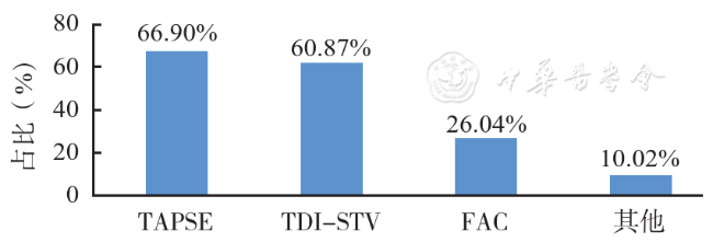
调研发现常规经胸超声心动图检查存图率超过80%的二维切面包括心尖四腔心切面(98.97%)、胸骨旁左心室长轴切面(98.65%)、胸骨旁大动脉短轴切面(96.66%)、胸骨旁肺动脉长轴切面(87.96%)、心尖五腔心切面(87.19%)、胸骨旁二尖瓣水平短轴切面(83.63%)。彩色多普勒超声包括心尖四腔心切面二尖瓣前向血流及频谱(94.45%)、心尖四腔心切面三尖瓣反流及反流频谱(92.55%)、心尖五腔心切面主动脉瓣前向血流及频谱(89.50%)、心尖四腔心切面二尖瓣反流及反流频谱(87.77%)、胸骨旁肺动脉长轴切面肺动脉瓣前向血流及频谱(86.26%)、心尖五腔心切面主动脉瓣反流及反流频谱(85.36%)、胸骨旁大动脉短轴切面(85.07%)、胸骨旁左心室长轴切面(83.63%),可基本满足显示心脏腔室结构及各瓣膜血流动力学评估的要求。组织多普勒存图率二尖瓣环间隔壁、二尖瓣环侧壁、三尖瓣环右心室游离壁分别为84.40%、64.75%、25.91%。反映出部分超声医师对左心室舒张功能以及右心室收缩功能的评估未引起足够重视。对经胸超声心动图检查存图及报告最多的建议是缺乏不同心脏疾病的经胸超声心动图存图及报告标准,且需规范化培训。
大规模调研为心脏质量控制工作的开展提供了重要依据,心脏亚专业超声质量控制组将从硬件设备、人才培训、心脏单病种质控及结构化报告模板等多方面同步开展工作。
王益佳 , 周青 , 曹省 , 袁芳洁 , 周妍 , 张梅 . 中国经胸超声心动图检查存图及报告质控现状分析[J]. 中华医学超声杂志(电子版), 2024 , 21(07) : 657 -663 . DOI: 10.3877/cma.j.issn.1672-6448.2024.07.003
To obtain the true status of quality control for transthoracic echocardiography image archiving and reports in medical institutions of all levels in various regions of China, in order to provide guidance for quality control work.
The "Cardiac Ultrasound Quality Control Working Group" conducted a nationwide survey questionnaire on the quality control of transthoracic echocardiography image archiving and reports.
The two-dimensional views with an image archiving rate of over 80% in conventional transthoracic echocardiography included apical four chamber view (A4C; 98.97%), parasternal long-axis view of the left ventricle (PLAX-LV; 98.65%), parasternal short-axis view of the aortic valve (PSAX-AV; 96.66%), parasternal long-axis view of the pulmonary artery (PLAX-PA; 87.96%), apical five chamber view (A5C; 87.19%), and parasternal short-axis view of the mitral valve (PSAX-MV; 83.63%). The Doppler sections with an image archiving rate of over 80% included the flow and spectrum of A4C-MV (94.45%), A4C-TR (92.55%), A5C-AV (89.50%), A4C-MR (87.77%), PV (86.26%), A5C-AR (85.36%), PSAX-AV (85.07%), and PLAX-LV (83.63%), which can meet the basic requirements for displaying the structure of heart cavities and evaluating the hemodynamics of each valve. The image archiving rates of tissue Doppler imaging for the septal wall of the mitral annulus, the lateral wall of the mitral annulus, and the right ventricular free wall of the tricuspid annulus were 84.40%, 64.75%, and 25.91%, respectively, reflecting that some ultrasound physicians did not pay enough attention to the evaluation of left and right ventricular systolic function. The most common suggestions for image archiving and reporting of transthoracic echocardiography were the lack of standards for image archiving and reporting of transthoracic echocardiography for different cardiovascular diseases, as well as the need for standardized training.
This large scale research provides an important basis for the implementation of quality control work. The "Cardiac Ultrasound Quality Control Working Group" will carry out work from multiple aspects such as hardware equipment development, clinical training, cardiac single disease ultrasound quality control, and structured report templates simultaneously.
图3 常规经胸超声心动图检查彩色多普勒切面存图率注:A4C-MV为心尖四腔心切面二尖瓣前向血流及频谱;A4C-TR为心尖四腔心切面三尖瓣反流及反流频谱;A5C-AV为心尖五腔心切面主动脉瓣前向血流及频谱;A4C-MR为心尖四腔心切面二尖瓣反流及反流频谱;PV为胸骨旁肺动脉长轴切面肺动脉瓣前向血流及频谱;A5C-AR为心尖五腔心切面主动脉瓣反流及反流频谱;PSAX-AV为胸骨旁大动脉短轴切面;PLAX-LV为胸骨旁左心室长轴切面;PR为胸骨旁肺动脉长轴切面肺动脉瓣反流及反流频谱;A5C-LVOT为心尖五腔心切面左心室流出道血流及频谱;SVC/IVC为剑突下双房切面上下腔静脉血流及频谱;RVOT为胸骨旁肺动脉长轴切面右心室流出道血流及频谱;DAO为胸骨上窝主动脉弓长轴切面降主动脉血流及频谱;A3C-AV为心尖三腔心切面主动脉瓣前向血流及频谱;A3C-AR为心尖三腔心切面主动脉瓣反流及反流频谱;A3C-LVOT为心尖三腔心切面左心室流出道血流及频谱 |
| 1 |
Guidelines for performing a comprehensive transthoracic echocardiographic examination in adults: recommendations from the American Society of Echocardiography [J]. J Am Soc Echocardiogr, 2019, 32(1): 1-64.
|
| 2 |
中华医学会超声医学分会超声心动图学组. 中国成年人超声心动图检查测量指南 [J].中华超声影像学杂志, 2016, 25(8): 645-666.
|
| 3 |
中华医学会超声医学分会超声心动图学组, 国家超声诊断专业医疗质量控制中心专家委员会. 经胸超声心动图检查规范化应用中国专家共识(2024版) [J]. 中华超声影像学杂志, 2024, 33(1): 1-13.
|
| 4 |
|
| 5 |
|
/
| 〈 |
|
〉 |


