
2024 , Vol. 21 >Issue 09: 843 - 851
DOI: https://doi.org/10.3877/cma.j.issn.1672-6448.2024.09.004
应用二维斑点追踪成像技术评估孕周及心尖方向对中晚孕期正常胎儿左心房应变的影响
Copy editor: 吴春凤
收稿日期: 2023-12-12
网络出版日期: 2024-10-16
版权
Impact of gestational age and apex orientation on left atrial strain assessed by two-dimensional speckle tracking imaging in normal fetuses during the second and third trimesters
Received date: 2023-12-12
Online published: 2024-10-16
Copyright
目的
探讨二维斑点追踪成像技术(2D-STI)在评估胎儿左心房应变3 个时相功能中的应用价值、左心房应变3 个时相功能与孕周的相关性以及该技术在胎儿心脏检查实际应用中对心尖方向是否存在角度依赖性。
方法
选取2021 年11 月至2022 年1 月在浙江大学医学院附属邵逸夫医院超声科接受胎儿超声心动图检查的正常妊娠中、晚孕期单胎胎儿180 例,共获取611 个不同心尖角度胎儿四腔心动态图像,应用2D-STI 分析获得左心房储存期峰值应变(LASr_ED)、管道期峰值应变(LAScd_ED)和收缩期峰值应变(LASct_ED),其中有11 例共计32 个动态图像2D-STI 分析心房收缩的起始点不能识别,无法区分心房收缩期与管道期。选取169 例四腔心切面心尖方向为10:30~1:30 能良好追踪并识别左心房3 个时相功能的病例与孕周进行线性回归分析。选取能良好追踪并识别左心房3 个时相的动态图像579 个,其中中孕期18~24 周的图像126 个、25~28 周的图像190个,晚孕期29~32 周的图像150 个、33~36 孕周的图像113 个;按不同心尖方向分成4 组,组1 心尖方向10:30~1:30,组2 心尖方向1:30~4:30,组3 心尖方向4:30~7:30,组4 心尖方向7:30~10:30;同一孕期不同心尖方向组间应变参数比较采用F 检验和LSD-t 检验。
结果
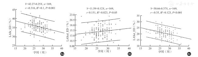
2D-STI 能够成功追踪并识别胎儿左心房3 个时相应变的比例为94.76%(579/611)。线性回归分析显示,LASr_ED 随孕周增加而减小(r =-0.32,P<0.001),LAScd_ED 随孕周增加而增大(r =0.15,P<0.05),LASct_ED 随孕周增加而减小(r =-0.35,P<0.001)。中孕期18~24 周、25~28 周不同心尖方向4 组左心房3 个应变参数比较,差异均无统计学意义(P 均>0.05);晚孕期29~32 周不同心尖方向4 组胎儿的LASr_ED 总体均数比较,差异存在统计学意义(F=2.759,P=0.044);晚孕期33~36 周不同心尖方向4 组胎儿的LASr_ED 及LAScd_ED 总体均数比较,差异存在统计学意义(F=2.919,P=0.037;F=4.234,P=0.007)。4 组间两两比较,同一孕期组1 和组3 的LASr_ED、LAScd_ED 及LASct_ED 比较,差异均无统计学意义(P 均>0.05),同一孕期组2 和组4 的LASr_ED、LAScd_ED 及LASct_ED 比较,差异均无统计学意义(P 均>0.05)。
结论
中晚孕期正常胎儿左心房应变参数LASr_ED、LASct_ED 与孕周呈弱负相关,LAScd_ED 与孕周呈极弱正相关。2D-STI 在正常妊娠中孕期胎儿心脏左心房应变定量分析中无心尖角度依赖性,在正常妊娠晚孕期胎儿心脏左心房应变定量分析中LASr_ED 及LAScd_ED 存在一定的心尖角度依赖性。选择轴向或横向四腔心切面作为统一图像采集标准,能更加准确地获得胎儿左心房应变参数,为胎儿心脏功能的定量评估和连续性监测提供依据。
杜祖升 , 赵博文 , 张帧 , 潘美 , 彭晓慧 , 陈冉 , 毛彦恺 . 应用二维斑点追踪成像技术评估孕周及心尖方向对中晚孕期正常胎儿左心房应变的影响[J]. 中华医学超声杂志(电子版), 2024 , 21(09) : 843 -851 . DOI: 10.3877/cma.j.issn.1672-6448.2024.09.004
Objective
To assess the application value of two-dimensional speckle tracking imaging(2D-STI) in the evaluation of fetal left atrial strain in three phases, and to explore the correlation between the three phases of left atrial strain and gestational age, and whether the technique has angular dependence on the apex orientation in fetal heart examination.
Methods
A total of 180 normal singleton fetuses in the second and third trimesters who underwent fetal echocardiography at the Department of Diagnostic Ultrasound & Echocardiography,Sir Run Run Shaw Hospital, Zhejiang University College of Medicine from November 2021 to January 2022 were selected. A total of 611 fetal four-chamber view video clips with different apex angles were obtained. 2D-STI analysis was used to obtain the peak storage strain (LASr_ED), peak pipeline strain (LAScd_ED), and peak systolic strain (LASct_ED) of the left atrium. 2D-STI could not identify the onset of atrial contraction in 11 fetuses (32 dynamic images), and could not distinguish the atrial contraction phase from the conduit phase. Linear regression analysis was used to analyze the relationship between gestational age and 2D-STI parameters in patients in whom the three phases of left atrial strain with the apex orientation of 10:30 to 1:30 in the four-chamber view could be well tracked and identified. A total of 579 dynamic images with good tracking and recognition of the three phases of left atrial strain were selected, including 126 during 18-24 gestational weeks,190 during 25-28, 150 during 29-32, and 113 during 33-36. The patients were divided into four groups according to the apex orientation: group 1(apex orientation: 10:30-1:30), group 2 (1:30-4:30), group 3 (4:30-7:30), and group 4 (7:30-10:30). The F test and LSD-t test were used to compare the left atrial strain parameters between different groups of normal fetuses in the same gestational weeks.
Results
2D-STI could successfully track and identify the three temporal variations of the left atrium in 94.76% (579/611) of fetuses. LASr_ED and LASct_ED decreased with the increase of gestational age(r=-0.32, P<0.001; r=-0.35, P<0.001), while LAScd_ED increased with the increase of gestational age (r=0.15,P<0.05). There was no statistical difference in the three strain parameters of the left atrium in different apex orientations among the four groups at 18-24 gestational weeks and 25-28 gestational weeks (all P>0.05). There were significant differences in LASr_ED among the four groups at 29-32 gestational weeks (F=2.759, P=0.044).There were significant differences in LASr_ED and LAScd_ED among the four groups at 33-36 gestational weeks(F=2.919, P=0.037; F=4.234, P=0.007). Pairwise comparisons were then made among the four groups. There were no significant differences in LASr_ED, LAScd_ED, and LASct_ED between group 1 and group 3 during the same gestational weeks (all P>0.05), and in LASr_ED, LAScd_ED, and LASct_ED between group 2 and group 4 during the same gestational weeks (all P>0.05).
Conclusion
LASr_ED and LASct_ED of normal fetuses in the second and third trimesters are weakly negatively correlated with gestational age. There is a very weakly positive correlation between LAScd_ED and gestational age in normal fetuses in the second and third trimesters. 2D-STI has no apex angle dependence in the practical application of fetal left atrial strain in the second trimester, but has certain apical angle dependence in the third trimester. Choosing axial or transverse four-chamber heart view as the unified image acquisition standard can help obtain fetal left atrial strain parameters more accurately, and provides a basis for quantitative assessment and continuous monitoring of fetal cardiac function.
Key words: Echocardiography; Fetus; Atrial function; Two-dimensional speckle tracking
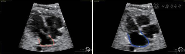
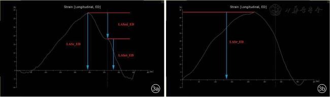
图2 胎儿左心房二维斑点追踪成像图像。感兴趣区从房室环的外侧经过心房顶一直追踪到房室环的间隔侧,心室终末舒张作为零应变。图2a:心脏机械活动二尖瓣关闭前一帧,即左心房收缩末期;图2b:下一个二尖瓣关闭前一帧,即左心房舒张末期 |
 ±s 表示。用简单线性回归方法评估左心房应变3 个时相功能与孕周的关系,服从正态分布用Pearson 相关系数评价,不服从正态分布用Spearman 秩相关系数评价;P<0.05为差异有统计学意义,说明自变量与应变量回归关系成立,以r 值表示,r 值为0.80~1.00 表示极强相关,0.60~0.79 表示强相关,0.40~0.59 表示中等程度相关,0.20~0.39 表示弱相关,0.0~0.19 表示极弱相关或无相关。同一孕期不同心尖方向组间应变参数比较采用F 检验,组间两两比较采用LSD-t 检验。采用ICC 评估变量在观察者间和观察者内的变异性,ICC<0.50 表示信度较差,>0.75 表示信度好。以P<0.05 为差异有统计学意义。
表1 中孕期18~24 周胎儿不同心尖方向组间左心房应变参数比较(%, |
| 组别 | 图像数 | LASr_ED | LAScd_ED | LASct_ED |
|---|---|---|---|---|
| 组1 | 46 | 37.14±4.39 | 15.14±4.24 | 21.99±4.24 |
| 组2 | 20 | 36.73±2.59 | 14.80±3.13 | 21.93±3.62 |
| 组3 | 38 | 35.89±3.04 | 14.15±2.83 | 21.76±3.13 |
| 组4 | 22 | 36.03±3.11 | 14.26±2.60 | 21.76±3.13 |
| F值 | 1.022 | 0.700 | 0.034 | |
| P值 | 0.385 | 0.554 | 0.992 |
表2 中孕期25~28 周胎儿不同心尖方向组间左心房应变参数比较(%, |
| 组别 | 图像数 | LASr_ED | LAScd_ED | LASct_ED |
|---|---|---|---|---|
| 组1 | 88 | 35.81±3.35 | 14.84±3.57 | 20.96±4.74 |
| 组2 | 34 | 34.62±2.79 | 13.95±3.49 | 20.67±3.88 |
| 组3 | 33 | 36.17±2.63 | 15.30±2.92 | 20.83±3.96 |
| 组4 | 35 | 35.56±3.50 | 13.81±3.39 | 21.73±4.35 |
| F值 | 1.565 | 1.645 | 0.405 | |
| P值 | 0.199 | 0.181 | 0.750 |
表3 晚孕期29~32 周胎儿不同心尖方向组间左心房应变参数比较(%, |
| 组别 | 图像数 | LASr_ED | LAScd_ED | LASct_ED |
|---|---|---|---|---|
| 组1 | 56 | 36.74±3.60 | 15.73±2.97 | 20.99±4.12 |
| 组2 | 42 | 34.79±3.58a | 14.97±3.40 | 20.05±5.01 |
| 组3 | 31 | 35.41±3.16 | 15.04±3.38 | 20.36±4.05 |
| 组4 | 21 | 35.50±3.10 | 16.72±4.24 | 18.78±4.54 |
| F值 | 2.759 | 1.540 | 1.333 | |
| P值 | 0.044 | 0.207 | 0.266 |
表4 晚孕期33~36 周胎儿不同心尖方向左心房应变参数比较(%, |
| 组别 | 图像数 | LASr_ED | LAScd_ED | LASct_ED |
|---|---|---|---|---|
| 组1 | 51 | 34.50±3.30 | 16.07±3.81 | 18.43±4.29 |
| 组2 | 24 | 33.22±3.10 | 13.25±3.51a | 19.97±3.93 |
| 组3 | 18 | 34.92±2.63 | 16.54±3.05b | 18.38±4.00 |
| 组4 | 20 | 32.59±2.73ac | 14.73±3.60 | 17.86±5.01 |
| F值 | 2.919 | 4.234 | 1.042 | |
| P值 | 0.037 | 0.007 | 0.377 |
| 1 |
张清凤, 王胰, 李文华, 等. 三维斑点追踪成像技术对高血压患者左心房功能的评价 [J/OL]. 中华医学超声杂志(电子版), 2022,19(2): 150-155.
|
| 2 |
Amundsen BH, Helle-Valle T, Edvardsen T, et al. Noninvasive myocardial strain measurement by speckle tracking echocardiography:validation against sonomicrometry and tagged magnetic resonance imaging [J]. J Am Coll Cardiol, 2006, 47(4): 789-793.
|
| 3 |
Miranda JO, Hunter L, Tibby S, et al. Myocardial deformation in fetuses with coarctation of the aorta: a case-control study [J].Ultrasound Obstet Gynecol, 2017, 49(5): 623-629.
|
| 4 |
Dahlbäck C, Gudmundsson S. Investigations on atrial function in fetuses with signs of impaired placental function [J]. Prenat Diagn,2015, 35(6): 605-611.
|
| 5 |
Patey O, Carvalho JS, Thilaganathan B. Perinatal changes in cardiac geometry and function in growth-restricted fetuses at term [J].Ultrasound Obstet Gynecol, 2019, 53(5): 655-662.
|
| 6 |
Van Mieghem T, Giusca S, DeKoninck P, et al. Prospective assessment of fetal cardiac function with speckle tracking in healthy fetuses and recipient fetuses of twin-to-twin transfusion syndrome [J]. J Am Soc Echocardiogr, 2010, 23(3): 301-308.
|
| 7 |
Patey O, Carvalho JS, Thilaganathan B. Perinatal changes in fetal cardiac geometry and function in iabetic pregnancy at term [J].Ultrasound Obstet Gynecol, 2019, 54(5): 634-642.
|
| 8 |
Cohen J, Binka E, Woldu K, et al. Myocardial strain abnormalities in fetuses with pulmonary atresia and intact ventricular septum [J].Ultrasound Obstet Gynecol, 2019, 53(4): 512-519.
|
| 9 |
Rato J, Vigneswaran TV, Simpson JM. Speckle-tracking echocardiography for the assessment of atrial function during fetal life[J]. J Am Soc Echocardiogr, 2020, 33(11): 1391-1399.
|
| 10 |
Thomas L, Marwick TH, Popescu BA, et al. Left atrial structure and function, and left ventricular diastolic dysfunction: JACC state-of-theart review [J]. J Am Coll Cardiol, 2019, 73(15): 1961-1977.
|
| 11 |
付晓燕, 闫瑞玲, 左思阳, 等. 二维斑点追踪技术评价左房功能应用进展 [J]. 心脏杂志, 2019, 31(3): 347-351.
|
| 12 |
叶晓光, 李一丹, 吕秀章, 等. 二维斑点追踪成像技术评价正常妊娠胎儿左心房时相功能的临床研究 [J]. 中华超声影像学杂志,2021, 30(6): 489-493.
|
| 13 |
Haeger C, Hammer K, Braun J, et al. Importance of frame rate for the measurement of strain and synchrony in fetuses using speckle tracking echocardiography [J]. J Perinat Med, 2021, 50(2): 176-184.
|
| 14 |
Eckersley L, Hornberger LK. Cardiac function and dysfunction in the fetus [J]. Echocardiography, 2017, 34(12): 1776-1787.
|
| 15 |
Badano LP, Kolias TJ, Muraru D, et al. Standardization of left atrial,right ventricular, and right atrial deformation imaging using twodimensional speckle tracking echocardiography: a consensus document of the EACVI/ASE/Industry Task Force to standardize deformation imaging [J]. Eur Heart J Cardiovasc Imaging, 2018, 19(6): 591-600.
|
| 16 |
Meister M, Axt-Fliedner R, Graupner O, et al. Atrial and ventricular deformation analysis in normal fetal hearts using two-dimensional speckle tracking echocardiography [J]. Fetal Diagn Ther, 2020, 47(9):699-710.
|
| 17 |
阿尔弗莱德·阿布汗默德, 拉宾·查欧里. 胎儿超声心动图实用指南:正常和异常心脏 [M]. 刘琳, 主译. 3 版. 北京: 北京科学技术出版社, 2017: 7.
|
| 18 |
Chang CH, Chang FM, Yu CH, et al. Systemic assessment of fetal hemodynamics by Doppler ultrasound [J]. Ultrasound Med Biol, 2000,26(5): 777-785.
|
| 19 |
Forsha D, Risum N, Rajagopal S, et al. The influence of angle of insonation and target depth on speckle-tracking strain [J]. J Am Soc Echocardiogr, 2015, 28(5): 580-586.
|
| 20 |
Semmler J, Day TG, Georgiopoulos G, et al. Fetal speckle-tracking:impact of angle of insonation and frame rate on global longitudinal strain [J]. J Am Soc Echocardiogr, 2020, 33(9): 1141-1146.
|
| 21 |
Sivesgaard K, Christensen SD, Nygaard H, et al. Speckle tracking ultrasound is independent of insonation angle and gain: an in vitro investigation of agreement with sonomicrometry [J]. J Am Soc Echocardiogr, 2009, 22(7): 852-858.
|
| 22 |
Steinhard J, Entenmann A, Valk EV, et al. The transverse four-chamber view for the assessment of atrial tissue deformation in the fetus [J].PLoS One, 2018, 13(7): e0199581.
|
| 23 |
Nichting TJ, Vet CM, Ven MV, et al. Angle independency of fetal speckle-tracking echocardiography: a commentary letter [J]. J Am Soc Echocardiogr, 2022, 35(7): 783-785.
|
/
| 〈 |
|
〉 |