
2024 , Vol. 21 >Issue 09: 872 - 876
DOI: https://doi.org/10.3877/cma.j.issn.1672-6448.2024.09.009
超声在多重肺部感染诊断中的临床应用价值
Copy editor: 吴春凤
收稿日期: 2024-03-03
网络出版日期: 2024-10-16
版权
Clinical value of ultrasound in diagnosis of multiple microbial pulmonary infections
Received date: 2024-03-03
Online published: 2024-10-16
Copyright
目的
探讨多重肺部感染(MMPI)肺病灶超声影像特征及超声引导经皮介入穿刺肺活检(UGPLB)的临床应用价值。
方法
选取2012 年8 月至2024 年2 月南宁市第四人民医院临床初诊疑为MMPI 的患者262 例,经胸部CT、超声检查后对周围型肺病灶成功实施UGPLB,获取肺病变标本作病理组织学诊断/病原体检测,依据确诊的感染病原体种类将入选患者分为MMPI 组与单一病原体肺部感染(SPPI)组;采用χ2 检验比较MMPI 组与SPPI 组患者超声表现的差异,对相关临床资料进行综合分析。
结果
所有肺病灶均成功实施UGPLB 获取肺病变标本行病理组织学诊断及病原体检测,262 例患者中,最终确诊为MMPI 者210 例,其中感染病原体组合主要为结核分枝杆菌+马尔尼菲篮状菌(59 例),其次是结核分枝杆菌+马红球菌(44 例)、结核分枝杆菌+隐球菌(37例)及结核分枝杆菌+肺炎克雷伯杆菌(36 例);确诊为SPPI 者52 例,感染病原体主要为结核分枝杆菌(22 例),其次是马尔尼菲篮状菌(14 例)。MMPI 组超声测量肺病灶三径均值≥50 mm 的患者比例大于SPPI 组[79.52%(167/210) vs 34.62%(18/52)],MMPI 组肺病灶内部呈实液混合回声的患者比例大于SPPI 组[62.86%(132/210) vs 21.15%(11/52)],差异均具有统计学意义(χ2=40.506、29.240,P 均<0.001);2 组肺病灶的边界、内部血流信号及合并胸腔积液方面比较,差异均无统计学意义(P均>0.05)。
结论
MMPI 患者胸部超声特征为肺病灶一般体积较大,内部大多呈实液混合回声,但无明显特异性;对临床高度疑诊MMPI 的患者,UGPLB 可以帮助临床精准诊断和鉴别诊断。
农云洁 , 黄小桂 , 黄裕兰 , 农恒荣 . 超声在多重肺部感染诊断中的临床应用价值[J]. 中华医学超声杂志(电子版), 2024 , 21(09) : 872 -876 . DOI: 10.3877/cma.j.issn.1672-6448.2024.09.009
Objective
To explore the ultrasonic imaging characteristics of pulmonary lesions in multiple microbial pulmonary infections (MMPI) and evaluate the clinical value of ultrasound-guided percutaneous lung biopsy (UGPLB) in these conditions.
Methods
Two hundred and sixty-two patients with suspected MMPI from August 2012 to February 2024 at the Fourth People's Hospital of Nanning were selected. After chest CT and ultrasound examinations, UGPLB was successfully implemented for peripheral lung lesions, and the obtained lung lesion samples were subjected to histopathological examination and pathogen detection. The patients were divided into either an MMPI group or a single pathogen pulmonary infection (SPPI) group according to the confirmed pathogen(s). The ultrasonic manifestations were compared between the MMPI group and SPPI group using the χ2 test, and the relevant clinical data were comprehensively analyzed.
Results
All lung lesions were successfully obtained by UGPLB for pathological diagnosis and pathogen detection. Among the 262 patients, 210 were finally diagnosed with MMPI, of which the pathogen combination was mainly Mycobacterium tuberculosis + Marneffei basket (59 cases), followed by Mycobacterium tuberculosis + Rhodococcus equi (44 cases), Mycobacterium tuberculosis + Cryptococcus(37 cases), and Mycobacterium tuberculosis + Klebsiella pneumoniae (36 cases); 52 patients were diagnosed with SPPI, and the main pathogen was Mycobacterium tuberculosis (22 cases), followed by Marneffei basket(14 cases). One hundred and sixty-seven cases (79.52%) in the MMPI group and 18 cases (34.62%) in the SPPI group has a mean lung lesion size of ≥50 mm as measured by ultrasound, and there was a significant difference between the two groups (χ2=40.506, P<0.001). There were 132 cases (62.86%) in the MMPI group and 11 cases (21.15%) in the SPPI group with mixed solid and liquid echo in the lung lesions, and there was a significant difference between the two groups (χ2=29.240, P<0.001). There was no significant difference in the boundary of lung lesions, internal blood flow signal, or complication with pleural effusion between the two groups (P>0.05).
Conclusion
The characteristics of chest ultrasound in patients with MMPI are that the volume of lung lesions is large, and most of them showed mixed echo of solid and liquid, but there was no obvious specificity. For patients with clincially highly suspected MMPI, UGPLB is effectively helpful for the precise and differential diagnosis of MMPI.
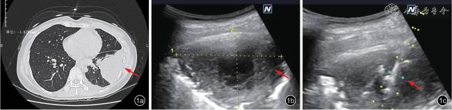
图1 艾滋病合并多重肺部感染患者,女性,36 岁,发热、咳嗽伴胸痛15 d。图a:左下肺病灶(箭头所示)CT 图;图b:肺病灶超声呈片状实液混合回声(箭头所示);图c:对肺病灶实施超声引导经皮介入穿刺肺活检并置管抽取脓液(箭头所示),病理/病原体检测结果为结核分枝杆菌+马红球菌肺部感染 |
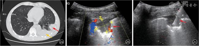
图2 艾滋病合并单一病原体肺部感染患者,男性,29 岁,反复咳嗽咳痰1 个月。图a:左下肺病灶(红箭头所示)并左胸腔积液(黄箭头所示)CT 图;图b:肺病灶超声呈实性低回声(箭头所示);图c:对肺病灶实施超声引导经皮介入穿刺肺活检(箭头所示),病理/病原体检测结果为马尔尼菲篮状菌肺部感染 |
表1 2 组肺部感染患者胸部超声影像特征比较[例(%)] |
| 超声表现 | MMPI组(n=210) | SPPI组(n=52) | χ2值 | P值 |
|---|---|---|---|---|
| 肺病灶三径均值 | 40.506 | <0.001 | ||
| <50mm | 43(20.48) | 34(65.38) | ||
| ≥50mm | 167(79.52) | 18(34.62) | ||
| 肺病灶边界 | 0.142 | 0.706 | ||
| 清楚 | 111(52.86) | 29(55.77) | ||
| 模糊 | 99(47.14) | 23(44.23) | ||
| 肺病灶内部回声 | 29.24 | <0.001 | ||
| 实性回声 | 78(37.14) | 41(78.85) | ||
| 实液混合回声 | 132(62.86) | 11(21.15) | ||
| 肺病灶血流信号 | 1.64 | 0.2 | ||
| 丰富 | 72(34.29) | 13(25.00) | ||
| 稀少 | 138(65.71) | 39(75.00) | ||
| 合并胸腔积液 | 2.61 | 0.106 | ||
| 有 | 196(93.33) | 45(86.54) | ||
| 无 | 14(6.67) | 7(13.46) |
表2 2组肺部感染患者超声引导经皮介入穿刺肺活检病原体检测结果 |
| 感染病原体 | 例数 | 占比(%) |
|---|---|---|
| MMPI组 | 210 | |
| 结核分枝杆菌+马尔尼菲篮状菌 | 59 | 28.10 |
| 结核分枝杆菌+马红球菌 | 44 | 20.95 |
| 结核分枝杆菌+隐球菌 | 37 | 17.62 |
| 结核分枝杆菌+肺炎克雷伯杆菌 | 36 | 17.14 |
| 结核分枝杆菌+马红球菌+真菌 | 15 | 7.14 |
| 肺炎克雷伯杆菌+真菌 | 19 | 9.05 |
| SPPI组 | 52 | |
| 结核分枝杆菌 | 22 | 42.30 |
| 马尔尼菲篮状菌 | 14 | 26.92 |
| 马红球菌 | 12 | 23.08 |
| 肺炎克雷伯杆菌 | 3 | 5.78 |
| 梅毒螺旋体 | 1 | 1.92 |
| 1 |
Mongodi S, Via G, Girard M, et al. Lung ultrasound for early diagnosis of ventilator-associated pneumonia [J]. Chest, 2016, 149(4): 969-980.
|
| 2 |
Wang H, Wang L, Luo Z, et al. Performance of rapid on-site evaluation of touch imprints of lung tissue biopsies for the diagnosis of pulmonary cryptococcosis in patients without HIV infection [J].Mycoses, 2022, 65(6): 635-642.
|
| 3 |
Lee SB, Kim MJ, Lee IJ. Assessment of diagnostic accuracy and complication rates of CT-guided percutaneous core-needle biopsy for lung lesion: difference between solid and sub-solid nodules based on propensity score matching analysis [J]. Clin Radiol, 2023, 78(9):e620-e626
|
| 4 |
陈桂荣, 夏凤鸣, 秦志强, 等. CT 引导经皮穿刺肺活检诊断肺感染性疾病的临床意义 [J]. 中国感染控制杂志, 2023, 22(6): 680-687.
|
| 5 |
中华医学会感染病学分会艾滋病丙型肝炎学组, 中国疾病预防控制中心.中国艾滋病诊疗指南(2021 年版) [J]. 中华内科杂志,2021, 60(12): 1106-1128.
|
| 6 |
Nong HR, Wu FY, Su N, et al. Ultrasound guided biopsy: a powerful tool in diagnosing AIDS complications [J]. Radiol Infect Dis, 2015,2(3): 123-127.
|
| 7 |
农恒荣, 苏楠. 艾滋病相关周边型肺结核超声影像分析 [J]. 中华超声影像学杂志, 2018, 27(5): 427-430.
|
| 8 |
Demi L, Wolfram F, Klersy C, et al. New international guidelines and consensus on the use of lung ultrasound [J]. J Ultrasound Med, 2023,42(2): 309-344.
|
| 9 |
Orso D, Guglielmo N, Copetti R. Lung ultrasound in diagnosing pneumonia in the emergency department: a systematic review and meta-analysis [J]. Eur J Emerg Med, 2018, 25(5): 312-321.
|
| 10 |
Audette LD, Parent MC. BET 3: Bedside lung ultrasound for the diagnosis of pneumonia in children [J]. Emerg Med J, 2016, 33(8):589-592.
|
| 11 |
石宝玉, 李枍恒, 阮婷, 等. 非实时超声支气管镜联合病原宏基因组二代测序在局灶性肺部感染性疾病诊断中的应用 [J]. 中国呼吸与危重监护杂志, 2022, 21(10): 725-730.
|
| 12 |
Limonta S, monge E, Montuori M, et al. Lung ultrasound in the management of pneumocystis pneumonia: a case series [J]. Int J STD AIDS, 2019, 30(2): 188-193.
|
| 13 |
Ko RE, Jeong BH, Huh HJ, et al. Clinical usefulness of fungal culture of EBUS-TBNA needle rinse fluid and core tissue [J]. Yonsei Med J,2020, 61(8): 670-678.
|
| 14 |
Jaliawala HA, Farooqui SM, Harris K, et al. Endobronchial ultrasoundguided transbronchial needle aspiration (EBUS-TBNA): technical updates and pathological yield [J]. Diagnostics, 2021, 11(12): 2331.
|
| 15 |
沙敏, 刘超, 汪泱, 等. 超声引导下经支气管针吸活检术获取组织标本培养在肺部感染性疾病诊疗中的应用观察 [J]. 山东医药,2023, 63(23): 24-28.
|
| 16 |
蔡存良, 张明强, 赵景全, 等. 肺穿刺活检联合组织培养在肺部感染性疾病中的诊断价值 [J]. 北京医学, 2020, 42(6): 504-509.
|
/
| 〈 |
|
〉 |