
2024 , Vol. 21 >Issue 10: 937 - 942
DOI: https://doi.org/10.3877/cma.j.issn.1672-6448.2024.10.002
超声心动图在孤立性左心室心尖发育不良疾病中的应用价值
Copy editor: 汪荣
收稿日期: 2024-07-25
网络出版日期: 2024-12-23
版权
Value of echocardiography in diagnosing isolated left ventricular apical dysplasia
Received date: 2024-07-25
Online published: 2024-12-23
Copyright
目的
探讨超声心动图在孤立性左心室心尖发育不良(ILVAH)疾病中的应用价值。
方法
收集2014年1月至2022年12月在中国医学科学院阜外医院就诊并最终确诊为ILVAH的患者。所有患者均接受心电图、经胸超声心动图及MRI检查。总结ILVAH患者的临床特征及超声心动图表现特征。
结果
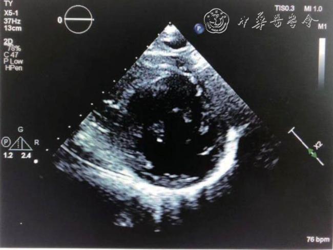
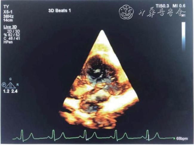
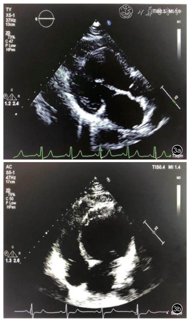
最终共收集10例确诊为ILVAH的患者,其中男性5例,女性5例,平均年龄(31.2±10.3)岁。ILVAH的超声心动图特点如下:10例均表现为乳头肌水平以下的左心室组织消失,乳头肌直接附着于左心室“心尖部”;10例均表现为左心室形态异常,左心室“心尖部”圆钝而不尖,长径缩短,横径扩大,左心室呈球形,室间隔呈弧形向右心室膨出,室间隔及左心室壁厚度不均;10例均表现为右心室形态异常,右心室狭长,向左后下方延伸,包绕短缩的左心室心尖部,整个心脏心尖部由右心室构成,由于室间隔向右膨凸,右心室中部内径偏小。
结论
ILVAH具有较为典型的超声心动图特征,结合患者临床特征及其他影像学检查手段,可以为该疾病提供更多诊断依据。
关键词: 超声心动图; 孤立性左心室心尖发育不良; 心室形态
李晓妮 , 卫青 , 孟庆龙 , 牛丽莉 , 田月 , 吴伟春 , 朱振辉 , 王浩 . 超声心动图在孤立性左心室心尖发育不良疾病中的应用价值[J]. 中华医学超声杂志(电子版), 2024 , 21(10) : 937 -942 . DOI: 10.3877/cma.j.issn.1672-6448.2024.10.002
Objective
To assess the value of echocardiography in diagnosing isolated left ventricular apical hypoplasia (ILVAH).
Methods
Patients who were diagnosed with ILVAH and treated at Fuwai Hospital, Chinese Academy of Medical Sciences from January 2014 to December 2022 were collected.All patients underwent electrocardiography (ECG), transthoracic echocardiography, and MRI examinations.The clinical characteristics and echocardiographic manifestations of patients with ILVAH were summarized.
Results
A total of 10 patients diagnosed with ILVAH were finally collected, including 5 males and 5 females,with an average age of (31.2 ± 10.3) years. The echocardiographic characteristics of ILVAH are as follows:In all the 10 cases, there was disappearance of left ventricular tissue below the papillary muscle level, and the papillary muscle was directly attached to the "apex" of the left ventricle; the left ventricle showed an abnormal morphology. The "apex" of the left ventricle was blunt rather than pointed. The long diameter was shortened,the transverse diameter was enlarged, the left ventricle was spherical, the ventricular septum protruded to the right ventricle in an arc shape, and the thickness of the ventricular septum and left ventricular wall was uneven; and the right ventricle also showed an abnormal morphology. The right ventricle was long and narrow and extended to the left and rear lower part, surrounding the shortened left ventricular apex. The apex of the entire heart was composed of the right ventricle. Due to the rightward bulge of the ventricular septum, the inner diameter of the middle part of the right ventricle was small.
Conclusion
ILVAH has relatively typical echocardiographic characteristics. Combined with the patient's clinical characteristics and other imaging examination methods, echocardiography can provide more diagnostic information for this disease.
表1 10例ILVAH患者的基本资料 |
| 序号 | 年龄(岁) | 性别 | 临床症状 | 心电图表现 | MRI检查结果 | 超声检查结果 |
|---|---|---|---|---|---|---|
| 1 | 23 | 男 | 无症状 | 电轴右偏,V5 R/S<1 | 左心室心尖发育不良,局部脂肪替代,估测EF约49% | 左房室增大,左心室心尖发育不良 |
| 2 | 31 | 女 | 乏力 | 心房颤动,ST-T改变 | 左心室心尖发育不良,局部脂肪替代,估测EF约56% | 左房室增大,左心室心尖发育不良 |
| 3 | 17 | 男 | 胸痛 | ST-T改变 | 左心室心尖发育不良,少量脂肪替代,左心室扩大,估测EF约50% | 左心室增大,左心室心尖发育不良 |
| 4 | 25 | 男 | 乏力 | 胸导R波递增不良,异常Q波,T波改变 | 左心室心尖发育不良,少量脂肪替代,左心室腔形态异常,估测EF约54% | 左心室增大,左心室心尖发育不良 |
| 5 | 36 | 男 | 劳累后气短 | 电轴右偏,胸导R波递增不良,ST-T改变 | 左心室心尖发育不良,少量脂肪替代,左心室扩大,估测EF约33% | 左房室增大,左心室心尖发育不良,左心室心尖部致密化不全 |
| 6 | 25 | 女 | 心悸 | 异常Q波 | 左心室心尖收缩减低并致密化不全,心尖部脂肪浸润,估测EF约55% | 左心室增大,左心室心尖发育不良 |
| 7 | 48 | 女 | 无症状 | 心律不齐,偶发房性早搏,部分T波改变 | 左心室心尖发育不良,局部脂肪替代,估测EF约53% | 左心室心尖发育不良 |
| 8 | 29 | 女 | 无症状 | 异常Q波 | 左心室心尖发育不良,局部脂肪替代,估测EF约41% | 左房室增大,左心室心尖发育不良,左心室收缩功能减低 |
| 9 | 53 | 女 | 胸闷 | 异常Q波,T波改变 | 左心室心尖发育不良,心尖局部脂肪浸润,估测EF约15% | 左心室心尖发育不良,三尖瓣少量反流,左右心功能减低 |
| 10 | 25 | 男 | 心慌、心悸 | 室性早搏,P波异常 | 左心室心尖发育不良,局部脂肪替代,估测EF约45% | 左房室增大,左心室心尖发育不良,二尖瓣中量反流,三尖瓣少中量反流,肺动脉高压 |
表2 10例ILVAH患者的超声参数测量结果 |
| 序号 | LV前后径(mm) | LV左右径(mm) | LV上下径(mm) | SI | LVEF(%) | LA前后径(mm) | RV前后径(mm) | RV左右径(mm) | RV上下径(mm) | |
|---|---|---|---|---|---|---|---|---|---|---|
| 1 | 63 | 58 | 72 | 1.24 | 61 | 38 | 19 | 29 | 80 | |
| 2 | 60 | 66 | 50 | 0.76 | 60 | 46 | 17 | 22 | 77 | |
| 3 | 71 | 60 | 46 | 0.77 | 58 | 34 | 15 | 18 | 51 | |
| 4 | 63 | 69 | 57 | 0.83 | 58 | 30 | 19 | 26 | 70 | |
| 5 | 62 | 59 | 60 | 1.01 | 54 | 40 | 14 | 28 | 87 | |
| 6 | 53 | 61 | 54 | 0.89 | 57 | 32 | 16 | 22 | 77 | |
| 7 | 58 | 53 | 52 | 0.98 | 57 | 32 | 18 | 29 | 66 | |
| 8 | 63 | 67 | 55 | 0.82 | 47 | 41 | 18 | 26 | 71 | |
| 9 | 33 | 46 | 67 | 1.46 | 33 | 26 | 26 | 30 | 81 | |
| 10 | 69 | 57 | 80 | 1.40 | 67 | 36 | 22 | 33 | 95 |
| 1 |
Fernandez-Valls M, Srichai MB, Stillman AE, et al. Isolated left ventricular apical hypoplasia: a new congenital anomaly described with cardiac tomography[J]. Heart, 2004, 90(5): 552-555.
|
| 2 |
Román R, Anchante H, Menacho K, et al. Isolated left ventricular apical hypoplasia: case report[J].Eur Heart J Case Rep, 2023, 7(2):ytad046.
|
| 3 |
Elias Neto J, Tonet J, Frank R, et al. Arrhythmogenic right ventricular cardiomyopathy/dysplasia (ARVC/D)—What we have learned after 40 years of the diagnosis of this clinical entity[J]. Arq Bras Cardiol, 2019,112(1): 91-103.
|
| 4 |
李建蓉, 孙欣, 徐楠. 超声心动图诊断左心尖发育不良[J]. 中国循环杂志, 2011, 26(8): 144-145.
|
| 5 |
Bassareo PP, Duignan S, James A, et al. Isolated left ventricular apical hypoplasia: systematic review and analysis of the 37 cases reported so far[J]. World J Clin Cases, 2023, 11(23): 5494-5503.
|
| 6 |
Mirdamadi A, Ashrafi S. Isolated left ventricular apical hypoplasia:reporting a case with mild manifestations and different echocardiography features[J]. Iran Red Crescent Med J, 2016, 18(8): e26065.
|
| 7 |
Şahin S, Özbülbül NI. Cardiac magnetic resonance imaging findings of isolated left ventricular apical hypoplasia[J]. J Cardiol Cases, 2023,28(5): 221-223.
|
| 8 |
Arbelo E, Protonotarios A, Gimeno JR, et al. 2023 ESC Guidelines for the management of cardiomyopathies [J]. Eur Heart J, 2023, 44(37):3503-3626.
|
| 9 |
任书堂, 黄云洲, 刘晓程. 左心室心尖发育不良的临床及超声诊断进展[J].中华超声影像学杂志, 2013, 22(9): 817-819.
|
| 10 |
闫朝武, 李建荣, 赵世华. 左心室心尖发育不良的临床和影像学特征[J]. 中华心血管杂志, 2012, 40(12): 1012-1015.
|
| 11 |
Vanhecke TE, Decker J, Leonowicz N, et al. Isolated left ventricular apical hypoplasia[J]. Congenit Heart Dis, 2011, 6(6): 646-649.
|
| 12 |
李剑明, 史蓉芳. 心肌致密化不全的诊断及影像学特征[J]. 中国医学影像技术, 2012, 28(7): 1411-1414.
|
| 13 |
Starmer G, Younger JF, Stewart P, et al. Multimodality imaging of isolated left ventricular apical hypoplasia[J]. Eur Heart J, 2012, 33(5):675.
|
| 14 |
Cannavale G, Francone M, Galea N, et al. Fatty images of the heart: spectrum of normal and pathological findings by computed tomography and cardiac magnetic resonance imaging[J]. Biomed Res Int, 2018, 2018: 5610347.
|
| 15 |
Motwani M, Witte KK, Plein S, et a1. Isolated left ventricular apical hypoplasia evaluated by cardiovascular magnetic resonance and gadolinium enhancement techniques[J]. J Am Coil Cardiol, 201l,58(22): 2355.
|
/
| 〈 |
|
〉 |