
2024 , Vol. 21 >Issue 11: 1011 - 1016
DOI: https://doi.org/10.3877/cma.j.issn.1672-6448.2024.11.002
女性盆腔内神经鞘肿瘤超声声像图及临床特征分析
Copy editor: 吴春凤
收稿日期: 2024-10-04
网络出版日期: 2025-01-24
基金资助
国家重点研发计划子课题骨干2022(2022YFB3804605)
版权
Ultrasonographic imaging and clinical features of pelvic nerve sheath tumors in female patients
Received date: 2024-10-04
Online published: 2025-01-24
Copyright
目的
探讨女性盆腔内神经鞘肿瘤的超声声像图及临床特征,以提高对神经鞘肿瘤的认识。
方法
选取2019年3月至2023年10月于南京大学医学院附属鼓楼医院妇产医学中心接受妇科超声检查、经手术治疗并经病理诊断为盆腔内神经鞘肿瘤的患者11例为研究对象,分析患者术前超声图像特征及临床资料,并进行随访。
结果
11例盆腔内神经鞘肿瘤中神经纤维瘤病1例,神经纤维瘤1例,神经鞘瘤9例,发病年龄29~68岁,多以无症状或体检偶然发现就诊,肿块大小差异大(最大直径为29~105 mm),内部回声以囊实性为主(5/9),形态多为规则(6/9),边界清晰(9/9),其中神经鞘瘤多见高回声包膜(7/9),彩色多普勒超声提示病灶内血流以I级为主(5/9)。
结论
女性盆腔内神经鞘肿瘤多表现为良性肿瘤的超声声像图特征,缺乏特异性征象,超声医师应提高对盆腔内神经鞘肿瘤的认识以提高术前诊断率。
赵静 , 荆秀娟 , 戴晨燕 . 女性盆腔内神经鞘肿瘤超声声像图及临床特征分析[J]. 中华医学超声杂志(电子版), 2024 , 21(11) : 1011 -1016 . DOI: 10.3877/cma.j.issn.1672-6448.2024.11.002
Objective
To investigate the ultrasonographic imaging and clinical features of pelvic nerve sheath tumors in female patients, in order to improve their recognition.
Methods
From March 2019 to October 2023, patients with pelvic nerve sheath tumors who underwent gynecological ultrasound, surgical treatment, and pathological examination at the Gynecologic Center Ultrasound Department of Nanjing Drum Tower Hospital Affiliated to Nanjing University were included. Their ultrasound findings and clinical data were reviewed retrospectively.
Results
A total of 11 cases were included, including 9 cases of schwannom and 1 case each of neurofibroma and neurofibromatosis, age of onset: 29-68 years old. Most of them were asymptomatic or found by physical examination. The size of the tumors varied (maximum diameter 29-105 mm), the masses were well-defined, the internal echo was mainly solid-cystic, the shape was mainly regular, and the presence of a hyperechogenic rim was mainly detected in schwannoa. Color Doppler showed that the blood flow in the lesions was mainly grade I.
Conclusion
Pelvic never sheath tumors in female patients have the characteristics of benign tumors on ultrasonographic images and lack typical imaging features. Sonographers should improve their understanding of pelvic never sheath tumors to further improve the accuracy of preoperative diagnosis.
Key words: Female; Pelvic; Nerve sheath; Neurilemmoma; Ultrasound
表1 盆腔内神经鞘肿瘤患者病灶声像图特性及临床特征总结 |
| 序号 | 年龄(岁) | 病史 | 超声表现 | 肿块位置 | 病理诊断 | 随访 | |||||
|---|---|---|---|---|---|---|---|---|---|---|---|
| 最大直径(mm) | 形态 | 内部回声 | 包膜 | 血流分级 | 术前超声 | 术中所见 | |||||
| 1 | 29 | 无 | 105 | 圆形 | 实性为主 | 可见 | I | 左侧盆腔 | 骶骨左侧,腰大肌后外侧 | 神经纤维瘤病 | 术后22个月多处皮下软组织内复发,病理检查恶性外周神经鞘膜瘤 |
| 92 | 椭圆形 | 实性,伴声影 | 可见 | 0 | 子宫后方 | 骶前间隙 | |||||
| 2 | 38 | 无 | 37 | 分叶状 | 实性,小囊性 | 无 | III | 左侧髂血管后方贴近盆壁 | 左侧髂内静脉下方 | 神经纤维瘤 | 无异常 |
| 3 | 68 | 下腹坠胀伴排尿困难 | 100 | 圆形 | 囊实性 | 可见 | III | 阴道后方 | 阴道后上方 | 神经鞘瘤 | 无异常 |
| 4 | 51 | 下腹坠胀 | 76 | 圆形 | 囊实性 | 可见 | III | 右侧附件区 | 右侧闭孔区 | 神经鞘瘤 | 术后15个月后于左侧髋周软组织内复发 |
| 5 | 37 | 无 | 81 | 分叶状 | 实性,小囊性灶 | 不明显 | II | 盆腔底部 | 右侧髂血管区 | 神经鞘瘤 | 无异常 |
| 6 | 34 | 下腹坠胀 | 63 | 圆形 | 实性,钙化斑 | 不明显 | I | 子宫右后方 | 右侧髂血管分叉处 | 神经鞘瘤 | 无异常 |
| 7 | 46 | 无 | 60 | 圆形 | 囊实性 | 可见 | I | 左侧附件区 | 左髂窝内 | 神经鞘瘤 | 无异常 |
| 8 | 54 | 无 | 29 | 圆形 | 实性 | 可见 | I | 左侧盆腔 | 左侧闭孔窝 | 神经鞘瘤 | 术后出现左下肢皮肤刺痛麻木,目前仍在康复中 |
| 9 | 68 | 无 | 55 | 圆形 | 囊实性 | 可见 | II | 左侧附件区 | 乙状结肠后方 | 神经鞘瘤 | 无异常 |
| 10 | 45 | 外阴不适 | 55 | 分叶状 | 囊实性 | 可见 | I | 子宫后方 | 子宫后方 | 神经鞘瘤 | 术后一过性左下肢疼痛 |
| 11 | 66 | 无 | 32 | 圆形 | 实性为主 | 可见 | I | 盆腔左后方 | 左侧盆壁 | 神经鞘瘤 | 无异常 |
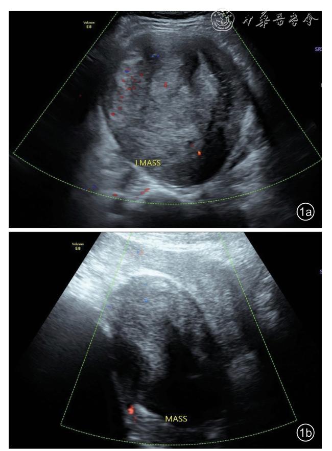
图1 神经纤维瘤病患者超声声像图表现。图a为经腹部超声探查获取位于骶骨左侧肿块声像图表现,肿块内回声以实性为主,形态规则,边界清晰,见包膜,血流分级I级;图b为经腹部超声探查获取位于子宫后方、骶前间隙肿块的声像图表现,肿块内回声以实性为主,呈椭圆形,见包膜,血流分级为0级注:LMASS为左侧盆腔肿块,MASS为肿块 |
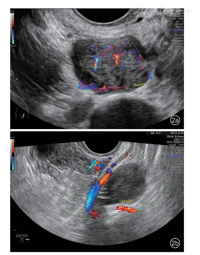
图2 神经纤维瘤患者超声声像图表现。图a为经腹部探查获取的肿块声像图,图b为经阴道探查获取的肿块声像图,肿块形态略呈分叶状,肿块内回声以实性为主,见多个小囊性病灶,边界清晰,血流分级为Ⅲ级,肿块贴近左侧髂血管注:LMASS为左侧盆腔肿块 |
| 1 |
Murphey MD, Kransdorf MJ. Staging and classification of primary musculoskeletal bone and soft-tissue tumors according to the 2020 WHO update, from the AJR special series on cancer staging [J]. AJR,2021, 217(5): 1038-1052.
|
| 2 |
Fischerova D, Santos G, Wong L, et al. Imaging in gynecological disease:clinical and ultrasound characteristics of benign retroperitoneal pelvic never sherth tumors [J]. Ultrasound Obstet Gynecol, 2023,62(5): 727-738.
|
| 3 |
Timmerman D, Valentin L, Bourne TH, et al. Terms, definitions and measurements to describe the sonographic features of adnexal tumors:a consensus opinion from the International Ovarian Tumor Analysis(IOTA) Group [J]. Ultrasound Obstet Gynecol, 2000, 16(5): 500-505.
|
| 4 |
张丹, 王茜, 王佳颖. IOTA共识与O-RADS共识指南的解读与分析 [J/OL]. 中华医学超声杂志(电子版), 2022, 19(2): 105-113.
|
| 5 |
许彩, 周苑, 赵胜, 等. 美国放射学会卵巢-附件报告和数据系统解读 [J/OL]. 中华医学超声杂志(电子版), 2022, 19(5): 391-395.
|
| 6 |
Adler DD, Carson PL, Rubin JM, et al. Doppler ultrasound color flow imaging in the study of breast caner:preliminary findings [J].Ultrasound Med Biol, 1990, 16(6): 553-559.
|
| 7 |
张敏, 范小波, 孙玉清, 等, 胃肠道及腹膜后神经鞘瘤的影像诊断及误诊分析 [J]. 医学影像学杂志, 2022, 32(9): 1539-1542
|
| 8 |
杨帆, 吴火林, 陈贤翔, 等. 神经鞘瘤内靶征的超声诊断价值及形成机制探讨 [J]. 中国超声医学杂志, 2015, 31(9): 824-826.
|
| 9 |
武奎宁, 张培培, 齐振红, 等. 腹膜后神经鞘瘤的超声诊断质量及其影响因素分析 [J/OL]. 中华医学超声杂志(电子版), 2023, 20(7):701-704.
|
| 10 |
杨帆, 陈贤翔, 吴灼金, 等. 周围神经鞘瘤的超声特征分析 [J]. 中华超声影像学杂志, 2015, 24(2): 151-154.
|
/
| 〈 |
|
〉 |