
2024 , Vol. 21 >Issue 11: 1042 - 1047
DOI: https://doi.org/10.3877/cma.j.issn.1672-6448.2024.11.006
CT/MRI引导下再次超声检查对首次超声漏诊肾肿瘤的再评价
Copy editor: 吴春凤
收稿日期: 2024-02-27
网络出版日期: 2025-01-24
版权
Re-evaluation of renal tumors missed by initial ultrasound by CT or MRI guided second-look ultrasound
Received date: 2024-02-27
Online published: 2025-01-24
Copyright
目的
探讨CT/MRI引导下再次超声检查对首次常规超声漏诊肾肿瘤的临床价值并分析超声漏诊肾肿瘤的原因。
方法
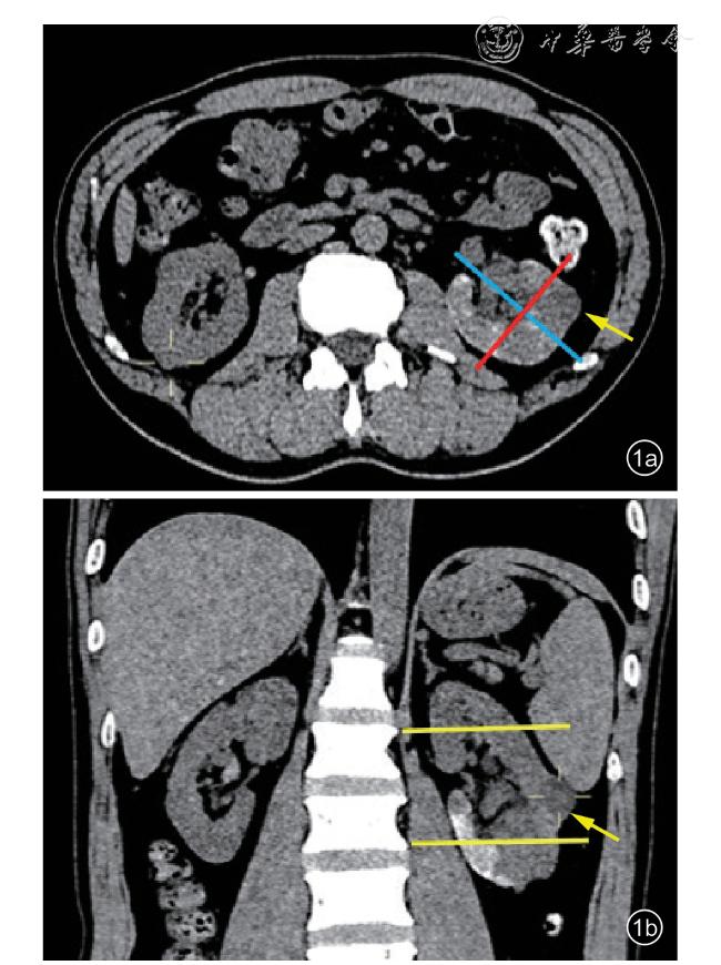
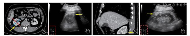
选择山西省肿瘤医院2013年1月至2022年12月间经泌尿外科手术切除且经病理证实为肾实质肿瘤的2354例患者,其中术前首次超声漏诊肾肿瘤患者30例(31个病灶),以CT/MRI病灶解剖学特征为参考标准,描述性分析首次超声漏诊原因;其中24例(25个病灶)患者在增强CT/MRI引导下行再次超声检查,具体地,侧卧位及俯卧位横切扫查获得肾超声横断面图像,侧卧位纵切扫查获得冠状面图像,俯卧位纵切扫查获得超声矢状面图像。根据再次超声是否检出肾肿瘤分为阳性组与阴性组,采用Fisher精确概率法比较2组病灶大小、侧别、极性、位置(深/浅)、生长模式等解剖学特征及病理类型方面的差异,分析影响病灶二次超声检出的因素。
结果
首次超声检查漏诊肾肿瘤30例(31个病灶),漏诊率为1.3%(30/2354),其中≤4 cm病灶27个(27/31,87.1%),≤1 cm病灶5个(5/31,16.1%);位置深病灶23个(23/31,74.2%);外凸率<50%病灶14个(14/31,45.2%)。再次超声检查共24例患者(25个病灶),阴性组7个病灶(7/25,28.0%),阳性组18个病灶(18/25,72.0%)。具体地,≤1 cm病灶共4个,均未检出,占阴性组的57.1%(4/7);>1~≤2 cm病灶11个,未检出2个,占阴性组的28.6%(2/7),检出9个,占阳性组的50.0%(9/18);>2~≤3 cm病灶6个,未检出1个,占阴性组14.3%(1/7),检出5个,占阳性组的27.8%(5/18);>3 cm病灶4个,均检出,占阳性组的22.2%(4/18)。阴性组与阳性组病灶大小比较,差异具有统计学意义(P=0.017),在侧别、极性、位置、生长模式、病理类型等方面2组差异均无统计学意义(P均>0.05)。
结论
直径≤4 cm、位置深(内侧/背侧)、外凸率<50%等肾肿瘤解剖学特征是常规超声检查漏诊肾肿瘤的主要原因。再次超声检查时,超声医师利用CT/MRI与超声图像进行认知融合,在脑海中构建肾肿瘤“三维”扫查思路,从肾横断面、冠状面及矢状面对病灶行实时动态的超声扫查,显示病灶三维解剖特征,可以有效提高肾肿瘤检出率。
郭馨阳 , 张妍 , 原韶玲 , 史泽洪 , 牛菁华 . CT/MRI引导下再次超声检查对首次超声漏诊肾肿瘤的再评价[J]. 中华医学超声杂志(电子版), 2024 , 21(11) : 1042 -1047 . DOI: 10.3877/cma.j.issn.1672-6448.2024.11.006
Objective
To assess the clinical value of CT (or MRI) guided second-look ultrasound examination for renal tumors missed by initial conventional ultrasound and analyze the causes of missed diagnosis.
Methods
A total of 2354 patients with pathologically confirmed renal parenchymal tumors who underwent urological surgery at Shanxi Cancer Hospital from January 2013 to December 2022 were selected. Among them,30 cases (31 lesions) had renal tumors missed by initial ultrasound. The anatomical characteristics indicated by CT/MRI were used as the reference standard to analyze the causes of missed diagnosis by initial ultrasound descriptively. In the above cases, 24 patients (25 lesions) underwent a second ultrasound examination under the guidance of contrast-enhanced CT/MRI. Cross sectional images of the kidneys were obtained through transverse scans of the waist and back, coronal images were obtained through longitudinal scans of the waist, and sagittal images were obtained through longitudinal scans of the back in the prone position. The patients were divided into a negative group and a positive group according to whether the renal tumor was detected. Tumor size, side, polarity, deep or shallow location, growth pattern, and pathological type of lesions were compared by the Fisher’s exact test between the two groups, and the factors affecting secondary detection were analyzed.
Results
Thirty cases (31 lesions) were missed by initial ultrasound examination with a missed diagnosis rate of 1.3% (30/2354), among which 27 lesions (27/31, 87.1%) were ≤ 4 cm,5 (5/31, 16.1%) were ≤ 1 cm, 23 (23/31, 74.2%) were deep, and 14 (14/31, 45.2%) had an exophytic rate<50%. Twenty-four cases (25 lesions) were examined by a second ultrasound examination, involving 7 lesions in the negative group (7/25, 28.0%) and 18 lesions in the positive group (18/25, 72.0%). All of the 4 lesions with a diameter ≤ 1 cm were missed, accounting for 57.1% (4/7) of the negative group. In 11 lesions with a diameter >1 but≤ 2 cm, there were 2 missed cases, accounting for 28.6% (2/7) of the negative group, and 9 detected cases, accounting for 50% (9/18) of the positive group. In 6 lesions with a diameter >2 but ≤ 3 cm, one lesion was missed, accounting for 14.3% (1/7) of the negative group, and 5 lesions were detected, accounting for 27.8% (5/18) of the positive group. All of 4 lesions with a diameter >3 cm were detected,accounting for 22.2% (4/18) of the positive group. There was a statistical difference in lesion size between the two groups (P<0.05), but not in lesion side, polarity, deep or shallow location, growth pattern, or pathological type (P>0.05).
Conclusion
Anatomical features of renal tumors, such as diameter ≤ 4 cm, deep position (medial or dorsal), and exophytic rate < 50%, are the main reasons for the missed diagnosis of renal tumors by conventional ultrasound. During second-look ultrasound examination, sonographers should perform cognitive fusion of CT/MRIand ultrasound images, and construct a three-dimensional concept of kidney tumor in the mind. Then, real-time dynamic three-dimensional ultrasound should be performed from the cross-sectional, coronal, and sagittal aspects of the kidney to display the three-dimensional anatomical characteristics of the lesions, which can effectively improve the detection rate of renal tumors.
表1 首次超声漏诊31个病灶解剖学特征、病理类型[个(%)] |
| 病灶特征 | 个数 |
|---|---|
| 大小 | |
| >4 cm | 4(12.9) |
| >3~≤4 cm | 2(6.5) |
| >2~≤3 cm | 9(29.0) |
| >1~≤2 cm | 11(35.5) |
| ≤1 cm | 5(16.1) |
| 侧别 | |
| 左肾 | 22(71.0) |
| 右肾 | 9(29.0) |
| 极性 | |
| 上极 | 9(29.0) |
| 体部 | 14(45.2) |
| 下极 | 8(25.8) |
| 位置 | |
| 深 | 23(74.2) |
| 浅 | 8(25.8) |
| 生长模式 | |
| 外凸率≥50% | 8(25.8) |
| 外凸率<50% | 14(45.2) |
| 完全内生 | 9(29.0) |
| 病理类型 | |
| 肾透明细胞癌 | 23(74.2) |
| 肾乳头状细胞癌 | 1(3.2) |
| 肾嫌色细胞癌 | 2(6.5) |
| 肾转移癌 | 1(3.2) |
| 肾血管平滑肌脂肪瘤 | 1(3.2) |
| 肾嗜酸细胞腺瘤 | 3(9.7) |
表2 阴性组及阳性组肾肿瘤患者病灶解剖学特征、病理类型比较[个(%)] |
| 病灶特征 | 阴性组(n=7) | 阳性组(n=18) | P值 |
|---|---|---|---|
| 大小 | 0.017 | ||
| >4 cm | 0(0) | 2(11.1) | |
| >3~≤4 cm | 0(0) | 2(11.1) | |
| >2~≤3 cm | 1(14.3) | 5(27.8) | |
| >1~≤2 cm | 2(28.6) | 9(50.0) | |
| ≤1 cm | 4(57.1) | 0(0) | |
| 侧别 | 0.640 | ||
| 左肾 | 4(57.1) | 13(72.2) | |
| 右肾 | 3(42.9) | 5(27.8) | |
| 极性 | 1.000 | ||
| 上极 | 3(42.9) | 6(33.3) | |
| 体部 | 3(42.9) | 8(44.4) | |
| 下极 | 1(14.3) | 4(22.2) | |
| 位置 | 0.637 | ||
| 深 | 6(85.7) | 13(72.2) | |
| 浅 | 1(14.3) | 5(27.8) | |
| 生长模式 | 0.618 | ||
| 外凸率≥50% | 1(14.3) | 5(27.8) | |
| 外凸率<50% | 3(42.9) | 9(50.0) | |
| 完全内生 | 3(42.9) | 4(22.2) | |
| 病理类型 | 0.754 | ||
| 肾透明细胞癌 | 5(71.4) | 12(66.7) | |
| 肾乳头状细胞癌 | 0(0) | 1(5.5) | |
| 肾嫌色细胞癌 | 0(0) | 2(11.1) | |
| 肾转移癌 | 0(0) | 1(5.5) | |
| 肾血管平滑肌脂肪瘤 | 0(0) | 1(5.5) | |
| 肾嗜酸细胞腺瘤 | 2(28.6) | 1(5.5) |
表3 再次超声检查2组肾肿瘤患者病灶大小与位置、生长模式情况 |
| 组别 | 个数 | 病灶位置及生长模式 |
|---|---|---|
| 阴性组 | ||
| >4 cm | 0 | |
| >3~≤4 cm | 0 | |
| >2~≤3 cm | 1 | 背内侧:1个,完全内生 |
| >1~≤2 cm | 2 | 腹内侧:1个,外凸率≥50%; 背内侧:1个,外凸率<50% |
| ≤1 cm | 4 | 腹内侧:2个,1个完全内生,1个外凸率≥50%; |
| 背外侧:1个,外凸率<50%; | ||
| 腹外侧:1个,完全内生 | ||
| 阳性组 | ||
| >4 cm | 2 | 腹内侧:1个,外凸率≥50%; 背外侧:1个,外凸率≥50% |
| >3~≤4 cm | 2 | 腹内侧:1个,外凸率<50%; 背外侧:1个,外凸率≥50% |
| >2~≤3 cm | 5 | 腹内侧:2个,外凸率均<50%; 背内侧:2个,外凸率均≥50%; 腹外侧:1个,外凸率<50% |
| >1~≤2 cm | 9 | 腹内侧:0个; 背内侧:1个,完全内生; 腹外侧:4个,2个外凸率<50%,2个完全内生; 背外侧:4个,3个外凸率<50%,1个完全内生 |
| ≤1 cm | 0 |
| 1 |
Campbell SC, Clark PE, Chang SS, et al. Renal mass and localized renal cancer: evaluation, management, and follow-up: AUA guideline:part I [J]. J Urol, 2021, 206(2): 199-208.
|
| 2 |
Stock KF, Slotta-Huspenina J, Kübler H, et al. Innovative ultraschalldiagnostik bei nierentumoren [J]. Der Urologe, 2019, 58(12):1418-1428.
|
| 3 |
Tamara JI, Juan GR, Patricia ZJ, et al. Diagnosis and treatment of small renal masses: where do we stand? [J]. Curr Urol Rep, 2022,23(6): 99-111.
|
| 4 |
Kang SK, Huang WC, Pandharipande PV, et al. Solid renal masses:what the numbers tell us [J]. AJR Am J Roentgenol, 2014, 202(6):1196-1206.
|
| 5 |
冯敏, 俞丽芳, 包凌云. 第二眼超声检查对乳腺肿块的诊断价值 [J].浙江医学, 2019, 41(16): 1782-1784.
|
| 6 |
王元元, 朱嘉宁, 李静波, 等. 常规超声及超声造影诊断小肾肿瘤的价值 [J]. 中国超声医学杂志, 2022, 38(7): 803-806.
|
| 7 |
Tao L, Fan J, Zhan W, et al. Contrast-enhanced ultrasound manifestations of renal masses undetectable on conventional ultrasound [J]. Front Oncol, 2022, 12: 943960.
|
| 8 |
Yates DR, Rouprêt M. Small renal mass and low-risk prostate cancer:any more for active surveillance? [J]. Eur Urol, 2011, 60(1): 45-47.
|
| 9 |
Yamauchi FI, Paiva OA, Mussi TC, et al. A comparative study of ultrasound and cross-sectional imaging for detection of small renal masses: anatomic factors and radiologist’s experience [J]. Einstein (Sao Paulo), 2020, 18: eAO5576.
|
| 10 |
Herts BR, Silverman SG, Hindman NM, et al. Management of the incidental renal mass on CT: a white paper of the ACR Incidental Findings Committee [J]. J Am Coll Radiol, 2018, 15(2): 264-273.
|
| 11 |
Coll DM, Smith RC. Update on radiological imaging of renal cell carcinoma [J]. BJU Int, 2007, 99(5 Pt B): 1217-1222.
|
| 12 |
Tsili AC, Andriotis E, Gkeli MG, et al. The role of imaging in the management of renal masses [J]. Eur J Radiol, 2021, 141: 109777.
|
| 13 |
卢畅, 赵佳琦, 张正委. 超声漏诊小肾癌1例并国内文献复习 [J].第二军医大学学报, 2021, 42(12): 1438-1443.
|
| 14 |
Rossi SH, Klatte T, Usher-Smith J, et al. Epidemiology and screening for renal cancer [J]. World J Urol, 2018, 36(9): 1341-1353.
|
| 15 |
Wylie B, Necas M, Heaney A. Visualisation of focal renal lesions on ultrasound: A review of 518 lesions with contrast CT correlation [J].Australas J Ultrasound Med, 2020, 23(4): 248-254.
|
| 16 |
Sienz M, Ignee A, Dietrich CF. Sonography today: reference values in abdominal ultrasound: aorta, inferior vena cava, kidneys [J]. Z Gastroenterol, 2012, 50(3): 293-315.
|
| 17 |
Ko SE, Lee MW, Lim HK, et al. The semi-erect position for better visualization of subphrenic hepatocellular carcinoma during ultrasonography examinations [J]. Ultrasonography, 2021, 40(2): 274-280.
|
| 18 |
David N, Horrow MM. Pitfalls in renal ultrasound [J]. Ultrasound Q,2020, 36(4): 300-313.
|
| 19 |
Li C, Lu Q, Huang B, et al. The value of contrast-enhanced ultrasound(CEUS) in detecting minute renal cell carcinoma [J]. Discov Med,2014, 18(99): 179-188.
|
/
| 〈 |
|
〉 |