
2025 , Vol. 22 >Issue 03: 191 - 196
DOI: https://doi.org/10.3877/cma.j.issn.1672-6448.2025.03.001
胎儿眼睑及睑裂的产前超声检查
Copy editor: 吴春凤
收稿日期: 2024-12-25
网络出版日期: 2025-06-10
版权
Prenatal ultrasonographic evaluation of fetal eyelids and palpebral fissures
Received date: 2024-12-25
Online published: 2025-06-10
Copyright
目的
探讨胎儿眼睑及睑裂的产前超声检查方法和声像图特征。
方法
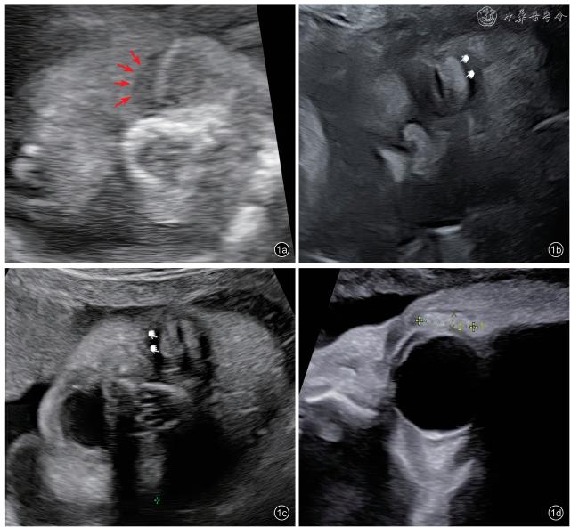
前瞻性采集2023年1月至2024年3月在深圳市龙华区妇幼保健院常规产前超声检查的不同孕周胎儿眼睑、睑裂图像,观察其显示切面(眼睑冠状切面和经眼睑中部矢状切面)及声像特征。采用χ2检验比较同孕周胎儿左右眼睑裂声像特征在矢状切面和冠状切面的差异,采用Fisher检验比较不同孕周胎儿左右眼睑裂声像特征在矢状切面和冠状切面的差异。追踪并随访胎儿娩出后眼部情况。
结果
本研究纳入15~40孕周正常胎儿125例,在胎儿眼睑冠状切面及经眼睑中部矢状切面观察250只眼,左眼125只,右眼125只。15周前的胎儿眼睑、睑裂回声大多无法显示。随孕周增加,眼睑从小叶片状中等回声逐渐过渡到高回声。胎儿睑裂冠状切面显示率为98.4%(左眼)、96.0%(右眼);经眼睑中部矢状切面显示率为96.8%(左眼)、94.4%(右眼)。睑裂呈线状高回声、低回声、带状无回声。20周前,上下眼睑未分离,睑裂呈线状高回声;20~28周,上下眼睑逐渐分离,睑裂可表现为线状高回声、线状低回声、带状无回声3种超声声像;28周后,睑裂仍可见线状高回声、线状低回声、带状无回声3种超声声像,其中线状低回声及带状无回声检出率较28周前增加。相同孕周,在矢状切面和冠状切面,胎儿左、右眼睑裂回声比较,差异均无统计学意义(P均>0.05)。不同孕周间比较,胎儿左、右眼睑裂超声声像的特征中不同切面的线状高回声及线状低回声表现差异均具有统计学意义(P均<0.05),不同切面的带状无回声表现在不同孕周间差异均无统计学意义(P均>0.05)。经娩出后证实,125例胎儿双侧眼睑、睑裂均正常。
结论
产前超声可以对胎儿眼睑、睑裂发育情况进行评估,有助于提高胎儿眼睑、睑裂发育异常的产前检出率。
廖佳音 , 文华轩 , 薛淑贞 , 黄慧 , 魏英妮 , 陈亚岩 . 胎儿眼睑及睑裂的产前超声检查[J]. 中华医学超声杂志(电子版), 2025 , 22(03) : 191 -196 . DOI: 10.3877/cma.j.issn.1672-6448.2025.03.001
Objective
To investigate prenatal ultrasonographic methods for evaluating fetal eyelids and palpebral fissures and their imaging characteristics.
Methods
Ultrasonographic images of fetal eyelids and palpebral fissures in fetuses at various gestational weeks were prospectively collected from January 2023 to March 2024 at Shenzhen Longhua Maternal and Child Health Hospital. The imaging planes (coronal plane of the eyelids and mid-eyelid sagittal plane) and sonographic characteristic were systematically analyzed. Chi-square tests were used to compare left-right eyelid fissure echoic patterns in sagittal and coronal planes within the same gestational week, and the Fisher’s exact test was used to compare differences across gestational weeks. Postnatal ocular outcomes were tracked.
Results
A total of 125 normal fetuses at 15-40 gestational weeks were included, with 250 eyes (125 left, 125 right) evaluated via coronal and mid-eyelid sagittal planes. Eyelid and palpebral fissure echogenicity was rarely detectable before 15 weeks. With advancing gestation, eyelid echogenicity transitioned from flake-like moderate to hyperechoic.Visualization rates for palpebral fissures were 98.4% (left) and 96.0% (right) in coronal planes, and 96.8%(left) and 94.4% (right) in sagittal planes. Palpebral fissures manifested as linear hyperechoic, hypoechoic,or band-like anechoic structures. Before 20 weeks, fused eyelids displayed linear hyperechoic fissures. From 20-28 weeks, gradual eyelid separation yielded three patterns: linear hyperechoic, hypoechoic, or band-like anechoic. After 28 weeks, hypoechoic and anechoic patterns increased in prevalence. No significant left-right differences were observed in echoic patterns within the same gestational week (P>0.05). Across gestational weeks, significant differences were noted in linear hyperechoic and hypoechoic patterns (P<0.05), while band-like anechoic patterns showed no statistical variation (P>0.05). Postnatal follow-up confirmed normal eyelid and fissure development in all 125 cases.
Conclusion
Prenatal ultrasound enables effective evaluation of fetal eyelid and palpebral fissure development, enhancing prenatal detection of abnormalities.
Key words: Ultrasonography; Prenatal; Fetus; Eyelid; Palpebral fissure
表1 125例正常胎儿眼睑的产前超声表现(只) |
| 孕周 | 只数 | 中等回声 | 高回声 |
|---|---|---|---|
| 15+1~ 20 周 | |||
| 左眼 | 10 | 10 | 0 |
| 右眼 | 10 | 10 | 0 |
| 20+1~ 28 周 | |||
| 左眼 | 39 | 29 | 10 |
| 右眼 | 39 | 29 | 10 |
| 28+1~ 40 周 | |||
| 左眼 | 76 | 0 | 75 |
| 右眼 | 76 | 0 | 72 |
表2 125例正常胎儿睑裂产前超声表现[只(%)] |
| 孕周 | 只数 | 眼睑冠状切面 | 经眼睑中部矢状切面 | ||||
|---|---|---|---|---|---|---|---|
| 线状高回声 | 线状低回声 | 带状无回声 | 线状高回声 | 线状低回声 | 带状无回声 | ||
| 15+1 ~ 20 周 | |||||||
| 左眼 | 10 | 10(100) | 0(0) | 0(0) | 9(100) | 0(0) | 0(0) |
| 右眼 | 10 | 10(100) | 0(0) | 0(0) | 9(100) | 0(0) | 0(0) |
| 20+1 ~ 28 周 | |||||||
| 左眼 | 39 | 27(69.2) | 7(17.9) | 5(12.8) | 29(74.3) | 6(15.4) | 4(10.3) |
| 右眼 | 39 | 27(69.2) | 8(20.5) | 4(10.3) | 30(76.9) | 5(12.8) | 4(10.3) |
| 28+1 ~ 40 周 | |||||||
| 左眼 | 76 | 8(10.8) | 54(73.0) | 12(16.2) | 8(11.0) | 53(72.6) | 12(16.4) |
| 右眼 | 76 | 11(15.5) | 51(71.8) | 9(12.7) | 10(14.3) | 51(72.9) | 9(12.9) |
| P值a | < 0.001 | < 0.001 | 0.508 | < 0.001 | < 0.001 | 0.439 | |
| P值b | < 0.001 | < 0.001 | 0.735 | < 0.001 | < 0.001 | 0.732 | |
注:冠状切面观察时,孕28+1~40周,左眼(2例),右眼(5例)远场眼睑因孕周及体位因素无法显示;经眼睑中部矢状切面观察时,其中1例孕15+6周(因胎儿孕周过小)和孕40周(因胎儿入盆,胎头姿势固定)双睑裂均显示不清;另有7例仅显示一侧近场睑裂(均在晚孕期37周后):远场有左眼(2例),右眼(5例)无法显示。a为左眼不同孕周超声特征差异比较的统计值,b为右眼不同孕周超声特征差异比较的统计值 |
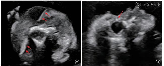
图2 胎儿睑裂的线状高回声表现。孕19周胎儿在上下眼睑未分离的情况下,由于睑裂无法直观显示,此时在超声影像中上下眼睑融合处表现为一条连续的高回声线。图a为经眼睑冠状切面图像,表现为上下眼睑融合,睑裂处形成一条高回声线;图b为经眼睑中部矢状切面图像:睑裂处为短线状高回声 |
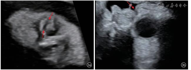
图3 胎儿睑裂的线状无回声表现。图a为孕27+3周胎儿经眼睑冠状切面超声图像,睑裂表现为上下眼睑已经分开但未睁开,睑裂呈线状低回声,可见高-低-高“三线征”。图b为经眼睑中部矢状切面超声图像,也可见睑裂表现为线状低回声 |
| 1 |
李胜利, 罗国阳. 胎儿畸形产前超声诊断学 [M]. 北京: 科学出版社, 2017: 721-722
|
| 2 |
Byun TH, Kim JT, Park HW, et al. Timetable for upper eyelid development in staged human embryos and fetuses [J]. Anat Rec(Hoboken), 2011, 294(5): 789-796.
|
| 3 |
Tawfik HA, Abdulhafez MH, Fouad YA, et al. Embryologic and fetal development of the human eyelid [J]. Ophthalmic Plast Reconstr Surg,2016, 32(6): 407-414.
|
| 4 |
Landau-Prat D, Kim DH, Bautista S, et al. Cryptophthalmos:associated syndromes and genetic disorders [J]. Ophthalmic Genet,2023, 44(6): 547-552.
|
| 5 |
Kabra M, Gulati S, Ghosh M, et al. Fraser-cryptophthalmos syndrome[J]. Indian J Pediatr, 2000, 67(10): 775-778.
|
| 6 |
罗丹丹, 文华轩, 陈曦, 等. 胎儿眼球及眼附属结构异常的产前超声研究及进展 [J/OL]. 中华医学超声杂志(电子版), 2023, 20(3): 343-355.
|
/
| 〈 |
|
〉 |