
2025 , Vol. 22 >Issue 03: 209 - 214
DOI: https://doi.org/10.3877/cma.j.issn.1672-6448.2025.03.004
经会阴超声检测肛提肌形态及功能对盆腔器官脱垂的诊断价值
Copy editor: 吴春凤
收稿日期: 2024-12-09
网络出版日期: 2025-06-10
基金资助
湖南省自然科学基金面上项目(2023JJ30920)
版权
Value of detecting morphology and function of the levator ani muscle by transperineal ultrasound in diagnosis of pelvic organ prolapse
Received date: 2024-12-09
Online published: 2025-06-10
Copyright
目的
研究经会阴超声检测肛提肌形态及功能对盆腔器官脱垂(POP)的诊断价值。
方法
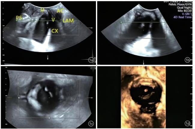
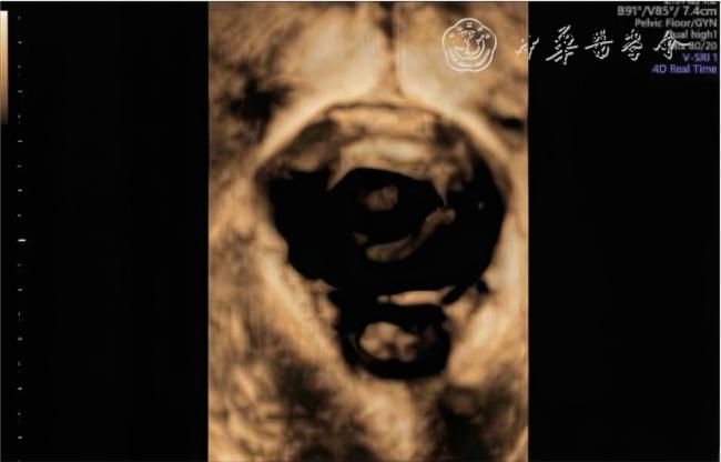
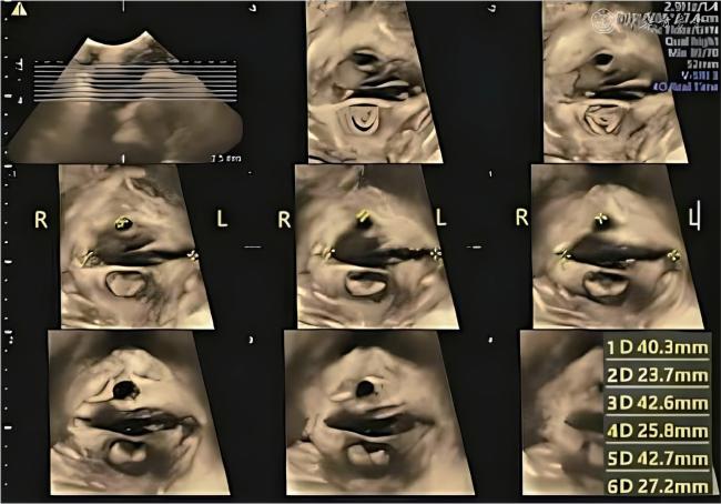
选取2023年10月至2024年5月中南大学湘雅医院收治的100例POP患者为研究组,同期100例非POP者为对照组。经会阴部扫查,启用三维模式,在静息及最大Valsalva状态下实时测量肛提肌裂孔的前后径、左右径及面积;启用断层成像技术,在盆底肌肉收缩状态下评估肛提肌的完整性,并测量左、右侧肛提肌尿道间隙。采用独立样本t检验比较组间肛提肌裂孔测值的差异,采用χ²检验比较组间肛提肌损伤率的差异。
结果
静息状态下,研究组肛提肌裂孔的前后径、左右径及面积均高于对照组[(57.6±4.1)mm vs (53.2±4.7)mm,(47.7±4.4)mm vs (40.8±4.1)mm,(18.12±2.15)cm2 vs (14.87±2.63)cm2],最大Valsalva状态下,研究组肛提肌裂孔的前后径、左右径及面积也高于对照组[(63.4±3.1)mm vs (53.5±3.2)mm,(51.3±4.7)mm vs (40.2±3.0)mm,(22.27±2.65)cm2 vs (16.46±2.71)cm2],差异均具有统计学意义(t=6.922、11.275、9.383、21.820、19.618、15.053,P均<0.001)。盆底肌收缩状态下,研究者肛提肌损伤率为22.1%,明显高于对照组(3.1%),差异具有统计学意义(χ²=16.067,P<0.001)。研究组左、右侧肛提肌尿道间隙均高于对照组[(23.92±3.87)mm vs (18.14±2.75)mm,(24.76±3.62)mm vs (18.72±2.76)mm],差异具有统计学意义(t=11.989、13.060,P均<0.001)。
结论
经会阴三维、实时三维超声联合断层成像检测肛提肌形态及功能对POP具有较高的诊断价值。
李沐宸 , 鲁蓉 . 经会阴超声检测肛提肌形态及功能对盆腔器官脱垂的诊断价值[J]. 中华医学超声杂志(电子版), 2025 , 22(03) : 209 -214 . DOI: 10.3877/cma.j.issn.1672-6448.2025.03.004
Objective
To investigate the diagnostic efficacy of transperineal ultrasound in detecting the morphology and function of the levator ani muscle in patients with pelvic organ prolapse (POP).
Methods
The clinical and ultrasonographic data of 100 POP patients and 100 non-POP patients from the Xiangya Hospital of Central South University from October 2023 to May 2024 were retrospectively analyzed in this study. In resting and maximal Valsalva state, three-dimensional transperineal ultrasound was used to measure the anteroposterior and left-to-right diameters and area of levator ani hiatus in real time. The integrity of the levator ani muscle was assessed during pelvic floor muscle contraction, and the levator-urethra gap for both sides was measured. Independent sample t-test was used to compare the differences in measurements of anal sphincter hiatus between groups, and Chi-square test was used to compare the differences in rates of anal sphincter injury between groups.
Results
In the resting state, the anteroposterior and left-toright diameters, and area of levator ani hiatus in the study group were significantly higher than those of the control group [(57.6±4.1) mm vs (53.2±4.7) mm, (47.7±4.4) mm vs (40.8±4.1) mm, and (18.12±2.15) cm2 vs (14.87±2.63) cm2; t=6.922, 11.275, and 9.383, respectively; P<0.001 for all]. In maximum Valsalva state, the anteroposterior and left-to-right diameters and area of levator ani hiatus in the study group were also significantly higher than those of the control group [(63.4±3.1) mm vs (53.5±3.2) mm, (51.3±4.7) mm vs (40.2±3.0) mm, and (22.27±2.65) cm2 vs (16.46±2.7) cm2; t=21.820, 19.618, and 15.053, respectively;P<0.001 for all]. Assessing the contraction status of pelvic floor muscles showed that the injury rate of the levator ani muscle in the study group was 22.1%, which significantly higher than that of the control group (3.1%;χ²=16.067, P<0.001). The left and right levator-urethra gaps in the study group were significantly higher than those in the control group [(23.92±3.87) mm vs (18.14±2.75) mm and (24.76±3.62) mm vs (18.72±2.76) mm; t=11.989 and 13.060, respectively; P<0.001 for both].
Conclusion
Three-dimensional and real-time three-dimensional transperineal ultrasound combined with tomographic ultrasound imaging can effectively assess the morphological changes and the functional status of levator ani muscles, which has high diagnostic value for POP.
 表示,采用两独立样本t检验比较组间差异。肛提肌损伤情况为计数资料,采用百分率(%)表示,采用χ²检验比较组间差异。以P<0.05为差异具有统计学意义。
表1 2组研究对象一般临床资料比较( |
| 组别 | 例数 | 年龄(岁) | 体质量指数(kg/m2) | 产次(次) |
|---|---|---|---|---|
| 研究组 | 95 | 51.2±8.5 | 23.9±2.5 | 1.5±0.6 |
| 对照组 | 98 | 53.6±7.3 | 24.0±2.3 | 1.6±0.4 |
| t值 | 0.923 | 0.623 | 0.575 | |
| P值 | 0.368 | 0.657 | 0.594 |
表2 静息与最大Valsalva状态下2组肛提肌裂孔的大小形态比较( |
| 组别 | 例数 | 静息状态 | 最大Valsalva 状态 | ||||
|---|---|---|---|---|---|---|---|
| 前后径(mm) | 左右径(mm) | 面积(cm2) | 前后径(mm) | 左右径(mm) | 面积(cm2) | ||
| 研究组 | 95 | 57.6±4.1 | 47.7±4.4 | 18.12±2.15 | 63.4±3.1 | 51.3±4.7 | 22.27±2.65 |
| 对照组 | 98 | 53.2±4.7 | 40.8±4.1 | 14.87±2.63 | 53.5±3.2 | 40.2±3.0 | 16.46±2.71 |
| t值 | 6.922 | 11.275 | 9.383 | 21.820 | 19.618 | 15.053 | |
| P值 | < 0.001 | <0.001 | < 0.001 | < 0.001 | < 0.001 | < 0.001 | |
表3 盆底肌收缩状态下2组盆腔器官脱垂患者肛提肌尿道间隙比较(mm, |
| 组别 | 例数 | 左侧 | 右侧 |
|---|---|---|---|
| 研究组 | 95 | 23.92±3.87 | 24.76±3.62 |
| 对照组 | 98 | 18.14±2.75 | 18.72±2.76 |
| t值 | 11.989 | 13.060 | |
| P值 | < 0.001 | < 0.001 |
| 1 |
中华医学会妇产科学分会妇科盆底学组. 盆腔器官脱垂的中国诊治指南(2020年版) [J]. 中华妇产科杂志, 2020, 55(5): 300-306.
|
| 2 |
中华医学会泌尿外科分会女性泌尿学组. 女性盆腔器官脱垂的风险预警及早期干预专家共识 [J]. 实用妇产科杂志, 2024, 40(7):532-537.
|
| 3 |
Schulten SFM, Claas-Quax MJ, Weemhoff M, et al. Risk factors for primary pelvic organ prolapse and prolapse recurrence: an updated systematic review and Meta-analysis [J]. Am J Obstet Gynecol, 2022,227(2): 192-208.
|
| 4 |
Yeung E, Malacova E, Maher C. Is levator ani avulsion a risk factor for prolapse recurrence? A systematic review and meta-analysis [J]. Int Urogynecol J, 2022, 33(7): 1813-1826.
|
| 5 |
Horcicka L, Krcmar M, Nemec M, et al. Appearance of levator ani muscle subdivision defects on level III vaginal support structures in women with and without pelvic organ prolapse: an MRI study [J]. Int Urogynecol J, 2023, 34(8): 1971-1982.
|
| 6 |
郭志洁, 陈炯权, 张元吉, 等. 全自动三维盆底超声测量肛提肌裂孔大小的临床应用 [J].中国医学影像学杂, 2023, 31(9): 962-966.
|
| 7 |
Van Gruting IMA, Stankiewicz A, Van Delft KWM, et al. Diagnostic test accuracy of magnetic resonance imaging and pelvic floor ultrasound for diagnosis of levator ani muscle avulsion [J]. Ultrasound Obstet Gynecol, 2022, 60(4): 559-569.
|
| 8 |
吴曼丽, 林欣, 王旭东, 等. 肛提肌裂孔与盆腔器官脱垂量化分期及脱垂症状的相关性分析 [J]. 中华超声影像学杂志, 2020, 29(8):700-705.
|
| 9 |
王睿丽, 朱兆领, 甘宜鑫, 等. 盆底超声临床实用规范化检查专家共识(2022版) [J]. 中国医学影像学杂志, 2023, 31(2): 97-100.
|
| 10 |
李多, 鲁蓉. 盆底超声评估女性肛提肌形态及功能的研究进展 [J].中南大学学报(医学版), 2023, 48(8): 1267-1273.
|
| 11 |
Dietz HP. Ultrasound in the investigation of pelvic floor disorders [J].Curr Opin Obstet Gynecol, 2020, 32(6): 431-440.
|
| 12 |
顾其凤, 陈思佳, 刘学彬, 等. 多模态超声评估产次对会阴体及肛提肌功能影响的研究[J]. 实用医院临床杂志, 2024, 21(3): 134-139.
|
| 13 |
He X, Du Q, Chang L, et al. Analysis of minimal levator ani hiatus area based on MRI in female adults without pelvic floor dysfunction at different age groups [J]. Arch Gynecol Obstet, 2024, 309(5): 2183-2191.
|
| 14 |
Pankaj G, Sushil D, Vipul D, et al. Yagnik anal fistula at roof of ischiorectal fossa inside levator-ani muscle (RIFIL): a new highly complex anal fistula diagnosed on MRI [J]. Abdom Radiol (NY),2021, 46(12): 5550-5563.
|
| 15 |
Xuan Y, Friedman T, Dietz HP. Does levator ani hiatal area configuration affect pelvic organ prolapse [J]. Ultrasound Obstet Gynecol, 2019, 54(1): 124-127.
|
| 16 |
Montaguti E, Cariello L, Dodaro MG, et al. The role of a new threedimensional ultrasound technique in the diagnosis of levator ani muscle avulsion [J]. Neurourol Urodyn, 2020, 39(1): 455-463.
|
| 17 |
叶婷婷, 李清莹, 陈华, 等. 全栈式自动盆底超声与手动方式获取并测量最小肛提肌裂孔平面的一致性评价 [J/OL]. 中华医学超声杂志(电子版), 2024, 21(8): 794-801.
|
| 18 |
赵春桃, 李锦秋, 王义成, 等. 基于经会阴三维超声下盆底解剖结构变化评估不同类型盆腔器官脱垂患者术后疗效 [J]. 中国优生与遗传杂志, 2022, 30(7): 1205-1209.
|
| 19 |
朱霞, 高艳多, 许彩, 等. 盆底三维超声评价产次及分娩方式对肛提肌裂孔面积的影响[J/OL]. 中华医学超声杂志(电子版), 2022,19(9): 920-925.
|
| 20 |
Sammarco AG, Sheyn D, Hong CX, et al. Pelvic cross-sectional area at the level of the levator ani and prolapse [J]. Int Urogynecol J, 2021,32(4): 1007-1013.
|
| 21 |
Sainz-Bueno JA, Bonomi MJ, Suárez-Serrano C, et al. Quantification of 3/4D ultrasound pelvic floor changes induced by postpartum muscle training in patients with levator ani muscle avulsion: a parallel randomized controlled trial [J]. Quant Imaging Med Surg, 2022, 12(4):2213-2223.
|
| 22 |
Van Gruting IMA, Stankiewicz A, Van Delft KWM, et al. Diagnostic test accuracy of magnetic resonance imaging and pelvic floor ultrasound for diagnosis of levator ani muscle avulsion [J]. Ultrasound Obstet Gynecol, 2022, 60(4): 559-569.
|
| 23 |
Rotstein E, Ullemar V, Starck M, et al. Three-dimensional endovaginal ultrasound assessment using the levator ani deficiency score in primiparas: a replication study [J]. Acta Obstet Gynecol Scand, 2023,102(9): 1236-1242.
|
| 24 |
Dietz HP, Chavez-Coloma L, Friedman T, et al. Pelvic organ prolapse in nulliparae [J]. Aust N Z J Obstet Gynaecol, 2022, 62(3): 420-425.
|
/
| 〈 |
|
〉 |