
肺静脉异位引流(anomalous pulmonary venous drainage,APVD)是指部分或全部肺静脉未直接与左心房相连,而与体静脉或右心房相连接,约占出生后先天性心脏病的5%~6%[1]。APVD分为部分型和完全型,部分型肺静脉异位引流(partial anomalous pulmonary venous drainage,PAPVD)指1~3支肺静脉未与左心房相连接,完全型肺静脉异位引流(total anomalous pulmonary venous drainage,TAPVD)指所有肺静脉均未与左心房相连接。TAPVD患儿自然预后非常差,仅有约50%可以存活超过3个月[2],一部分PAPVD患儿由于肺部感染、肺动脉高压等症状也需要手术治疗。但是由于胎儿时期肺静脉较细,分支较多,二维超声不易显示,右心系统扩大不明显,因此,APVD在产前筛查诊断中极易漏诊[3],本研究旨在探讨区域血流追踪法在超声诊断胎儿APVD中的价值。
资料与方法
一、对象
回顾性选取2015年1月至2019年12月在河北生殖妇产医院进行胎儿超声心动图检查,诊断为APVD的胎儿,排除合并其他复杂心内畸形者,共纳入41例。孕妇年龄为(27.75±4.92)岁(范围19~39岁),诊断孕龄为(26.70±3.16)周(范围22~34周)。所有病例均由产后超声心动图、手术结果或引产后尸检证实。
二、仪器与方法
1. 仪器:采用Philips EPIQ7、TOSHIBA Aplio 500、GE Voluson E10彩色多普勒超声诊断仪,选用C5-1凸阵探头、C9-2凸阵探头、C10-3凸阵探头,探头频率1~10 MHz(依据孕妇及胎儿条件选择使用);设置胎儿超声心动图条件,图像局部放大。
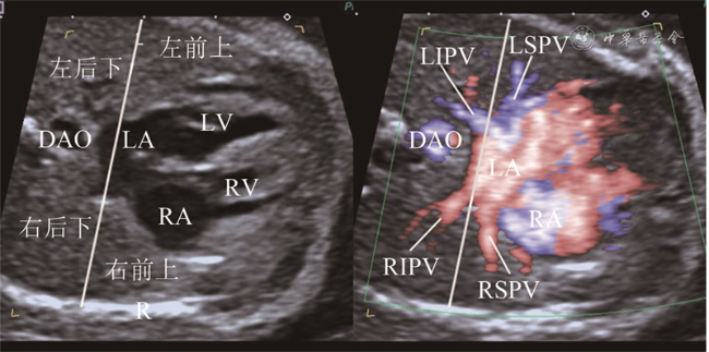
2. 方法:依据《中国胎儿心脏超声检查指南》[4]的要求,先明确胎位及胎儿左右,然后对胎儿心脏进行序列检查。检查胎儿肺静脉时,应用区域血流追踪法,即以横向四腔心切面为基础动态上下扫查肺组织,将肺组织大致分为4个区域,左侧前上部1/2肺野、左侧后下部1/2肺野、右侧前上部1/2肺野、右侧后下部1/2肺野,应用彩色血流技术,沿肺静脉血收集途径由4个区域内肺静脉远端分支向近心端追踪,观察其与左心房的关系及最终回流部位(图1 ,动态图1 )。彩色血流技术使用对低速血流敏感的彩色多普勒血流显像技术,如高级动态血流成像(advanced dynamic flow,ADF)、高分辨率血流成像技术(high-definition imaging,HDFI)、能量多普勒等,调节速度标尺在10~20 cm/s,调节合适彩色增益,使肺野内肺静脉分支血流能够显示得既饱满又不产生过多外溢。
结果
2015至2019年共完成19 372例胎儿的超声心动图检查,检出41例(0.2%)APVD胎儿(不合并其他心内畸形)。41例APVD胎儿中,TAPVD 36例(心上型25例,心内型7例,心下型2例,混合型2例),PAPVD 5例。在胸部横切面上,41例胎儿肺静脉均显示,显示率为100%;左侧前上部1/2肺野、左侧后下部1/2肺野、右侧前上部1/2肺野、右侧后下部1/2肺野肺静脉远端分支分别引流入左上肺静脉、左下肺静脉、右上肺静脉、右下肺静脉。
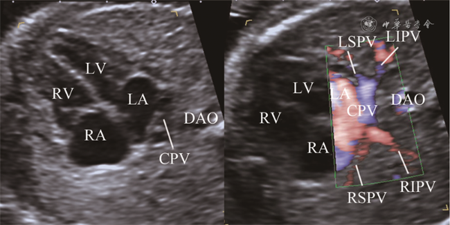
追踪36例TAPVD胎儿肺组织4大区域回流显示4条肺静脉均未汇入左心房,34例肺静脉近心端呈现左心房后方汇聚征(图2 ,动态图2 ),包括心上型25例,心内型7例,心下型2例。心上型TAPVD胎儿,其中20例4条肺静脉于左心房后方汇聚成肺总静脉腔,然后向头侧走行,最终汇入上腔静脉;3例胎儿显示3条肺静脉引流3大区域,于左心房后方汇聚成肺总静脉腔,肺总静脉腔向头侧走行过程中收集剩余区域肺静脉1条共同入上腔静脉。心内型TAPVD胎儿7例,其中6例4条肺静脉于左心房后下部汇聚成肺总静脉腔回流入冠状静脉窦,1例4条肺静脉于左心房后方汇聚成肺总静脉腔,直接回流入右心房。心下型TAPVD胎儿2例,4条肺静脉均于左心房后方汇聚成肺总静脉腔,然后向足侧走行汇入门静脉窦。混合型TAPVD 2例,1例左侧上肺及下肺静脉汇合后向头侧走行汇入上腔静脉,右侧上肺及下肺静脉汇合后向足侧走行入下腔静脉;1例左侧上肺及下肺静脉汇合后向头侧走行入上腔静脉,右侧上肺及下肺静脉汇合后直接回流入右心房。
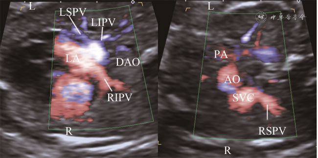
5例PAPVD胎儿部分区域肺静脉未回流入左心房,其中1例右上肺静脉直接回流入右心房,2例右上肺静脉回流入上腔静脉(图3 ),1例右上、右下肺静脉直接回流入右心房,1例左肺静脉变异为3支,最上支经无名静脉回流入上腔静脉。
讨论
一、区域血流追踪法与胎儿肺静脉分支定位
临床工作中,鉴别胎儿肺静脉回流正常还是异常,诊断胎儿APVD是完全型还是部分型,首先需要对肺静脉分支进行辨认。既往研究对肺静脉分支的超声定位多以正常成人肺静脉近心端为观察点。何怡华等[5]应用超声观察正常成人右上肺静脉开口紧邻房间隔,近端与上腔静脉相邻;右下肺静脉相对于右上肺静脉稍远离房间隔,位于右侧;左上肺静脉紧邻左心耳;左下肺静脉相对于左上肺静脉远离左心耳,靠近胸降主动脉。但是由于胎儿体位多变、肺静脉分支变异等因素,仅使用近心端位置定位肺静脉分支往往产生错误的判断,导致肺静脉分支不能得到全面的检查,继而发生漏诊、误诊;另外APVD胎儿肺静脉近心端位置常常发生变化,有时不能用来对分支进行定位。胎儿肺组织内没有气体,因此为超声观察肺静脉远心端提供了良好的透声窗,使用彩色血流尤其是对低速血流敏感的彩色多普勒血流显像技术如e-Flow、ADF、HDFI、能量多普勒等[6, 7, 8],能够使肺静脉远心端显像。本研究应用区域血流追踪法观察肺静脉,将肺组织大致分为4个区域,主要依据为肺静脉分支解剖。通常人体有4根肺静脉从心脏后部汇入左心房,即左上肺静脉、左下肺静脉、右上肺静脉、右下肺静脉;左上肺静脉收集左肺上叶的静脉血,属支包括尖后段、前段和舌段静脉干;左下肺静脉收集左肺下叶的静脉血,由上段和底段总静脉构成;右上肺静脉收集右肺上叶和中叶的静脉血,由上叶和中叶静脉汇合而成,右上叶静脉属支包括尖段、前段及后段静脉,右中叶静脉属支包括外侧段和内侧段静脉;右下肺静脉收集右肺下叶的血液,由上段和底段总静脉汇合而成[9]。因此,应用彩色血流技术可以观察到41例APVD胎儿肺组织4大区域(左侧前上、左侧后下、右侧前上、右侧后下部1/2肺野)肺静脉血流分别引流入左上肺静脉、左下肺静脉、右上肺静脉、右下肺静脉,这与正常胎儿是相同的。应用区域血流追踪法观察肺静脉分支回流区域,使得胎儿期肺静脉分支定位更加准确、更加全面,不容易受肺静脉近心端变异或引流部位异常的影响。不足的是,由于胎儿肺静脉细小,肺静脉分支属支不能完全显示,因此没有对其属支进行细致的划分,目前仅做了大致的区域划分,但其可作为今后研究的方向。
二、区域血流追踪法与APVD胎儿超声诊断
应用区域血流定位好肺静脉分支后,继续追踪其是否回流入左心房,用于鉴别正常与APVD胎儿。本研究中36例TAPVD胎儿呈现左心房后方汇聚征,而正常胎儿汇入左心房的声像图表现呈螃蟹征,如同4条“腿”插入左心房壁。二者区别在于正常胎儿4条肺静脉插入左心房壁之间,且分支间距较大,而TAPVD胎儿肺静脉向左心房后方共同肺静脉腔汇聚,分支间距较小,在彩色血流辨别不清时,需结合二维超声对比观察。本研究中3例TAPVD胎儿3条肺静脉汇入共同肺静脉腔,通过区域血流追踪发现剩余1条肺静脉在共同肺静脉腔移行为垂直静脉的过程中汇入,这也使得这3例胎儿最终诊断为TAPVD。对共同肺静脉腔的血流进一步追踪,可显示肺静脉向上、向下、向冠状静脉窦或直接向右心房回流,从而对TAPVD的分型做进一步的判断。
应用区域血流追踪法沿肺静脉血回流4个区域追踪胎儿肺静脉分支能够提高PAPVD的检出率,本研究发现了5例PAPVD,这在以往很难诊断[10]。其中2例胎儿右上肺静脉回流入上腔静脉,这2例在检查过程中发现右侧前上部1/2肺野的肺静脉血没有回流入左心房,继续追踪其回流入上腔静脉。其中1例胎儿左心房可见4个肺静脉分支开口,但在区域血流追踪过程中,发现左肺上部肺野部分血流没有回流至左心房,而是经无名静脉回流入上腔静脉,即左肺静脉有3个分支,最上支异位引流。
综上所述,应用区域血流追踪法诊断APVD需注意以下几点:首先要调整好彩色血流量程和增益使肺静脉远心端能很好显示,由于肺静脉血流速度低,速度标尺调整为10~20 cm/s为佳;仔细观察肺静脉分布4大区域(左侧前上部、左侧后下部、右侧前上部、右侧后下部1/2肺野)血液回流情况;仔细观察肺静脉分支与左心房的关系,是否有左心房后方汇聚征等特殊征象,彩色血流需结合二维图像对比观察。总之,应用区域血流追踪法可使肺静脉分支检查更全面,定位更加精确,有助于提高胎儿APVD的诊断准确性。