
资料与方法
一、对象
本组研究对象共73例,为2017年10月至2019年10月北京协和医院中孕期系统产前超声筛查或普通中、晚孕期超声检查中发现胎儿小脑延髓池增宽(>10 mm)并进行了胎儿MRI检查的病例,孕妇年龄(28.87±4.13)岁(范围18~41岁),孕周为(27.64±6.18)周(范围19~37周),均为单胎妊娠,小脑延髓池宽度范围为11~25 mm。
二、仪器与方法
1. 超声检查:超声检查使用的仪器为SAMSUNG WS80A型和GE Voluson E10型彩色多普勒超声诊断仪,常规使用凸阵探头,探头频率为1~5 MHz。部分病例同时使用三维容积探头及腔内探头。
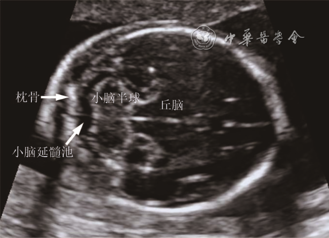
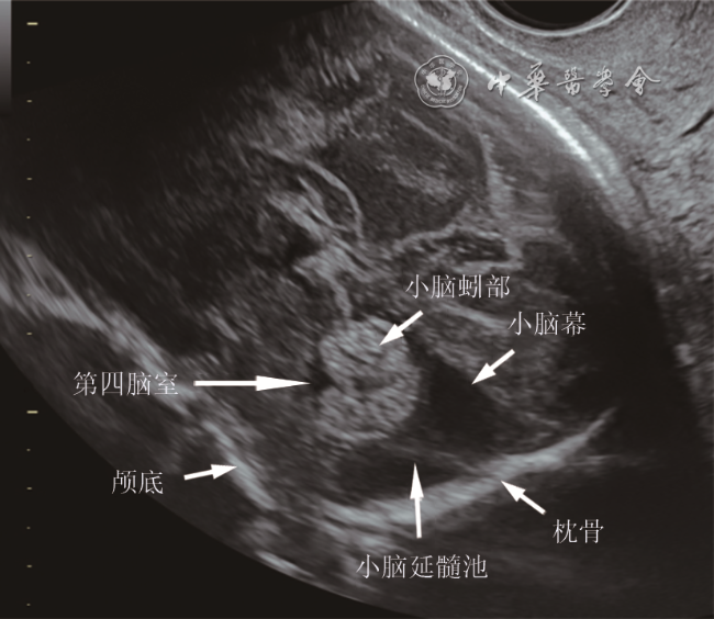
按照北京市产前超声筛查与诊断规范,对胎儿及其附属物进行系统性检查。对胎儿颅后窝进行多切面的超声扫查,对小脑半球、小脑蚓部、第四脑室、小脑延髓池、小脑幕等结构的位置、形态、大小进行观察及测量(图1 、2 )。测量方法及标准参考北京市产前超声筛查及诊断规范,所有病例均由具备产前诊断资格的超声医师完成。
2. MRI检查:MRI检查均在系统超声检查后3日内进行,所用仪器为GE公司1.5 T光纤MRI系统。检查采用快速成像序列,对胎儿头部二次定位后行横断面、冠状面、矢状面扫描,扫描时间35~45 s。小脑延髓池增宽标准为>10 mm。MRI诊断结果由2名放射科颅脑专业医师诊断并审核。
3. 观察指标:统计超声诊断胎儿异常情况,参照MRI检查结果,计算超声诊断符合率。
结果
一、胎儿后颅窝异常的MRI与超声诊断对比结果
本组胎儿小脑延髓池增宽病例中,与颅脑MRI结果比较,超声对后颅窝异常的诊断符合率为84.9%(62/73;表1 )。后颅窝病变共19例(26%,19/73),超声诊断符合率为63.2%。其中Dandy-Walker畸形(图3 )、小脑发育不良(图4 )、小脑蚓部发育不良(2例)的超声诊断符合率均为100%。颅后窝蛛网膜囊肿8例(图5 ),超声正确诊断2例,1例未定性,5例诊为单纯小脑延髓池增宽。Blake's囊肿1例,超声诊断为小脑蚓部发育不良。无明确后颅窝病变的单纯小脑延髓池增宽54例(74%,54/73),超声正确诊断50例,诊断符合率为92.6%;2例未定性,2例诊断为后颅窝蛛网膜囊肿。
表1 胎儿后颅窝异常的MRI与超声诊断结果对比 |
| MRI诊断结果 | 例数 | 超声正确诊断[例(%)] | 孕周(周) |
|---|---|---|---|
| 后颅窝病变 | 19 | 12(63.2) | 28.9(19.0~36.3) |
Dandy-Walker畸形 | 4 | 4(100) | 22.9(19.0~25.4) |
小脑发育不良 | 4 | 4(100) | 28.6(23.1~36.3) |
小脑蚓部发育不良 | 2 | 2(100) | 31.6(31.0~32.1) |
颅后窝蛛网膜囊肿 | 8 | 2(25.0) | 31.8(29.7~35.3) |
Blake's囊肿 | 1 | 0(0) | 23.3 |
| 单纯小脑延髓池增宽 | 54 | 50(92.6) | 27.2(19.4~36.9) |
| 合计 | 73 | 62(84.9) | 27.6(19.0~36.9) |
注:孕周表示为为平均值和起始周数 |

图3 胎儿Dandy-Walker畸形超声声像图(孕23+6周)。经小脑横切面:无小脑蚓部,小脑延髓池与第四脑室相通,小脑半球呈“八”字形分离注:CM为小脑延髓池,C为小脑半球 |
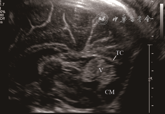
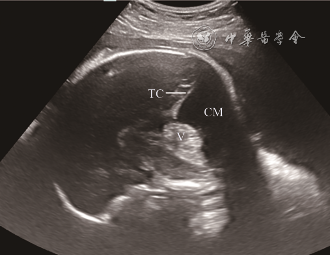
图4 胎儿小脑蚓部发育不良超声声像图(孕34周)。颅脑正中矢状面:小脑蚓部上旋,下蚓部部分缺失,小脑幕位置正常,小脑延髓池增大注:V为小脑蚓部,TC为小脑幕,CM为小脑延髓池 |
二、合并其他颅内异常情况
本组73例超声筛查小脑延髓池增宽病例中,MRI诊断合并其他颅内异常者为17例(23.3%,17/73),其中超声正确诊断13例(76.5%,13/17)。小脑发育不良伴菱脑融合、胼胝体部分发育不良1例,超声未诊断胼胝体病变。单纯小脑延髓池增宽伴侧脑室增宽13例,超声漏诊1例。单纯小脑延髓池增宽伴双侧室管膜下少量出血1例、双侧侧脑室旁缺血缺氧改变(囊肿)2例,超声均未诊断。
三、合并颅外异常情况
本组病例中超声检查发现颅外异常15例(20.5%,15/73),具体如下:Dandy-Walker畸形4例均合并颅外异常,分别为单脐动脉1例,婴儿型多囊肾1例,胆囊不显示、脊柱排列不规则1例,法洛四联症、心包腔积液1例。小脑发育不良4例中3例合并颅外异常,分别为双足内翻1例,双肾轻度积水1例,脐带增粗、华氏胶水肿1例。小脑蚓部发育不良1例合并法洛四联症、外生殖器形态异常、单脐动脉。后颅窝蛛网膜囊肿1例合并胎儿颈椎排列异常、脐静脉发育异常。单纯小脑延髓池增宽中6例(11.1%,6/54)合并颅外异常,分别为右位主动脉弓、迷走左锁骨下动脉1例,左肾体积小1例,心内膜垫缺损伴肺动脉狭窄及婴儿型多囊肾1例,致死性软骨发育不良1例,隔离肺1例,单侧唇腭裂1例。
讨论
本组中超声检查出现假阳性或假阴性病例为小脑蚓部发育不良、Blake's囊肿、后颅窝蛛网膜囊肿和单纯小脑延髓池增宽。小脑蚓部发育不良超声表现为颅脑正中矢状面上小脑蚓部部分缺失(以下蚓部缺失为主),蚓部上旋,小脑幕不抬高。Blake's囊肿的超声表现为颅脑正中矢状面小脑蚓部形态正常,位置上抬,第四脑室后方囊状突入小脑延髓池内,小脑幕不抬高[9]。后颅窝蛛网膜囊肿的超声表现为小脑延髓池占位性囊肿,向前可压迫小脑蚓部,向上可抬高小脑幕。单纯小脑延髓池增宽超声表现仅为小脑延髓池测值增大,后颅窝各结构大小、形态、位置等均无异常[10]。在前述几种情况的鉴别诊断中,由于超声较难量化评价小脑蚓部发育,如颅脑正中矢状面显示不满意,则鉴别诊断困难。本组中上述病例发现时期以晚孕期为主,颅骨声影明显,胎儿体位较固定,虽然合并使用了经阴道超声及三维超声[11, 12, 13, 14],部分病例仍鉴别困难。因此,当发现不易定性的小脑延髓池增宽、小脑蚓部显示不清,应建议行MRI检查以明确诊断[15]。
本组病例中,MRI同时发现17例合并颅内其他部位病变,其中13例为侧脑室增宽,侧脑室和小脑延髓池均为脑脊液循环的组成部分,因此影响脑脊液循环的因素均有可能同时累及上述两部位[16];此外,超声漏诊4例,分别为部分性胼胝体发育不良、室管膜下出血及囊肿,因此超声筛查发现小脑延髓池增宽时应进行颅内结构的全面检查,结合MRI有助于全面了解颅内发育情况,避免颅内病变的漏误诊。
本组病例中,15例合并颅外异常,其中,单纯小脑延髓池增宽病例中11.1%合并颅外结构异常,提示超声筛查发现小脑延髓池增宽时,除了对颅内结构进行系统的超声检查外,对胎儿全身结构也需要进行全面系统检查,以提供准确而全面的诊断信息。
综上所述,对于胎儿小脑延髓池增宽病例,超声总体诊断准确率较高,其中对Dandy-Walker畸形及小脑发育不良等严重疾病诊断准确性高,对于小脑蚓部发育不良、Blake's囊肿、颅后窝蛛网膜囊肿和单纯小脑延髓池增宽的鉴别诊断存在一定困难,需要通过MRI检查确诊。小脑延髓池增宽可能是多种畸形在颅内的声像表现,也可能是胎儿的正常变异,需根据每个病例的具体情况仔细分析,应建议孕妇做详尽的产前检查以明确诊断。