
盆底功能障碍(pelvic floor dysfunction,PFD)严重影响女性的日常工作、生活及社交活动,是女性身心健康的慢性疾病之一,部分PFD患者甚至会出现低落、自卑等情绪。早期发现与诊治有助于防范PFD的发生风险,对提高产妇生活质量具有重大意义。妊娠和分娩是PFD的独立危险因素,但阴道分娩与剖宫产相比,更容易发生孕产妇盆底筋膜支持结构的损伤,引发盆腔脏器移位,导致PFD的发生[1]。肛提肌作为盆底结构的主要支撑,其功能及盆膈裂孔的形态异常在PFD的发病中起着重要作用,其中耻骨直肠肌(puborectalis,PR)是其最为重要组成部分。与传统临床检查方法相比,经会阴盆底超声可通过测量盆底各径线及盆膈裂孔面积,直观动态地观察PR形态及损伤,间接评估肛提肌收缩功能[2]。既往研究[3]表明超声弹性成像可有效评估PR弹性方面的生物学特性,获取组织的杨氏模量值,量化评估PR肌力,对PFD的诊断具有一定价值。本研究拟通过构建二分类Logistic回归模型,对经阴道分娩者产后PFD的临床参数、经会阴超声及杨氏模量值进行综合分析,筛选出具有鉴别诊断意义的特征变量,为临床预防和治疗PFD提供科学依据。
资料与方法
一、对象
选取2019年1月至12月海南医学院附属海南医院收治的经阴道分娩的PFD患者42例(PFD组)和同期经阴道分娩的健康志愿产妇52例(正常对照组),PFD组患者年龄为(38.9±7.0)岁(范围26~56岁)。对照组产妇年龄为(43.1±8.8)岁(范围28~57岁)。纳入标准:所有受检产妇产前均无妊娠期并发症;近3个月未使用过激素类药物;根据病史、妇科检查及压力试验等确诊盆底功能障碍(压力性尿失禁、性功能障碍、排便障碍、子宫脱垂等)的患者;对照组盆脏器官脱垂分度均为stage 0。排除标准:合并盆腔巨大包块、神经系统病史、泌尿生殖系统急性炎症、盆腔手术史及长期慢性咳嗽等病史;肛提肌不能完成有效收缩者。所有入组研究对象均接受经会阴盆底超声和弹性成像检查。本研究经海南医学院附属海南医院医学伦理委员会批准,所有研究对象均签署知情同意书。
二、仪器与方法
(一)仪器
声触诊组织成像定量(virtual touch tissue imaging quantification,VTIQ)成像使用Siemens Acuson Sequoia 512型超声诊断仪,10L4线阵探头,探头频率为4~10 MHz;盆底超声检查采用GE Voluson E8超声诊断仪,RAB4-8-D实时三维容积探头,探头频率范围为4~8 MHz,4D View软件。探头二维发射角度70°,实时三维摆动角度85°。
(二)方法
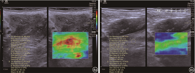
1. VTIQ检查:受检者取截石位,排空大小便,将线阵探头被覆避孕套行经会阴超声扫查,盆底旁矢状切面清晰显示低回声区域内带状高回声PR,使PR在屏幕上呈水平走行,深度为2.0~3.5 cm,并清楚显示其与耻骨下支附着处,成像时避免通过探头对会阴部施加压力。进入VTIQ模式,依次显示VTIQ速度及质量模式图,双幅实时观察二维及VTIQ速度及质量模式图,分别采集图像。VTIQ速度模式下,可获取二维空间分布的剪切波弹性成像图,图像中剪切波速度(shear wave velocity,SWV)由高至低分别呈现红色、黄色、绿色、蓝色。自高至低调整SWV量程大小(最大10 m/s),将感兴趣区(region of interest,ROI;大小3 mm×3 mm)分别置于肛提肌肌腹处,测量3组PR静息状态、最大缩肛状态的SWV值及杨氏模量值,并计算PR静息状态与最大缩肛状态杨氏模量值的差值。VTIQ质量模式可监控所获得图像的弹性分布质量,质量由高到低分别表示为绿色、黄色、红色,选定有效的SWV及杨氏模量值测量的区域(VTIQ质量控制图上色彩均匀分布且呈现为绿色的区域)。取3~5次测量的平均值进行分析。在重复测量时,尽量获取同一平面,ROI大小及放置位置相同。
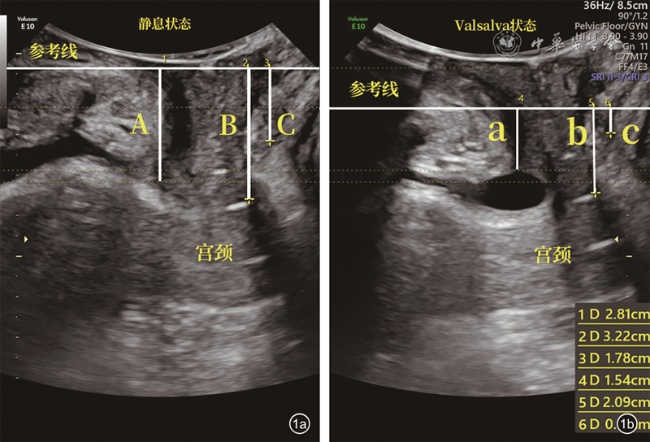
2.盆底超声检查:受检者取截石位,排空大小便,将三维容积探头被覆避孕套行经会阴超声扫查,盆底二维正中矢状面扫查,清晰地显示耻骨联合和肛提肌之间的尿道、膀胱、阴道、直肠等结构,分别在静息状态和最大Valsalva状态下测量数据并采集相应图像,以经耻骨联合内下缘的水平线作为参考线,脏器位于耻骨联合上方时以正数记录,位于下方时以负数记录。二维模式下测量膀胱颈移动度(bladder neck descent,BND)、膀胱尿道后角(posterior urethrovesical angle,PUVA)、尿道旋转角度(urethral rotation angle,URA)、子宫颈外口移动度(cervical orifice descent,COD)、直肠壶腹部移动度(rectal ampulla descent,RAD)。启动四维成像模式,重建出最大Valsalva状态盆膈裂孔图像,容积数据的离线分析使用4D View软件,盆膈裂孔调整至最大Valsalva状态最小裂孔标准水平,平面能够显示前方耻骨联合、两侧耻骨内脏肌、尿道、直肠、阴道,测量盆膈裂孔面积(levator hiatal area,LHA)。
3.测量指标定义:BND为静息状态下与Valsalva状态下膀胱颈下移距离差值的绝对值;PUVA为Valsalva状态下测量膀胱后壁与近端尿道的夹角;URA为静息状态下与Valsalva状态下尿道倾斜角旋转的角度;COD为子宫颈外口最低点下移距离差值的绝对值;RAD为直肠壶腹部位置下移距离差值的绝对值;LHA为即最大Valsalva状态下耻骨联合与耻骨内脏肌内侧缘间覆盖的面积。
三、统计学分析
数据统计采用SPSS 19.0软件,所有资料采用Kolmogrov-Smirnov检验是否符合正态分布,正态分布数据以
±s表示;单因素分析计量资料两组间比较采用独立样本t检验,非正态分布的数据以M(QR)表示,2组间比较采用Mann-Whitney U检验。P<0.05为差异具有统计学意义。应用受试者操作特征(receiver operating characteristic,ROC)曲线计算PR不同状态杨氏模量值及其差值对阴道分娩后盆底功能疾病预测价值,计算曲线下面积、敏感度、特异度,并得出最佳诊断截断值。ROC曲线下面积比较采用MedCalc 18.2统计软件。选出有统计学意义的因素进行二分类Logistic回归分析,变量筛选使用向前似然比法,定义X1为年龄,X2为体质量指数,X3为产次,X4为新生儿体质量,X5为BND,X6为PUVA,X7为URA,X8为COD,X9为RAD,X10为LHA,X11为静息状态PR杨氏模量值,X12为收缩状态PR杨氏模量值,X13为PR杨氏模量值差值。模型系数检验使用Wald χ2检验,模型预测概率值与单个数据一同进行ROC曲线分析。

结果
一、2组产妇一般临床资料与盆底超声参数的单因素分析结果
一般临床资料中,PFD组产次及新生儿体质量均值均高于对照组,差异均有统计学意义(P均<0.05),2组年龄及体质量指数比较差异均无统计学意义(P均>0.05);正常对照组和PFD组盆底超声参数BND、PUVA、URA、COD、RAD及LHA比较,差异均有统计学意义(P均<0.05,表1 ,图1 , 2 , 3 )。
二、VTIQ检查结果及诊断价值分析
正常对照组和PFD组最大缩肛状态杨氏模量值及杨氏模量值差值比较,差异均有统计学意义(P均<0.05),2组静息状态杨氏模量值比较差异无统计学意义(P>0.05,表2 ,图4 )。
ROC曲线显示,最大缩肛状态杨氏模量值区分正常对照组与PFD组的曲线下面积、诊断界值、敏感度、特异度及约登指数分别为0.750(95%CI:0.648~0.834,P<0.05)、73 kPa、71.34%、69.23%及0.407;VTIQ技术测得杨氏模量值差值区分正常对照组与PFD组的曲线下面积、诊断界值、敏感度及特异度分别为0.865(95%CI:0.799~0.927,P<0.001)、28 kPa、71.43%、92.31%及0.637。两者区分正常对照组与PFD组的曲线下面积比较,VTIQ技术测得杨氏模量值差值大于最大缩肛状态杨氏模量值(Z=2.844,P<0.001,图5 )。
表1 2组产妇一般临床资料与盆底超声参数的单因素分析 |
| 组别 | 例数 | 年龄(岁, ±s) | 体质量指数(kg/m2, ±s) | 产次(次, ±s) | 新生儿体质量(g, ±s) | BND[mm,M(QR)] | PUVA[°,M(QR)] | URA[°,M(QR)] | COD[mm,M(QR)] | RAD[mm,M(QR)] | LHA(cm2, ±s) |
|---|---|---|---|---|---|---|---|---|---|---|---|
| 正常对照组 | 52 | 41.3±8.8 | 21.5±2.5 | 1.27±0.53 | 3260.7±301.0 | 17(10,25) | 137(122,144) | 29(16,40) | 6(3,10) | 6(3,13) | 17.4±3.1 |
| PFD组 | 42 | 38.9±7.0 | 22.3±2.0 | 1.62±0.86 | 3621.3±346.6 | 31(27,38) | 149(132,158) | 48(41,65) | 12(5,24) | 13(9,21) | 28.4±6.8 |
| 统计值 | t=1.448 | t=-1.859 | t=2.305 | t=-5.396 | U=5.590 | t=3.033 | t=5.662 | t=3.210 | t=3.948 | t=-8.059 | |
| P值 | 0.151 | 0.066 | 0.021 | <0.001 | <0.001 | 0.002 | <0.001 | 0.001 | <0.001 | <0.001 |

注:PFD为盆底功能障碍;BND为膀胱颈移动度;PUVA为膀胱尿道后角;URA为尿道旋转角度;COD为子宫颈外口移动度;RAD为直肠壶腹部移动度;LHA为盆膈裂孔面积 |
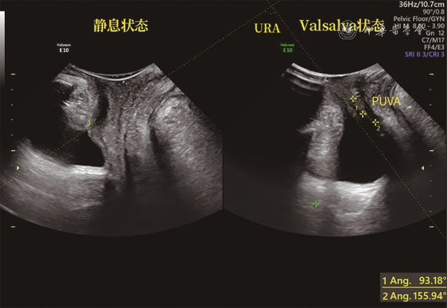
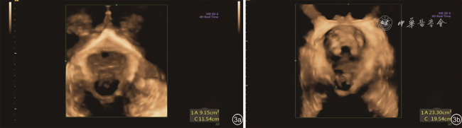
表2 2组产妇不同状态下杨氏模量值及其差值的比较(kPa) |
| 分组 | 例数 | 静息状态( ±s) | 最大缩肛状态[M(QR)] | 弹性值差值[M(QR)] |
|---|---|---|---|---|
| 正常对照组 | 52 | 34.39±15.10 | 83(65~105) | 47(41~61) |
| PFD组 | 42 | 38.09±13.33 | 63(54~82) | 23(20~34) |
| 统计值 | t=-1.234 | U=-4.155 | U=-6.072 | |
| P值 | 0.217 | <0.001 | <0.001 |

注:PFD为盆底功能障碍 |
三、阴道分娩后PFD危险因素的二分类Logistic回归分析及ROC曲线结果
以正常对照组与PFD组2组作为因变量,以单因素分析中对鉴别PFD组有意义的变量:产次(X3)、新生儿体质量(X4)、BND(X5)、PUVA(X6)、URA(X7)、COD(X8)、RAD(X9)、LHA(X10)及杨氏模量值差值(X13)等因素(P<0.05)为自变量,将其带入二分类Logistic回归模型中,经向前似然比法筛选变量。结果显示新生儿体质量(X4)、BND(X5)及LHA(X10)是阴道分娩后PFD的独立危险因素,杨氏模量值差值(X13)是阴道分娩后PFD的独立保护因素(表3 )。Logistic回归模型为:Logit(P)=-60.011+0.010X4+0.599X5+1.202X10-0.416X13。上述方程经似然比检验,差异有统计学意义(χ2=84.534,P<0.001)。
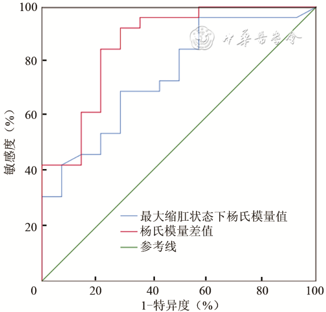
表3 经阴道分娩后盆底功能障碍危险因素的二分类Logistic回归模型分析结果 |
| 自变量 | B值 | SE值 | Wald值 | P值 | OR值 | 95%CI |
|---|---|---|---|---|---|---|
| 新生儿体质量 | 0.010 | 0.004 | 4.872 | 0.027 | 1.010 | 1.001~1.018 |
| 膀胱颈移动度 | 0.599 | 0.279 | 4.617 | 0.032 | 1.821 | 1.054~3.145 |
| 弹性值差值 | -0.416 | 0.177 | 5.514 | 0.019 | 0.659 | 0.466~0.933 |
| 盆膈裂孔面积 | 1.202 | 0.526 | 5.217 | 0.022 | 3.328 | 1.186~9.336 |
| 常量 | -60.011 | 26.031 | 5.315 | 0.021 | 0.000 |
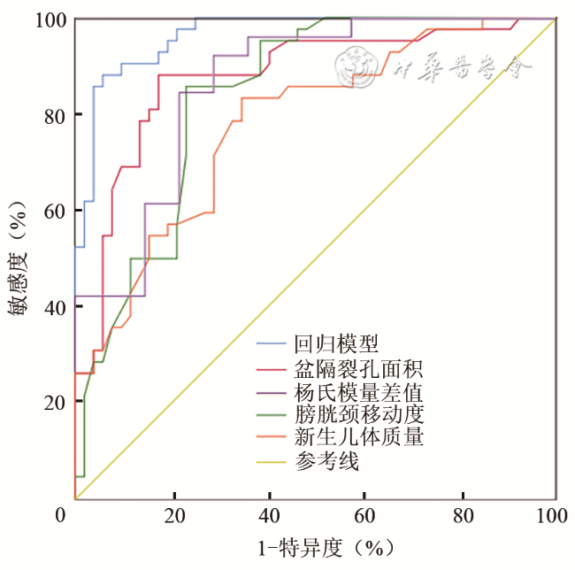
ROC曲线结果显示新生儿体质量(X4)、BND(X5)及LHA(X10)、杨氏模量值差值(X13)以及各指标联合检测预测阴道分娩后PFD的ROC曲线下面积分别为0.779、0.836、0.876、0.865、0.996,其中各指标联合预测的ROC曲线下面积明显高于各指标单独检测的结果(P均<0.05),其余单个指标曲线下面积比较,差异均无统计学意义(P均>0.05)。说明该模型对预测阴道分娩后PFD具有更佳的诊断效能。当回归模型预测概率为0.5454时,约登指数最大为0.8233,其敏感度为88.10%,特异度为94.23%(图6 ,表4 )。
讨论
盆底肌群、筋膜、韧带及神经等盆底主动与被动的复杂支持结构主要用于维持盆腔脏器正常解剖学位置,任何结构的缺陷及损伤都会导致PFD的发生,盆腔脏器的承托更多依赖盆底肌群。其中肛提肌作为盆底肌群重要组成部分,参与形成围绕尿道、阴道、直肠结构的“U”形吊带,通过主动收缩可关闭肛提肌裂孔,有研究表明肛提肌可以进行非手术锻炼治疗,防治PFD的发生[4]。
表4 各参数对盆底功能障碍性疾病的预测分析 |
| 参数 | 最佳截断值 | 曲线下面积 | P值 | 95%CI | 敏感度 | 特异度 |
|---|---|---|---|---|---|---|
| 新生儿体质量 | 3342 | 0.779 | <0.001 | 0.686~0.872 | 83.33 | 65.38 |
| 膀胱颈移动度 | 25 | 0.836 | <0.001 | 0.756~0.917 | 85.71 | 76.92 |
| 弹性值差值 | 28 | 0.865 | <0.001 | 0.791~0.939 | 71.43 | 92.31 |
| 盆膈裂孔面积 | 21.4 | 0.876 | <0.001 | 0.802~0.950 | 88.10 | 82.69 |
| 回归模型联合预测 | 0.545 | 0.966a | <0.001 | 0.906~0.992 | 88.10 | 94.23 |
注:a与新生儿体质量、膀胱颈移动度、弹性值差值及盆膈裂孔面积的曲线下面积比较,差异具有统计学意义(P<0.001、<0.001、=0.006、=0.013) |
流行病学数据显示,女性产后PFD的发生取决于多种复杂的影响因素,包括种族和遗传因素、分娩和妊娠、年龄、新生儿体质量、产次等,其中妊娠和分娩被认为是PFD公认的首要影响因素[5]。尤其在阴道分娩时,激素水平变化、重力作用、胎头下降、机械牵拉及会阴切开均可使盆底组织受压迫及过度牵拉扩张,盆底组织出现去神经化,盆底肌力及肌张力逐渐减弱,导致产后PFD的发生。但仍有部分阴道分娩者甚至是多胎分娩的女性并未出现PFD,因此,探讨哪些因素及诊断指标在阴道分娩后PFD的临床科研工作中具有重要意义。近年来,盆底超声及弹性成像在肛提肌形态及生物力学方面的研究成为国内外热点[6, 7, 8],为其在鉴别PFD中提供了丰富的诊断指标。VTIQ作为一种新型技术,其质量模式图可提高弹性测量准确性,避免了过多无效测量,而最新VTIQ技术不仅可以获取组织SWV,还可直获取组织的杨氏模量值,更直观地评估组织肌肉硬度及弹性方面的信息。
本研究采用VTIQ技术对42例PFD组及52名正常对照者进行了分析。对照组最大缩肛状态杨氏模量值及杨氏模量值差值均大于PFD组,2组患者静息状态杨氏模量值差异无统计学意义。通过构建的ROC曲线显示,最大缩肛状态杨氏模量值和VTIQ技术测得的杨氏模量值差值诊断PFD的ROC曲线下面积分别为0.750和0.865,杨氏模量值差值曲线下面积大于最大缩肛状态杨氏模量值(Z=2.844,P<0.001),当杨氏模量值差值为28 kPa时,敏感度及特异度为71.43%、92.31%,杨氏模量值差值的特异度高于最大缩肛状态杨氏模量值,两者的敏感度相差不大,相比之下杨氏模量值差值在阴道分娩者产后PFD的预测更有价值,杨氏模量差值的改变更能体现盆底肌肌力功能的减弱。
本研究应用单因素分析以及二分类Logistic回归对阴道分娩者产后PFD的相关因素进行综合分析并建立回归模型,包括临床参数、经会阴超声检查参数及杨氏模量值。将单因素分析中对诊断PFD有意义的变量纳入方程,结果显示新生儿体质量、BND和LHA是阴道分娩后PFD的独立危险因素,杨氏模量值差值是阴道分娩后PFD的独立保护因素。本研究中LHA的OR值为3.328,相对于其他特征变量,是准确性最高的特征参数,在临床工作中应对此予以足够重视,这与Dietz等[9]的研究结果一致,他们认为PR撕脱伤和LHA的扩张是盆腔脏器脱垂的独立危险因素。王秋静[10]也发现LHA与产后压力性尿失禁的发生密切相关,均高于其他二维超声诊断参数,OR值为10.513。有研究发现阴道分娩时,为使胎儿正常通过,孕妇肛提肌可拉长至原来的2.5倍[11],造成LHA的扩大。本研究还发现杨氏模量值差值是阴道分娩后PFD的独立保护因素,其差值越大,阴道分娩后越不可能出现PFD。Handa等[12]研究发现肛提肌肌力与盆腔脏器脱垂成反比,肛提肌肌力每增加5 cm H2O(1 cm H2O=0.098 kPa),盆腔脏器脱垂发生的OR值为0.87。牛旺等[13]在肛提肌弹性成像技术研究中也证实了这点,他们认为PFD患者肛提肌成分中肌小节内结蛋白和波形蛋白表达减低、成纤维细胞增多导致纤维化修复增生,会导致盆底肌肌力及肌张力逐渐减弱,造成主动收缩的杨氏模量值变小,PR成分的改变会造成静息状态下杨氏模量值变大,从而使杨氏模量差值出现差异性。本研究中虽然BND的OR值较LHA低,但比盆底超声其他二维参数对阴道分娩后PFD的相关性更大。Laterza等[14]研究发现,阴道分娩后肛提肌损伤的女性1年后仅有泌尿系统的症状而无其他PFD的相关症状,但并未对原因作出过多解释。Nordin和Frankel[15]研究骨骼肌基本生物力学发现,弹性变形是由外力引起的可逆过程,如不超过阈值,一旦外力不再作用,弹性变形就会回到初始状态,笔者认为PR在最大缩肛状态下,PR的弹性变形对直肠、子宫的活动限制更大,牵拉脏器向腹内侧运动,而膀胱颈部的限制相对较小,从而解释了BND较中后盆腔COD、RAD对阴道分娩后PFD的相关性更大,对阴道分娩后更容易出现泌尿系统症状的原因。新生儿体质量作为阴道分娩后PFD的独立危险因素之一,当新生儿体质量大于3342 g时诊断效能较高,其敏感度和特异度为83.33%和65.38%,这与杨彩霞和唐淑稳[16]的研究结果一致。
本研究进一步通过绘制ROC曲线模型分析上述4种指标与回归模型的诊断效能,发现回归模型的曲线下面积为0.996,明显高于各指标单独检测的结果,单个指标中虽然LHA的曲线下面积最大,但其余各个指标的曲线下面积相互比较,差异均无统计学意义,说明建立二分类Logistic回归模型可以提高阴道分娩者产后PFD的诊断准确性。
综上所述,VTIQ技术联合盆底超声建立的回归模型对阴道分娩者产后PFD有较高的诊断价值,在指导临床的工作中起着重要作用。但本研究存在一定的局限性,如样本量偏少、地域性人群差异及未对阴道分娩时器械助产及会阴切开等影响因素进行分析,仍需加大样本、进行多中心临床研究,建立更加综合、有效的回归模型进一步验证。