
近年来,关于女性盆底功能障碍(pelvic floor dysfunction,PFD)的研究大量开展,越来越多的研究在关注妊娠和分娩对盆底功能的影响,尤其是分娩方式的选择及产次对女性盆底结构功能的影响[1, 2, 3, 4, 5, 6]。肛提肌裂孔(levator hiatus,LH)是人体最大的潜在疝口,所有盆腔器官脱垂(pelvic organ prolapse,POP)都经过此孔脱出[7]。因此,LH的大小与肛提肌的完整性一样,对盆腔器官的支撑作用很重要[8]。盆底三维超声测量LH面积的可重复性及其与MRI的一致性已得到证实[9, 10]。本研究旨在通过盆底三维超声检查评估不同分娩方式及不同产次对LH大小的影响及其与POP的关系。
资料与方法
一、对象
收集2019年4月至12月在湖北省妇幼保健院行产后42~60 d常规检查的产妇250例,进行问卷调查及盆底超声检查。其中初次经阴道分娩(first vaginal delivery,FVD)组150例;二次经阴道分娩(second vaginal delivery,SVD)组50例,两胎均经阴道分娩;剖宫产(cesarean section,CS)组50例,为剖宫产初产妇。排除标准:(1)有早产及引产史;(2)有腹部手术史;(3)有巨大盆腔包块者;(4)有糖尿病及高血压等慢性疾病者。对照组120例,为同期本院初次人工流产术后15 d复查的未分娩女性,排除标准同上。所有研究对象,根据是否有POP分为POP组(167例)和无POP组(203例)。
二、仪器与方法
1. 仪器:迈瑞Resona8彩色多普勒超声诊断仪,腔内容积探头,频率为3~9 MHz,容积扫查角度为120°;腹部容积探头,频率为2~8 MHz,容积扫查角度为85°。
2. 问卷调查:对入选对象进行一般问卷调查,调查内容包括年龄、身高、本次孕前及分娩前体质量、盆底超声检查时体质量(即分娩后42~60 d体质量)、分娩次数及方式、新生儿出生体质量、有无排便异常及既往史(慢性咳嗽、高血压、糖尿病、盆腔手术、早产及引产史等)等。测定体质量指数(body mass index,BMI)。
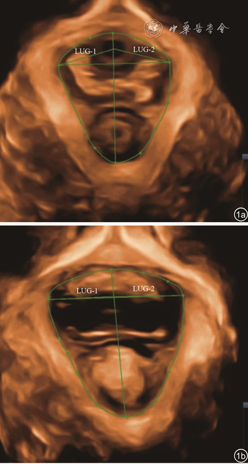
3. 检查方法及测量指标:检查前嘱受检者排空大便,膀胱容量为50~100 ml,正中矢状面清晰显示耻骨联合、膀胱、尿道、阴道、肛管和肛直肠角等结构,然后启动三维图像采集系统,将感兴趣区取样框置于耻骨联合内下缘及直肠肛管角区域,分别在静息状态及最大Valsalva状态下取得清晰三维重建图像,沿肛提肌内侧缘测量LH面积(图1 )。
4. POP超声诊断标准:盆底超声参考线采用经耻骨联合后下缘的人体水平线,即经耻骨联合后下缘与耻骨联合中轴线呈135°的直线。膀胱后壁或尿道突入阴道,最低点达参考线水平诊断为阴道前壁脱垂;子宫颈最低点距参考线水平<3.0 cm,或最大Valsalva状态时较静息时移动度>2.0 cm,诊断为子宫脱垂;超声显示直肠壶腹部膨出高度>0.5 mm,诊断为直肠膨出。
三、统计学分析
采用SPSS 17.0统计学软件对数据进行分析。孕前、分娩前及产后BMI、新生儿体质量、LH面积为计量资料,以
±s表示,分析SVD组、FVD组、CS组和对照组组间差异采用方差分析,各组间两两比较采用t检验。静息状态和Valsalva状态、有POP组和无POP组间LH面积比较采用t检验;POP超声检出率为计数资料,采用χ2检验分析SVD组、FVD组、CS组和对照组组间差异。P<0.05为差异具有统计学意义。

结果
一、一般临床资料比较
SVD组、FVD组及CS组各组间,孕前、分娩前、产后BMI及新生儿体质量比较,差异均无统计学意义(P>0.05)。各组间年龄比较,SVD组>FVD组>CS组>对照组,除FVD组与CS组间差异无统计学意义,其余各组间比较,差异均具有统计学意义(P均<0.05,表1 )。
表1 各组研究对象一般临床资料比较( |

| 组别 | 例数 | 年龄(岁) | 孕前BMI(kg/m2) | 分娩前BMI(kg/m2) | 产后BMI(kg/m2) | 新生儿体质量(kg) |
|---|---|---|---|---|---|---|
| SVD组 | 50 | 33±3.66 | 21.40±2.25 | 26.49±2.21 | 23.47±1.82 | 3.41±0.43 |
| FVD组 | 150 | 29±3.48a | 21.11±2.36 | 26.13±2.51 | 22.99±2.39 | 3.33±0.37 |
| CS组 | 50 | 28±3.75b | 20.92±2.62 | 26.30±2.30 | 22.82±2.02 | 3.43±0.56 |
| 对照组 | 120 | 23±2.97c | - | - | - | - |
| F值 | 124.68 | 0.52 | 0.44 | 1.22 | 1.37 | |
| P值 | <0.001 | 0.598 | 0.645 | 0.297 | 0.257 |
注:BMI为体质量指数,SVD为二次经阴道分娩组,FVD为初次经阴道分娩组,CS为剖宫产组;a与SVD组比较,差异具有统计学意义(t=6.95,P<0.001),b与SVD组比较,差异具有统计学意义(t=6.75,P<0.001),c与SVD组比较,差异具有统计学意义(t=18.64,P<0.001),a与c比较,差异具有统计学意义(t=15.01,P<0.001),b与c比较,差异具有统计学意义(t=9.23,P<0.001);-表示无数据 |
二、各组间静息状态及最大Valsalva状态下LH面积比较
静息状态及最大Valsalva状态LH面积均为:SVD组>FVD组>CS组>对照组,各组组间差异均具有统计学意义(P均<0.05,表2 )。各组组内比较,相比静息状态,最大Valsalva状态下LH面积明显增大,差异均具有统计学意义(P均<0.05,表2 )。
表2 各组研究对象间肛提肌裂孔面积测值比较( |
| 组别 | 例数 | 静息状态 | Valsalva状态 | t值 | P值 |
|---|---|---|---|---|---|
| SVD组 | 50 | 16.59±3.12 | 22.80±8.37 | 4.92 | <0.001 |
| FVD组 | 150 | 15.34±3.50a | 18.46±6.63a | 5.10 | <0.001 |
| CS组 | 50 | 13.09±2.78b | 15.58±3.91b | 3.67 | <0.001 |
| 对照组 | 120 | 11.09±2.47c | 13.11±5.30c | 3.78 | <0.001 |
| F值 | 31.89 | 11.14 | |||
| P值 | <0.001 | <0.001 |
注:SVD为二次经阴道分娩组,FVD为初次经阴道分娩组,CS为剖宫产组;a与SVD组比较,差异具有统计学意义(t=2.25、3.74,P=0.026、<0.001),b与SVD组比较,差异具有统计学意义(t=5.92、5.53,P均<0.001),c与SVD组比较,差异具有统计学意义(t=12.21、9.07,P均<0.001),a与b比较,差异具有统计学意义(t=4.13、2.91,P<0.001、=0.004),a与c比较,差异具有统计学意义(t=11.25、7.19,P均<0.001),b与c比较,差异具有统计学意义(t=4.63、2.97,P<0.001、=0.003) |
三、POP组和无POP组间比较
静息状态及最大Valsalva状态下LH面积,POP组均大于无POP组,差异均具有统计学意义(P均<0.05,表3 )。
表3 POP组与无POP组间肛提肌裂孔面积比较( |
| 组别 | 例数 | 静息状态 | Valsalva状态 |
|---|---|---|---|
| 无POP组 | 203 | 12.38±2.17 | 15.59±3.50 |
| POP组 | 167 | 14.88±3.49 | 20.93±6.53 |
| t值 | 8.06 | 9.50 | |
| P值 | <0.001 | <0.001 |
注:POP为盆底器官脱垂 |
对照组、CS组、FVD组及SVD组的POP超声检出率分别为17.5%(21/120)、26.0%(13/50)、60.0%(90/150)及86.0%(43/50)。CS组及对照组间超声检出率比较,差异无统计学意义,其余各组间比较差异均具有统计学意义(SVD组vs FVD组:χ2=11.38,P<0.001;SVD组vs CS组:χ2=36.53,P<0.001;SVD组vs对照组:χ2=70.55,P<0.001;FVD组vs CS组:χ2=17.36,P<0.001;FVD组vs对照组:χ2=49.74,P<0.001)。所有研究对象中,仅发现1例初次经阴道分娩的女性伴肛提肌撕裂。
讨论
妊娠及分娩均会导致盆底结构功能发生改变,是引起PFD的两个独立危险因素[1, 2, 3, 4, 5, 6,11]。但分娩方式对盆底结构功能的影响报道不一,剖宫产对女性盆底结构功能的完整性有无保护作用尚有争议[12, 13, 14, 15]。王丽珍和冯才敏[12]及刘瑞芳[13]的研究认为阴道分娩及剖宫产在盆底功能的影响上差异不大,盆底功能不应成为孕产妇选择分娩方式考虑的重要因素。Horak等[14]研究发现与二胎剖宫产相比,二胎经阴道分娩者Valsalva动作后膀胱膨出下降程度及LH面积无差异,膀胱颈移动度虽有增大,但差异无统计学意义;亦认为二胎的分娩方式对膀胱支持结构和(或)肛提肌没有明显影响。而杨慧燕等[15]对两胎分娩方式均相同的二胎经阴道分娩产妇和二胎剖宫产产妇于产后6~8周行盆底实时三维超声检查,发现二胎经阴道分娩组最大Valsalva状态下的LH面积高于二胎剖宫产组。本研究结果显示在静息状态和最大Valsalva状态下,CS组LH面积均介于对照组和阴道分娩组间,各组间比较差异有统计学意义,表明经阴道分娩及剖宫产对LH面积均有影响,且产后早期经阴道分娩较剖宫产对LH面接的影响更明显。经阴道分娩组与CS组间的差异,可能与阴道分娩继发的肛提肌损伤有关;而CS组与对照组间的差异,可能与妊娠本身对盆底结构产生的不可逆影响有关,且这种不可逆影响不受分娩方式的影响[5]。故剖宫产可以保护女性免受阴道分娩继发的肛提肌损伤的影响,但不能保护女性免受妊娠引起的对盆底不可逆的影响。
研究认为经产妇的盆底结构功能改变较初产妇更为明显,二次经阴道分娩女性最大Valsalval动作时LH面积、前后径、左右径均较初次经阴道分娩女性明显增大;子宫脱垂、尿道内口漏斗化、会阴体活动过度发生率亦均高于初次经阴道分娩女性[2,16, 17]。不仅阴道分娩多产次组明显高于单产次组,剖宫产多产次组亦明显高于单产次组[2]。且LH面积随产次增多而增大,产次是LH的独立影响因素。本研究结果显示,在静息状态和最大Valsalva状态下,SVD组LH面积均大于FVD组,与上述观点一致。而Horak等[14]对二胎女性的研究发现,与一胎分娩产后4.2个月相比,二胎分娩产后3个月最大Valsalva动作后膀胱膨出下降程度及LH面积未见明显差异,认为二次妊娠分娩对膀胱支持结构和(或)肛提肌没有明显影响。分析原因可能为李载红等[2]、徐英姿等[17]以及本研究的研究对象均为产后42 d女性,分析的是产次对盆底结构的近期影响,而Horak等[14]分析的是远期影响。亦可能与病例分组有关,Horak等[14]的研究中初次妊娠分娩组和二次妊娠分娩组仅根据分娩次数分组,2组中分娩方式既包括经阴道分娩也包括剖宫产,而本研究同时按照分娩次数和分娩方式进行分组,初次分娩分为FVD组和CS组,SVD组两胎均经阴道分娩。
肛提肌撕裂是指耻骨直肠肌从耻骨支附着点撕脱,是肉眼可见的损伤,会引起LH面积增大和肌肉收缩力减弱,导致POP的发生[18]。但是至少一半的阴道分娩女性的肛提肌没有肉眼可见的损伤,本组所有研究对象中,仅发现1例初次经阴道分娩者肛提肌撕裂。但是在阴道分娩时LH可能会过度膨胀[19],发生肌肉功能解剖上宏观不可见的“微创伤”。“微创伤”是指LH不可逆的过度扩张,即与产前相比,Valsalva状态下,产后LH面积增加超过20%,这种“微创伤”在阴道分娩的女性中很常见[19]。研究报道,90%二胎经阴道分娩女性及57.78%二胎剖宫产女性产后LH面积>20 cm2[15]。LH是人体最大的潜在疝口,所有POP都经过此孔脱出[7],LH出现“微创伤”面积增大,会导致POP的风险增加,且POP的发生风险随LH面积增大而增加[20]。本研究结果显示,静息状态及最大Valsalva状态下POP组LH面积均大于无POP组,验证了上述观点。
总之,不同分娩方式和产次对LH面积的影响不同,阴道分娩较剖宫产影响大,二次分娩较单次分娩影响大;LH面积越大,POP风险越大。与分娩方式及产次有关的LH面积增大可能是POP风险增加的原因。实时三维超声能够清楚呈现产后女性不同状态下的LH,并准确测量其面积,可早期发现盆底支持结构的改变。