
心室-动脉耦联的提出已经有30多年的历史,该机制认为心脏和动脉系统在解剖学及功能学上都存在一定的内在关联,耦联则是强调承担动脉系统压力变化时心室的泵功能反应,由此可将心室系统和动脉系统相互联系,进而了解心脏做功和能量储备。左心室-主动脉耦联的研究已经比较成熟,在高血压、心力衰竭、冠心病及代谢性疾病中的价值已被深入探讨[1]。随着右心系统的结构和功能逐渐受到重视,对右心室-肺动脉耦联的相关研究也在不断开展,并在心力衰竭、肺动脉高压、危重症医学等方面逐渐发挥重要作用。本文即总结右心室-肺动脉耦联的相关机制,同时回顾超声心动图指标在评价右心室-肺动脉耦联方面的研究进展,展望该领域的研究意义和方向。
一、右心室-肺动脉耦联的生理机制及有创评价
根据Frank-Starling曲线,心室功能会不断适应机体血流动力学的变化,即前负荷增加时,心室的收缩能力随之增强,进而增加心输出量,该曲线的整体斜率代表了心室收缩力,但其无法评估动脉系统的压力情况,因此就无法表征心室-动脉耦联关系。1983年,Sunagawa等[2]首次提出心室压力-容积曲线的概念,该曲线在表征心室收缩能力的同时也可以评价有效动脉弹性,综合了心室和动脉两个水平的特征,可以用来监测心室-动脉耦联。评估的具体方法是利用下腔静脉内充气球囊不断改变心脏前负荷,得到一系列不同前负荷条件下的心室压力-容积曲线,每条曲线的收缩末期点进行线性回归形成收缩末期压力容积关系线(end-systolic pressure-volume relationship,ESPVR),生理状态下其大致呈直线,该直线的斜率便称为收缩末期弹性(end-systolic elastance,Ees),用以表征心室收缩力,Ees本身对后负荷变化不敏感,但是对心肌收缩力敏感,其大小也会随之变化。与此同时,在单心动周期的压力-容积曲线中收缩末期点与舒张末期点间形成另外一条直线,其斜率即收缩末期压力与每搏量之间比值,称为有效动脉弹性(effective arterial elastance,Ea),用以表征动脉弹性特征,Ea对后负荷变化敏感,当心室后负荷增加时,心室收缩末期压力会升高,每搏量下降,Ea会增大,心室就需要加强收缩以维持机体需要。两者比值Ees/Ea便可将心室收缩能力与动脉弹性状态联合在一起,用于监测心室-动脉耦联状态[3](图1 )。这一指标在左右心均适用,是评估右心室-肺动脉耦联的重要生理参数和金标准。
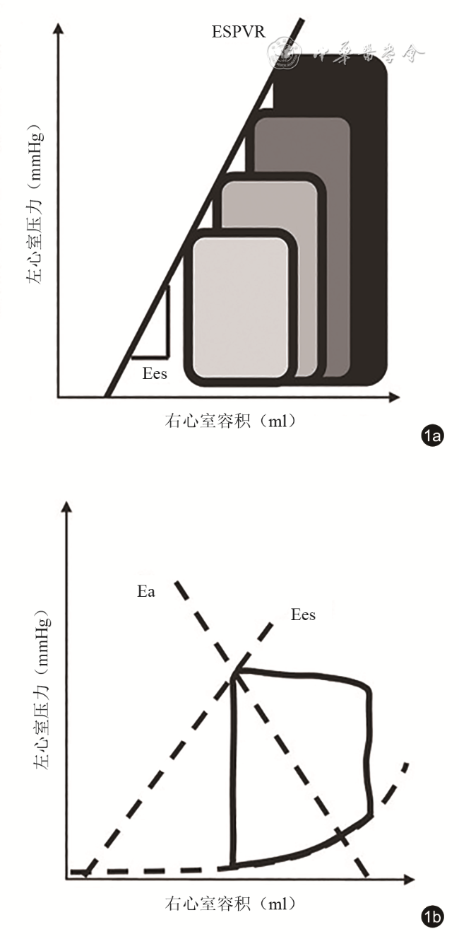
图1 右心室-肺动脉耦联的心导管评估方法。图a显示改变右心室前负荷后所得到的不同前负荷条件下的压力-容积曲线,每条曲线的收缩末期点形成右心室收缩末期压力容积关系线(ESPVR),其斜率即代表收缩末期弹性(Ees);图b显示在不改变前负荷的条件下,单个心动周期的压力-容积曲线(图中黑色闭环实线)中收缩末期点与舒张末期点连接形成的直线的斜率即有效动脉弹性(Ea),此时两条直线交汇点(Ees/Ea)即表征右心室-肺动脉耦联[图片改自:Ikonomidis I, Aboyans V, Blacher J, et al. The role of ventricular-arterial coupling in cardiac disease and heart failure: assessment, clinical implications and therapeutic interventions. A consensus document of the European Society of Cardiology Working Group on Aorta & Peripheral Vascular Diseases, European Association of Cardiovascular Imaging, and Heart Failure Association. Eur J Heart Fail, 2019, 21(4): 402-424.]注:1 mmHg=0.133 kPa |
然而,多次改变前负荷的监测方式其临床应用相对受限,因此目前多利用单一负荷状态的方法,即不再改变患者前负荷,在单个压力-容积环中进行Ees/Ea的估算,此时Ees仅近似为收缩末期压力(end-systolic pressure,ESP)与收缩末期容积(end-systolic pressure,ESV)的比值,Ea仍为收缩末期压力与每搏量的比值,这种估算方法操作相对简单,经过验证与标准测量方式的一致性很好[4],被大多数临床医师认可,也是目前进行临床研究的主要手段。Ees/Ea不仅可以判断右心室相对于肺动脉压力变化所做出的功能调整,还可以评估心室做功的效率,按照生理学要求,心室的整体收缩力不可能都用来克服动脉压力负荷,因此正常人静息状态下Ees/Ea通常大于1,其达到最佳能量转换时,数值一般是1.5~2[5]。例如当肺动脉压力负荷增加时,右心室就要牺牲自身的机械效率来维持正常的输出量,如果肺动脉压力负荷进一步升高,便会出现右心室-肺动脉解耦联的情况,导致一系列病理损害,心室做功的效率也会随之下降。
二、右心室-肺动脉耦联的超声相关评估指标
超声心动图在评价右心室-肺动脉耦联方面具有无创、简便、可重复性好的优势。截至目前,已有多种超声心动图指标被研究者发现可用于评价右心室-肺动脉耦联,通过这些参数,临床医师可以较快捷地预判疾病状态并做出决策,改善甚至降低患者的死亡风险。
(一)三尖瓣环收缩期位移与肺动脉收缩压的比值
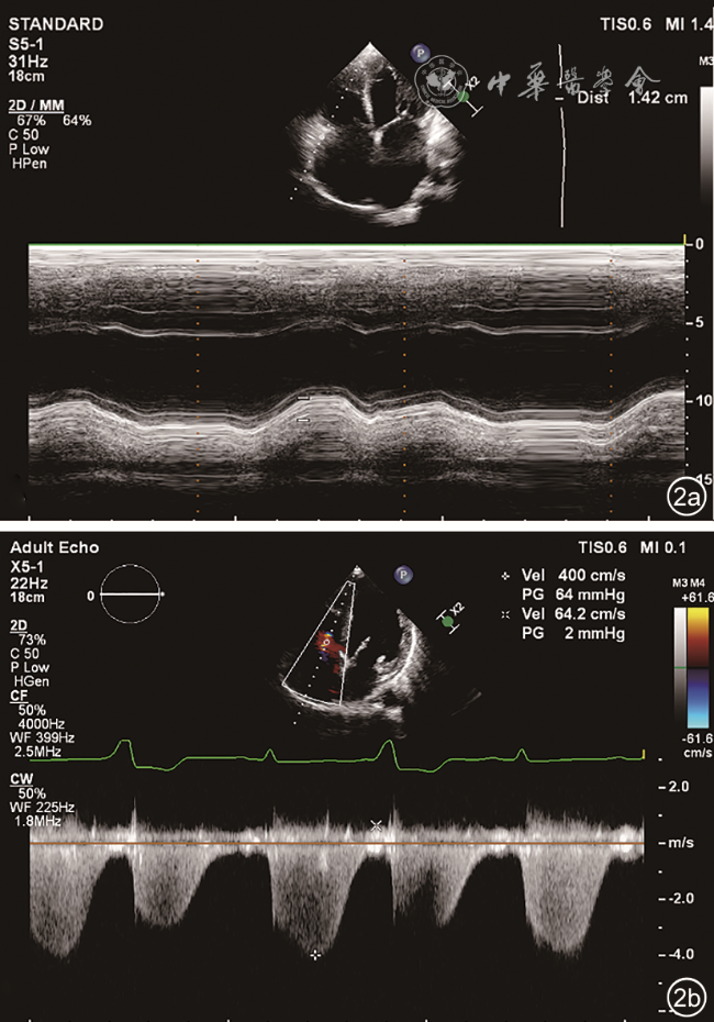
三尖瓣环收缩期位移与肺动脉收缩压的比值(tricuspid annular plane systolic excursion / pulmonary arterial systolic pressure,TAPSE/PASP)是由意大利研究者Guazzi等于2013年首次提出的[6],是目前最为常用的超声心动图领域用于表征右心室-肺动脉耦联的指标。心导管所得有创指标Ees/Ea是利用表征心室收缩能力的Ees,与表征动脉系统特性的Ea,两者相比而获得的。基于超声心动图方法,目前较常用且简便的评价右心室收缩功能的参数即为TAPSE,评价肺动脉压力的参数即为PASP,因此研究者创造性地引申出这一比值参数,来间接替代Ees/Ea(图2 )。后续研究表明,TAPSE/PASP与Ees/Ea的相关性较好,测量结果的组内及组间一致性也较高,而且其相较于TAPSE和PASP均能更好反映右心系统的功能特征[7]。
(二)每搏量与收缩末期容积的比值
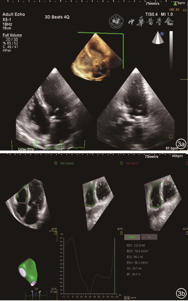
每搏量与收缩末期容积的比值(stroke volume/end-systolic ventricular volumes,SV/ESV)的概念是在一项MRI研究中提出来的。如前述,在不改变前负荷的监测方式下,Ees可近似为ESP/ESV,Ea则由ESP/SV获得,两者比值即为SV/ESV,因此其原理也是基于Ees/Ea的近似推导[8]。由于右心结构的独特性,MRI是目前判定右心室容积优选的无创检查方式,其能够更准确地获得SV/ESV,但是MRI检查复杂耗时,而且要保证患者不存在心律失常,否则所得数据不能完全对应心动周期,一定程度限制了其临床应用。随着近些年三维超声心动图的迅猛发展,特别是对于右心室的容积定量开始成熟,加之超声心动图具有简便、快捷等优势,SV/ESV作为一个新的评价右心室-肺动脉耦联的超声指标逐渐进入研究者的视线(图3 ),其评估效能也在多项研究中得到验证。
(三)其他指标
除以上2个常用的评价右心室-肺动脉耦联的超声指标以外,还有一部分研究和应用并不深入的参数,均基于对上述2个指标的延伸和修正而产生的,其中包括右心室整体纵向应变与肺动脉收缩压的比值(global right ventricle longitudinal strain / pulmonary arterial systolic pressure,RVGLS/PASP)、右心室面积变化率与平均肺动脉压的比值(fractional area change / mean pulmonary artery pressure,FAC/mPAP)、三尖瓣环收缩期位移与右心室舒张末期内径的比值(tricuspid annular plane systolic excursion / right ventricular end-diastolic diameter,TAPSE/RVEDD)等。这些新兴参数,部分已通过对比研究发现其有效性及临床价值高于传统指标,也有部分指标的优劣性尚未得到验证,其临床实用性仍需进一步探索[9, 10]。
三、右心室-肺动脉耦联的超声相关指标临床应用进展
(一)心力衰竭
一直以来,心力衰竭患者的右心系统评估都是临床医师需要关注且感到较为棘手的问题,研究证实无论是右心室功能障碍还是肺动脉血流动力学异常,都是心力衰竭演变框架中的重要环节,由于存在压力超负荷,严重的心力衰竭会出现明显的右心室扩大、肺动脉高压以及严重的三尖瓣反流,一旦出现右心衰竭,患者多预后不良[11],因此密切观察右心系统的功能变化至关重要。但是既往研究都是对右心室收缩功能及肺动脉系统进行独立讨论,获得的结果在不同层面上也不完全相同。Kjaergaard等[12]发现,TAPSE下降与心力衰竭患者住院死亡率升高明显相关,当TAPSE数值增加一倍时,患者住院死亡率可下降26%;而Mohammed等[13]则指出,表征右心室收缩功能的各项指标与心力衰竭患者全因死亡及再次住院率并无明显关联,而肺动脉压力则是重要的预测因子,因此近年来利用联合指标TAPSE/PASP对心力衰竭患者进行评估成为讨论的热点。这方面的研究在射血分数保留的慢性心力衰竭患者中具有较重要的临床意义,大规模临床观察指出,射血分数保留的慢性心力衰竭患者出现右心功能不全及肺循环障碍的概率要低于射血分数减低的心力衰竭患者,这增加了及早识别的难度,但是这类患者的远期再住院风险却极高,因此进行早期判断并有效预测不良事件的发生尤为重要。利用TAPSE/PASP对该类患者进行分组研究发现,当TAPSE/PASP越小时,患者出现心房颤动、肾功能异常等不良事件的可能性增大,其预测能力要高于既往得到验证的相关参数,被看作是对该类患者进行危险分层的有效手段[14],Santas等[15]研究甚至发现当该比值低于0.36时可以预测所有的临床终点事件,展现了较好的临床实用性。除此之外,TAPSE/PASP还可联合心肺运动实验、负荷超声等检查手段,对这类心力衰竭患者进行综合评估,预测其相关结局[16, 17],目前联合研究仍在不断尝试,相信可以更好地指导这类患者的急诊处理和后续诊治。
急性右心衰竭是导致危重患者死亡率升高的重要原因,其诊断也具有挑战性,患者通常会由于不能及时发现而导致治疗不足,因此获得比较简便、有效的评估指标是亟待解决的问题。一项对于极危重患者的前瞻性研究发现,右心室-肺动脉耦联参数Ees/Ea与右心收缩功能参数如右心室射血分数( right ventricular ejection fraction,RVEF)具有良好一致性,而且可以用来预测急性右心衰竭的发生,凸显出耦联机制在这方面的意义所在[18]。但是鉴于急性心力衰竭的紧急性和严重性,相关的临床研究以及动物实验仍然处于初步探索阶段,其中的机制是什么、如何更好地将超声心动图参数应用于这类人群进行预测和指导,更多、更细致的内容还需要进一步挖掘。
(二)肺动脉高压
在肺动脉高压患者中,随着肺循环压力的不断升高,右心室功能开始下降并逐渐与肺循环“脱钩”,这是病情恶化的转折点。然而,肺动脉高压患者右心室功能的评估本身就具有挑战性,因为右心室对后负荷依赖性较强,常用的负荷依赖性指标,如右心室面积变化率、三尖瓣环收缩期位移及右心室射血分数等可能不能完全反映右心室固有的功能障碍。但是右心室-肺动脉耦联却可以反映心肌收缩力与肺血管后负荷之间的复杂相互作用,对检测隐匿性右心室功能障碍具有良好的敏感度,因此其在肺动脉高压的诊断、预后评估及诊疗协助方面可能会更具优势。中长期队列研究已经指出,心导管指标Ees/Ea对于肺动脉高压患者的主要临床终点事件的预测能力明显优于常规生化或机械参数,其为该领域的研究奠定了理论基础[19]。Tello等[20]随后研究证实,在肺动脉高压患者中TAPSE/PASP与侵入性指标Ees/Ea具有较好的一致性,被认为是较简便的评价该类患者右心室-肺动脉耦联的超声参数,并通过回顾性研究发现TAPSE/PASP对原发性肺动脉高压患者的10年内总死亡率具有较好的预测能力,该比值越低,患者生存率越低,从而肯定了这一超声指标在原发性肺动脉高压中的应用价值。同时,关于继发性肺动脉高压的研究也强调了TAPSE/PASP的作用,如在一项关于系统性红斑狼疮合并肺动脉高压的观察中,研究者认为TAPSE/PASP能够改善这类患者的临床危险分层、增强其他指标的临床效能,同时其与患者的全因死亡率以及临床恶化密切相关[21],可为提早进行干预和治疗提供新的靶点。
除此之外,SV/ESV在肺动脉高压领域的意义也不断被发掘。Vanderpool等[22]首先以原发性肺动脉高压患者为研究对象,明确心脏磁共振测量获得的SV/ESV与心导管所得的Ees/Ea具有良好的一致性,Aubert等[23]同时验证,由三维超声心动图所获得的容积比值参数SV/ESV与心脏磁共振所得结果具有很高的一致性,与心导管所得Ees/Ea之间的相关性也较好,由此确立了在肺动脉高压领域利用SV/ESV进行右心室-肺动脉耦联评估的理论基础;研究同时指出当肺动脉压力越高、心肌收缩能力下降时,SV/ESV会明显减低,提示右心室-肺动脉解耦联,也预示着出现不良预后的可能性增加。对于因肺动脉高压行肺移植术后的患者,SV/ESV还可用于评价术后右心室对动脉负荷变化的适应性[24],帮助临床医师辨别治疗效果的差异以及可能存在的风险。
(三)危重症管理
右心室-肺动脉解耦联现象在危重症患者中并不少见,这种解耦联主要来源于右心室功能障碍以及肺动脉压力不匹配两个方面。原发性右心室功能障碍在败血症、右心室心肌梗死以及心肺手术中很常见,而肺动脉压力不匹配通常与急性呼吸窘迫综合征(acute respiratory distress syndrome,ARDS)、肺栓塞等有关,当两者之一出现异常时,就会影响整个右心系统的功能和效率[25]。以ARDS为例,ARDS相关的肺血管功能障碍增加了右心室后负荷,为克服增大的后负荷,右心室收缩力代偿性增加可以暂且维持耦联状态,当病程逐渐进展时,耦联状态便被破坏,能量的转换随之受到影响,这对于危重症患者可能是致命性的,因此诊治过程中不仅仅要进行右心室功能的改善,更要关注肺循环情况,两者同时进行纠正对于优化管理和救治更为重要。近期Alto等[26]对新型冠状病毒引发的ARDS患者进行右心室-肺动脉耦联状态的监测,验证了失耦联现象与ARDS的发生明显相关,而且TAPSE/PASP可以独立预测患者治疗结局,肯定了床旁超声在新型冠状病毒流行期间患者救治方面的独特价值。与此同时,在急性肺动脉栓塞领域也有研究在进行,有学者指出TAPSE/PASP在预测急性肺栓塞患者的不良预后方面具有独特优势,其可以改善患者危险分层并及时发现可能出现短期内恶化的患者[27]。然而,鉴于危重症医学领域的独特性,利用超声心动图来评价这类患者的右心室-肺动脉耦联的研究才刚刚展开,而且无创性的超声检查可能会更加受到临床医师的青睐,是新兴的热点方向,这些都需要循证医学证据的不断积累,也相信未来超声心动图在这一领域的价值会逐渐显现。
四、局限性与展望
纵观目前的研究进展,TAPSE/PASP作为应用最广泛的评价右心室-肺动脉耦联的超声指标,适用领域在不断扩展,具有良好的应用前景。然而,TAPSE/PASP本身也具有一定局限性,从指标本身而言,TAPSE仅代表右心室前壁的纵向运动情况,同时测量受到角度的影响,不能完全代表整个右心室功能;而PASP也是基于三尖瓣反流速度及右心房压力的估算值,并不能准确代表肺动脉系统的负荷状态,因此其临床应用价值不可被过分夸大。面对这样的局限性,有研究者将右心室应变指标引入来评价右心室-肺动脉耦联,指出新参数RVGLS/PASP在慢性心力衰竭患者中对远期死亡的预测能力要优于TAPSE/PASP[28],但是其测量简便性又受到限制,因此新的探索仍在继续。此外,TAPSE/PASP的正常范围及异常区间一直未被确定且受到患者年龄、机体容量状态等因素的影响较大[29],这也是阻碍其临床推广的原因之一。因此,TAPSE/PASP在评价右心室-肺动脉耦联方面尚存在较多问题需要解决。除此之外,SV/ESV也存在固有缺陷,这主要体现在Ees计算时存在简化流程,即不再改变前负荷而仅仅依靠单一状态下的压力-容积关系获得,且比值参数忽略了收缩末期压力的作用,成为了单纯与容积有关的指标,一定程度上限制了其临床应用范围[30]。目前,关于SV/ESV的超声心动图研究依然很少,在心力衰竭、先天性心脏病领域均无明显突破,随着三维超声心动图对右心系统评价的进步,更多的研究必将不断出现以验证并扩展其应用价值。
对这些超声指标的产生过程进行分析发现,其大多是根据Ees/Ea进行推论或衍生所得,且均经历2个主要研究过程,其一是验证与心导管测压的金标准Ees/Ea之间一致性如何,其二即观察相较于现有参数是否存在优势,如若效用更好,那么就要关注其在不同疾病谱中有怎样的变化特征,在诊断治疗等方面有怎样的价值和意义。这也为后续的研究指明了部分思路和方向。但是每项单独的指标不可随意组合,仍然需要研究者建立在已经获得的实验或数据基础上进行分析和解释,明确每个新事物的局限性和适用范围,这样才能获得可靠性高、有价值的参数。随着心脏疾病诊疗精细化及无创化要求的提高,尤其是对右心功能及其相关疾病的不断重视,对于右心室-肺动脉耦联的精准评价已逐渐被提上日程,超声心动图作为其中重要的辅助检查方式,伴随自身技术的逐步提高,其如何更好地反映右心室-肺动脉耦联的变化、如何进行有效准确的监测、如何确定在不同疾病中对诊断和治疗的价值,都需要更多的研究进一步探索。