
涎腺病变种类繁多,主要包括炎性病变、自身免疫性病变及肿瘤病变,以良性病变多见。近年来,涎腺肿瘤的发病率及检出率呈上升趋势,越来越多的小肿瘤被发现,涎腺恶性肿瘤发生率逐渐增加[1]。涎腺良恶性病变术前准确的诊断对治疗方式的选择以及预测术后复发率、面瘫发生率有重要的临床意义[2, 3]。高分辨率超声常作为涎腺疾病首选检查方法,其可显示病灶的位置、大小、形态、回声和淋巴结受累情况,但术前对病变良恶性进行评估仍存在一定的困难[4]。超声弹性成像及超声造影(contrast enhanced ultrasound sonography,CEUS)等新技术的发展,为诊断提供了更多的信息。目前,应用多种超声技术的多模态超声研究主要集中在乳腺、前列腺等疾病中[5, 6],尚无应用于鉴别涎腺良恶性局灶性病变性质的研究,本研究分析不同超声诊断模式对涎腺局灶性病变的诊断效能,探讨多模态超声鉴别涎腺局灶性病变良恶性的诊断价值。
资料与方法
一、对象
回顾性分析2018年1月至2020年12月在解放军总医院第一医学中心收治的128例经手术或穿刺病理确诊的涎腺病变患者的临床资料。其中男性74例,女性54例,年龄19~81岁,平均年龄(51.73±3.3)岁。所有患者手术或穿刺前均进行了常规超声检查(ultrasound,US)、实时超声弹性成像(real-time ultrasound elastography,RTE)及CEUS。排除标准为:(1)超声显示涎腺腺体呈弥漫性改变的病灶;(2)因各种原因RTE图像显示不佳者;(3)超声显示为纯囊性病变;(4)患者资料不全或有器质性病变不适合超声造影者。本研究获解放军总医院第一医学中心伦理委员会批准(伦理批件号:S2021-583-01),所有患者均签署知情同意书。
二、仪器与方法
1.仪器:US及CEUS采用迈瑞Resona 7型彩色多普勒超声诊断仪,探头型号为L11-3,频率3~11 MHz。CEUS均采用低机械指数(0.06~0.08),造影剂采用意大利Bracco公司生产的声诺维(SonoVue),成分为六氟化硫微泡。
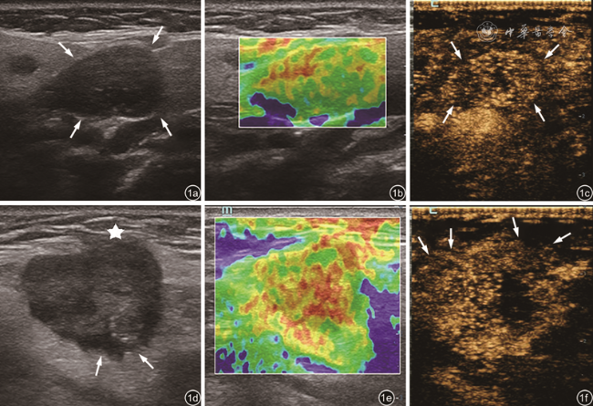
2.方法:患者平卧位,头部朝健侧微转,充分暴露检查区域。(1)US检查:记录病灶大小、形态(规则、不规则)、边界(清晰、不清晰)、内部回声情况(均匀、不均匀)以及血流情况。血流情况参考Martinoli分级标准[7]分为4级:Ⅰ级,病灶内未见血流信号;Ⅱ级,病灶内见1~2个点状或短棒状血流信号;Ⅲ级,病灶内见3~4根短棒状血流信号或有管壁清晰的线状血管穿过肿块;Ⅳ级,病灶内见大于5条短棒状或线状血流信号。(2)RTE检查:选择病灶常规超声纵切面,固定探头,启用实时双幅弹性成像模式,显示器同时显示实时弹性图像和常规灰阶图像。部分较大病灶采用单幅弹性模式。调节感兴趣区域,使其尽量同时包含病灶及部分正常的腺体组织,避开大血管和骨骼。嘱患者保持平稳呼吸,将探头垂直放置于皮肤表面,仅触及皮肤、匀速轻压,观察压力弹性标示达4~5级绿色并保持5 s以上时,冻结图像。回放并选取清晰的图像,根据病灶的弹性图像进行分型。(3)CEUS检查:选择病灶血流最丰富切面(同时包括部分正常腺体组织),固定探头,切换至CEUS模式,经肘部浅静脉团注2.5 ml造影剂,推注造影剂的同时开始计时,连续采集120 s动态图像并保存。逐帧回放图像,观察病灶的CEUS特征:①增强均匀程度:均匀、不均匀增强;②增强强度:比较病灶内造影剂达峰时与周围正常组织的增强程度,分为高增强、等增强、低增强;③增强后病灶边界:清晰、不清晰。根据病灶声像图特征对病灶进行分型(图1 )。
3. 评判标准:(1)US诊断[8, 9]:当病灶出现边界不清的征象时判断为恶性,无此征象则判断为良性。(2)RTE诊断[10, 11]:0级,病灶以囊性成分为主,表现为红蓝相间或蓝绿红相间;Ⅰ级,病灶与周围组织呈均匀的绿色;Ⅱ级,病灶以绿色为主,红色占少部分(绿色区域面积>50%);Ⅲ级,病灶呈杂乱的蓝绿相间分布,以红色为主,绿色占少许(红色区域面积>50%);Ⅳ级:病灶几乎为红色所覆盖。0~Ⅱ级判定为良性,Ⅲ~Ⅳ级判定为恶性。(3)CEUS诊断[12]:Ⅰ型,弥漫性均匀性高增强;Ⅱ型,不均匀增强(IIa:边界清晰,增强面积>50%;Ⅱb:呈散在点状增强或边界清晰,增强面积<50%;Ⅱc:呈边界不清的非均匀性增强);Ⅲ型,无或等增强。Ⅱc型判定为恶性,Ⅰ型、Ⅱa型、Ⅱb型以及Ⅲ型判断为良性。(4)多模态超声诊断:①US+RTE:当US、RTE中任意1种或以上模式判定为恶性时,多模态超声诊断为恶性;②US+CEUS:当US、CEUS中任意1种或以上模式判定为恶性时,多模态超声诊断为恶性;③US+RTE+CEUS:当US、RTE、CEUS中任意2种或以上模式判断为恶性时,多模态超声诊断为恶性。由2名主治医师进行图像判读,若两者判读不一致时,由副主任医师以上级别的医师会诊得出结果。
三、统计学分析
采用SPSS 24.0统计软件进行数据分析。计量资料以
±s表示,良性组与恶性组比较采用独立样本t检验或校正t检验。计数资料以例(%)表示,良性组与恶性组比较采用χ2检验或校正χ2检验。以病理结果为金标准,绘制受试者工作特征(receiver operating characteristic,ROC)曲线,分析US、US+RTE、US+CEUS、US+RTE+CEUS诊断涎腺局灶性病变的诊断效能,计算4种超声成像模式的曲线下面积(area under curve,AUC)。不同超声成像模式与病理金标准方法的一致性及差异性采用配对χ2检验进行分析。统计推断均为双侧检验,以P<0.05为差异有统计学意义。

结果
一、病理结果及一般情况
病理结果提示,128例涎腺局灶性病变中,良性病灶85例(良性组),其中包括36例混合瘤,19例腺淋巴瘤,9例基底细胞腺瘤,5例炎性病变,7例IgG4相关性疾病,3例嗜酸性粒细胞增生性淋巴肉芽肿,神经源性肿瘤、嗜酸性细胞腺瘤和结核各2例;恶性病灶43例(恶性组),包括9例腺样囊性癌,6例涎腺导管癌,9例淋巴瘤,3例低度恶性腺瘤,7例转移性肿瘤,黏液表皮样癌、肌上皮癌和低分化癌各2例,嗜酸性腺瘤伴恶变、多形性低度恶性腺瘤和腺泡细胞癌各1例。良性和恶性组患者平均年龄分别为(52.06±1.72)岁和(51.09±2.33)岁,病灶最大径分别为(2.56±0.13)cm和(3.16±0.25)cm,差异均无统计学意义(P均>0.05)。
二、良性组与恶性组多模态超声特征比较
1. US特征参数比较:恶性组中,病灶边界清晰的比例为58.1%(25/43)、形态规则的比例为18.6%(8/43),均明显低于良性组,差异均有统计学意义(P均<0.001);2组病灶的内部回声和血流分级比较,差异均无统计学意义(P均>0.05,表1 )。
表1 良性与恶性涎腺局灶性病变的常规超声特征比较[例(%)] |
| 组别 | 例数 | 边界 | 形态 | 内部回声 | 血流分级 | |||||
|---|---|---|---|---|---|---|---|---|---|---|
| 清晰 | 不清晰 | 规则 | 不规则 | 均匀 | 不均匀 | Ⅰ级 | Ⅱ~Ⅲ级 | Ⅳ级 | ||
| 恶性组 | 43 | 25(58.1) | 18(41.9) | 8(18.6) | 35(81.4) | 4(9.3) | 39(90.7) | 4(9.3) | 21(48.8) | 18(41.9) |
| 良性组 | 85 | 78(91.8) | 7(8.2) | 50(58.8) | 35(41.1) | 18(21.2) | 67(78.8) | 7(8.2) | 41(48.2) | 37(43.5) |
| χ2值 | 20.542 | 18.639 | 2.829 | 0.058 | ||||||
| P值 | <0.001 | <0.001 | 0.093 | 0.971 | ||||||
2. RTE分级特征比较:良性组与恶性组分级特征比较,差异有统计学意义(P<0.001,表2 )。恶性组主要以Ⅲ级为主(20/43,46.5%),良性组仅16.5%(14/85)表现为Ⅲ级,大部分表现为Ⅱ级(48/85,56.5%)。
表2 良性与恶性涎腺局灶性病变RTE分级结果比较[例(%)] |
| 组别 | 例数 | Ⅰ级 | Ⅱ级 | Ⅲ级 | Ⅳ级 |
|---|---|---|---|---|---|
| 恶性组 | 43 | 0 | 16(37.2) | 20(46.5) | 7(16.3) |
| 良性组 | 85 | 20(23.5) | 48(56.5) | 14(16.5) | 3(3.5) |
| χ2值 | 27.879 | ||||
| P值 | <0.001 | ||||
注:RTE为实时超声弹性成像 |
3. CEUS分型特征比较:CEUS分型特征在良、恶性组间比较差异有统计学意义(P<0.05,表3 ),恶性组Ⅱc型的比例(72.1%,31/43)明显高于良性组(11.8%,10/85)。
表3 良性与恶性涎腺局灶性病变CEUS分型结果比较[例(%)] |
| 组别 | 例数 | Ⅰ型 | Ⅱ型 | Ⅲ型 | ||
|---|---|---|---|---|---|---|
| Ⅱa | Ⅱb | Ⅱc | ||||
| 良性组 | 85 | 17(20.0) | 40(47.1) | 9(10.6) | 10(11.8) | 9(10.6) |
| 恶性组 | 43 | 2(4.7) | 6(14.0) | 3(7.0) | 31(72.1) | 1(2.3) |
| χ2值 | 9.086 | |||||
| P值 | 0.011 | |||||
注:CEUS为超声造影 |
三、多模态超声的诊断效能
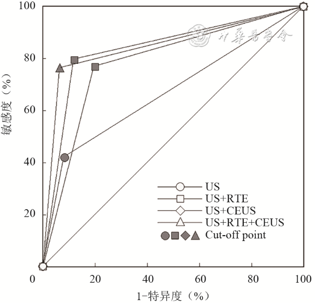
以病理结果为“金标准”,US+RTE+CEUS中任意2项或以上阳性即诊断为恶性,其诊断涎腺局灶性病变良恶性的敏感度、特异度、准确性、阳性预测值和阴性预测值分别为0.767、0.941、0.883、0.868、0.889,AUC和约登指数分别为0.854、0.708;US+CEUS任意1项阳性即诊断恶性,其诊断涎腺局灶性病变良恶性的敏感度、特异度、阴性预测值和阳性预测值分别为0.791、0.882、0.893、0.773,AUC和约登指数分别为0.837、0.673;US+RTE任意1项阳性即诊断恶性,其诊断涎腺局灶性病变良恶性的敏感度、特异度分别为0.767、0.800,AUC为0.784;US显示病灶边界不清即诊断恶性,其诊断涎腺局灶性病变良恶性的特异度、敏感度为0.918、0.419,AUC为0.669(图2 ,表4 )。
表4 不同超声成像模式诊断涎腺病变良恶性的效能 |
| 超声成像模式 | AUC | 敏感度 | 特异度 | 准确性 | 约登指数 | 阳性预测值 | 阴性预测值 |
|---|---|---|---|---|---|---|---|
| US | 0.669 | 0.419 | 0.918 | 0.750 | 0.337 | 0.720 | 0.757 |
| US+RTE | 0.784 | 0.767 | 0.800 | 0.789 | 0.567 | 0.660 | 0.872 |
| US+CEUS | 0.837 | 0.791 | 0.882 | 0.852 | 0.673 | 0.773 | 0.893 |
| US+RTE+CEUS | 0.854 | 0.767 | 0.941 | 0.883 | 0.708 | 0.868 | 0.889 |
注:US为常规超声;RTE为实时超声弹性成像;CEUS为超声造影;AUC为ROC曲线下面积 |
四、不同超声成像模式与病理金标准方法的一致性及差异性检验
1.关联性检验(一致性检验):不同超声成像模式与病理金标准方法之间关联性(一致性)均有统计学意义(P均<0.001)。
2.差异性检验:仅US与病理金标准之间差异性检验有统计学意义(P<0.05),US+RTE、US+CEUS、US+RTE+CEUS与病理金标准方法差异性检验均无统计学意义(P均>0.05,表5 )。
表5 不同超声成像模式诊断结果与病理诊断结果的对比分析 |
| 超声诊断结果 | 病理结果 | 关联性检验 | 差异性检验 | |||
|---|---|---|---|---|---|---|
| 恶性(n=43) | 良性(n=85) | χ2值 | P值 | χ2值 | P值 | |
| US | ||||||
| 恶性 | 18 | 7 | 18.459 | <0.001 | 9.031 | 0.003 |
| 良性 | 25 | 78 | ||||
| US+RTE | ||||||
| 恶性 | 33 | 17 | 36.279 | <0.001 | 1.333 | 0.248 |
| 良性 | 10 | 68 | ||||
| US+CEUS | ||||||
| 恶性 | 34 | 10 | 54.396 | <0.001 | 0.000 | 1.000 |
| 良性 | 9 | 75 | ||||
| US+RTE+CEUS | ||||||
| 恶性 | 33 | 5 | 65.338 | <0.001 | 1.067 | 0.302 |
| 良性 | 10 | 80 | ||||
注:US为常规超声;RTE为实时超声弹性成像;CEUS为超声造影 |
讨论
RTE技术作为鉴别涎腺病变性质的一种新技术,显示了感兴趣区域的相对组织弹性,以病灶区域颜色的变化反映病灶的硬度信息,病灶的软硬度与其病理组织关系密切[15, 16]。本研究结果显示,良性病灶的弹性分级明显低于恶性病灶,提示涎腺恶性病变平均硬度大于良性病变,与以往报道[10]一致。涎腺良性病变内部组织相对疏松,组织硬度较小,恶性病变由于细胞和微血管过度增生或出现钙化灶,间叶组织纤维化程度增高,部分病灶边缘甚至与周围组织出现不同程度的粘连,活动性差,导致病灶硬度增加[10,17]。Cortcu等[18]研究表明,超声弹性成像有助于涎腺良恶性病变的鉴别诊断,可提高涎腺肿块的无创诊断准确性。Altinbas等[19]使用弹性评分来确定RTE在腮腺肿瘤鉴别诊断中的价值,结果显示RTE较常规超声具有更好的诊断价值。另有研究认为,RTE在鉴别涎腺恶性肿瘤方面比良性肿瘤更有效,而US与之相反,这两种技术联合使用可使诊断更准确[13]。本研究联合US和RTE进行诊断,其诊断效能较US单独应用有明显提高,诊断涎腺恶性病变的敏感度从0.419提升至0.767,AUC从0.669提升至0.784。
依据以往研究结果,US和RTE虽然可以对涎腺良恶性病变进行鉴别诊断,但其诊断效能仍不够理想,因此有必要寻找更有效的补充检查手段。CEUS鉴别涎腺良恶性病变的应用目前已有大量文献报道[15,20, 21],其能够动态显示组织血流灌注情况,通过判断病变的增强模式、增强后病灶边缘情况等鉴别涎腺良恶性肿瘤。本研究恶性组病灶中72.1%超声造影后表现为边界不清,明显高于常规超声的41.86%。以往较多研究[8,22, 23]报道,边界是鉴别涎腺良恶性病变的重要指标。病理研究认为良性肿瘤呈膨胀性生长,多数有完整的包膜,表现为清晰的边界,而恶性肿瘤常呈侵袭性生长,容易破坏肿瘤包膜和其周边正常腺体组织,导致肿瘤的边界欠清晰[8,20]。但US仅可发现30%的恶性病变边界表现不清晰[4,24],而CEUS可显著提高恶性肿瘤边界不清的显示率[21],本研究结果与既往研究结果基本一致。蒋丽萍等[21,25]在一项纳入82例涎腺肿块的研究中指出,以增强后边界不清鉴别诊断涎腺良恶性病变的敏感度、特异度及准确性分别为70.59%、87.83%、84.61%。本研究将涎腺恶性局灶性病变CEUS后增强模式表现为边界不清的不均质增强(Ⅱc型)纳入多模态评估中,结合US恶性特征,当两者任一结果为阳性即诊断为恶性,其诊断的敏感度和AUC值较US+RTE联合多模态诊断进一步提升,分别为0.791、0.837。本研究进一步联合病灶US恶性征象、病灶内部组织超声弹性成像信息和病灶CEUS特征用于评估病灶性质,结果显示,任意2种或2种以上方式结果为阳性即诊断为恶性,其诊断效能最佳,AUC为0.854,较之于另外两种多模态联合方法,这种方法有最高的特异度、准确性和阳性预测值(分别为94.1%、88.3%和86.8%),且其敏感度仍高于US,为0.767。由US、RTE及CEUS共同构成的多模态超声,其结合了各项技术的优势,为病灶性质的判断提供更多信息,提高了诊断的准确性,降低了漏诊率。
本研究中仍有9例涎腺恶性病变被漏诊,其中8例US、RTE和CEUS均未见恶性特征,包括低度恶性腺瘤、腺泡细胞癌、黏液表皮样癌各1例和5例淋巴瘤,可能原因为:(1)部分病灶恶性程度低或处于癌变初期,体积较小,生长较缓慢,未浸润周围正常腺体组织[8,26];(2)部分病灶内部囊变区域较大,硬度减低,超声造影后仅周边和(或)内部带状增强,边界相对清晰;(3)一些低度和特殊类型恶性病灶与良性病变鉴别困难,如淋巴瘤。淋巴瘤起源于脏器的间质,多以肿瘤细胞增生为主,较少出现坏死及钙化,大部分质地相对较软[27, 28],且无周围正常结构破坏现象,表现为非侵入性的生长模式,CEUS后边界亦相对清晰[29]。因此,对于特殊类型病变,其组织结构、硬度和微循环改变不明显时,还需结合CEUS的其他增强特征及临床病史做出诊断,如淋巴瘤可结合其独特的暴风雪式增强方式以及病灶迅速增大的病史,必要时进行穿刺活检。
本研究尚存在一定局限性:(1)由于涎腺局灶性病变病理组织类型复杂,纳入病例构成比例及数量的差异可能会导致研究结果的偏差。(2)本研究将腮腺和颌下腺局灶性病变均纳入研究对象,两者虽均属涎腺,但其血供、邻近组织结构以及组织病理存在一定差异,可能会造成研究结果的偏差。(3)本研究排除了部分弹性图像显示不满意的患者,本研究尚未对这部分患者进行针对性总结及研究。
综上所述,多模态超声可以弥补常规超声的不足,有助于提高超声对涎腺病变良恶性的诊断价值。