
肝细胞腺瘤(hepatocellular adenoma,HCA)是一种极少见的肝良性肿瘤,常为单发。近年来随着口服雌激素及类固醇药物的增多及肥胖发病率的升高,HCA的发病率也呈逐年上升趋势[1]。既往文献报道[2],由于HCA病理组织成分复杂,常规二维超声对其检查特异度不高。既往认为HCA女性多发,其超声表现与经典型肝血管平滑肌脂肪瘤(classic hepatic angiomyolipoma,CAML;肝血管平滑肌血管瘤主要由平滑肌细胞、脂肪细胞和畸形的厚壁血管按不同比例构成,按照3种成分的构成比例及分布不同,可分为混合型、脂肪型、肌瘤型、血管瘤型4种病理分型,除肌瘤型外其余类型统称为CAML[3, 4])难以鉴别,部分甚至误诊为肝细胞癌(hepatocellular carcinoma,HCC)。HCA具有一定的恶变倾向并伴有破裂出血风险,其治疗策略、预后与其他肝内占位性病变存在明显差异,鉴别诊断具有非常重要的意义。超声造影技术临床应用越来越多,其在肝内局限性病变的诊断及鉴别诊断中被证明具有重要的作用,它具有可实时动态观察病灶、无辐射、无肾毒性等优点[5]。本研究回顾性分析经病理证实的HCA的临床表现、二维超声表现及超声造影特点,并与CAML及HCC进行对比,以期寻找具有鉴别诊断价值的特征,提高疾病诊断能力。
资料与方法
一、对象
回顾性分析中山大学附属第三医院数据库中2008年1月至2021年5月就诊的10例经穿刺活检或术后病理证实的HCA患者(HCA组)及17例CAML患者(CAML组,除外上皮型)的临床、影像及病理资料。同期从本院病理数据库中检索并随机抽取40例甲胎蛋白阴性(alpha-fetoprotein negative,AFP-)的HCC患者[HCC(AFP-)组]作为对照。HCA组及CAML组中病灶常在体检中偶然发现。HCA组患者中男性5例,女性5例,年龄为(36.40±16.68)岁(范围17~74岁),均无肝炎病史。CAML组患者中男性6例,女性11例,年龄为(42.59±9.79)岁(范围25~56岁),既往乙型肝炎病毒感染者1例,无合并肝硬化者。HCC(AFP-)组患者中男性36例,女性4例,年龄为(56.15±11.87)岁(范围33~78岁),既往乙型肝炎病毒感染者40例,合并肝硬化者23例。HCC(AFP-)组患者病灶常在乙型肝炎患者常规随访中发现。本研究整个方案经中山大学附属第三医院伦理审查委员会批准通过(伦理编号:[2021]-02-532)。
二、仪器与方法
由于HCA及CAML是少见的肝良性肿瘤,患者的纳入和资料收集时间较长。使用了多个配备实时低频(2.0~4.0 Hz)对比特异性成像软件的超声系统,包括IU-22,Philips;MyLab 90/Twice,Esaote;Logic E9,US;Aplio 500,Japan;迈瑞Resona 7S等。超声造影采用Bracco公司造影剂SonoVue(使用前以5 ml生理盐水稀释,充分震荡后抽取1.5~2.4 ml经肘前静脉团注,注射造影剂后即用5 ml生理盐水冲洗管)。每例患者在超声造影前均进行了肝常规超声检查。使用SonoVue后,对病灶和邻近肝组织进行实时对比扫描,在超声造影模式下连续观察360 s,增强时相为动脉期(0~30 s)、门静脉期(31~120 s)、延迟期(121~300 s),进行三期对比增强连续观察。
三、图像分析
由2名具有5年以上肝超声造影诊断经验的医师在不知病理结果的情况下通过图片存档和传输系统完成数据收集并独立分析图像。意见分歧以协商一致解决。超声观察内容包括:病灶数目、位置、大小、形态、边界、内部回声、彩色血流、出血、癌栓以及超声造影增强模式。彩色血流信号诊断标准参考Adler标准。回声水平诊断标准:整体肿瘤中高回声所占比例≥50%为高回声肿瘤,余者为低回声肿瘤。通过分析存储在系统中的动态图,确定超声造影增强模式。
四、统计学分析
使用SPSS 26.0进行统计分析。年龄为符合正态分布的计量资料,以
±s表示,采用t检验分别比较HCA组与CAML组和HCC组的差异。病灶大小为不符合正态分布的计量资料,以M(QR)表示,采用非参数检验(曼-惠特尼检验)分别比较HCA组与CAML组和HCC组的差异。其余资料为计数资料,采用例数表示,采用χ2检验或Fisher精确概率检验分别比较HCA组与CAML组和HCC组的差异。P<0.05为差异具有统计学意义。

结果
一、3组患者临床特征
HCA组、CAML组间患者性别、年龄、合并乙型肝炎病毒感染及合并肝硬化情况比较,差异均无统计学意义(P均>0.05)。在HCA组与HCC(AFP-)组间患者性别、合并乙肝病毒感染和合并肝硬化情况比较,差异均具有统计学意义(P均<0.05),HCC(AFP-)组患者中男性居多,且大部分HCC继发于乙肝病毒感染和(或)合并肝硬化。HCA组、CAML组间及HCC(AFP-)3组间雌激素及类固醇药物服用史、体质量指数及糖尿病患病率方面比较,差异均无统计学意义(P均>0.05,表1 )。
表1 3组患者人口统计学和临床特征比较 |
| 特征 | HCA(n=10) | CAML(n=17) | HCC(AFP-)(n=40) | HCA vs CAML | HCA vs HCC(AFP-) | ||
|---|---|---|---|---|---|---|---|
| 统计值 | P值 | 统计值 | P值 | ||||
| 年龄(岁, ±s) | 36.40±16.68 | 42.59±9.79 | 56.15±11.87 | t=-1.222 | 0.191 | t=4.329 | 0.355 |
| 性别(男/女,例) | 5/5 | 6/11 | 36/4 | χ2=0.564 | 0.687 | χ2=8.672 | 0.010 |
| 合并乙肝感染(是/否,例) | 0/10 | 1/16 | 40/0 | χ2=0.611 | 1.000 | χ2=50.000 | <0.001 |
| 合并肝硬化(是/否,例) | 0/10 | 0/17 | 23/17 | - | - | χ2=10.648 | 0.001 |
| 临床表现(例) | χ2=0.318 | 0.683 | χ2=8.290 | 0.016 | |||
| 腹痛或不适 | 4 | 5 | 11 | ||||
| 体检发现 | 6 | 12 | 11 | ||||
| 乙肝随访发现 | 0 | 0 | 18 | ||||
| 雌激素及类固醇药物服用病史(有/无,例) | 1/9 | 0/17 | 1/39 | χ2=1.765 | 0.370 | χ2=1.172 | 0.363 |
| 体质量指数≥24(例) | 2 | 2 | 4 | χ2=0.338 | 0.613 | χ2=0.758 | 0.586 |
| 糖尿病(有/无,例) | 0/10 | 1/16 | 3/37 | χ2=0.611 | 1.000 | χ2=0.798 | 1.000 |

注:-为无相应统计值;HCA为肝细胞腺瘤;CAML为经典型肝血管平滑肌脂肪瘤;HCC(AFP-)为甲胎蛋白阴性的肝细胞癌 |
二、常规二维超声和超声造影表现
1. 常规二维超声表现:HCA组与CAML组、HCA组与HCC(AFP-)组病灶数目、大小、形态、边界、血流差异均无统计学意义(P均>0.05,表2 )。HCA组与HCC(AFP-)组在病灶位置上差异具有统计学意义(P<0.05,表2 )。HCA组病灶回声可表现为高回声/低回声/其他回声(4/4/2,40%/40%/20%),而HCC(AFP-)组病灶回声(高回声/低回声/其他回声:4/34/2,10%/85%/5%)主要表现为低回声,两者间差异具有统计学意义(P<0.05,表2 )。
表2 HCA、CAML、HCC(AFP-)组常规二维超声及超声造影表现 |
| 特征 | HCA (n=10) | CAML(n=17) | HCC(AFP-)(n=40) | HCA vs CAML | HCA vs HCC(AFP-) | ||
|---|---|---|---|---|---|---|---|
| χ2值 | P值 | χ2值 | P值 | ||||
| 二维超声特征(例) | |||||||
| 位置(左肝/右肝/其他) | 3/6/1 | 10/6/1 | 29/10/1 | χ2=2.329 | 0.325 | χ2=6.694 | 0.042 |
| 数目(单/多发) | 9/1 | 16/1 | 28/12 | χ2=0.156 | 1.000 | χ2=1.663 | 0.258 |
| 大小[mm,M(QR)] | 31(17-53.25) | 40.5(21-112.125) | 25.25(16.25-50.13) | Z=-0.929 | 0.353 | Z=-1.577 | 0.115 |
| 形态(规则/不规则) | 7/3 | 10/7 | 22/18 | χ2=0.337 | 0.692 | χ2=0.739 | 0.488 |
| 边界(清晰/不清晰) | 8/2 | 13/4 | 30/10 | χ2=0.110 | 1.000 | χ2=0.110 | 1.000 |
| 回声(高/低/其他) | 4/4/2 | 11/3/3 | 4/34/2 | χ2=2.011 | 0.430 | χ2=8.404 | 0.011 |
| 回声(均匀/不均匀) | 6/4 | 13/4 | 36/4 | χ2=0.819 | 0.415 | χ2=5.357 | 0.041 |
| 血流(丰富/不丰富) | 3/7 | 10/7 | 3/37 | χ2=2.095 | 0.236 | χ2=3.835 | 0.086 |
| 超声造影表现(例) | |||||||
| 动脉期(高/等/低) | 8/2/0 | 15/2/0 | 39/1/0 | χ2=0.338 | 0.613 | χ2=4.344 | 0.098 |
| 门静脉期(高/等/低) | 1/6/3 | 6/10/1 | 0/4/36 | χ2=3.682 | 0.121 | χ2=15.106 | <0.001 |
| 延迟期(高/等/低) | 0/6/4 | 4/10/3 | 0/1/39 | χ2=3.236 | 0.216 | χ2=12.500 | 0.002 |
| 合并门静脉癌栓 | 0 | 0 | 7 | - | - | ||
| 合并肿瘤内出血 | 1 | 0 | 3 | - | - | ||
| 随访癌变 | 1 | 0 | - | - | - | ||
注:HCA为肝细胞腺瘤;CAML为经典型肝血管平滑肌脂肪瘤;HCC(AFP-)为甲胎蛋白阴性的肝细胞癌;-表示无相应统计值 |
2. 超声造影表现:HCA组与CAML组间、HCA组与HCC(AFP-)组间动脉期均表现为高增强,组间差异均无统计学意义(P均>0.05)。HCA组与CAML组间门静脉期及延迟期增强表现比较,差异均无统计学意义(P均>0.05),而HCA组与HCC(AFP-)组间门静脉期及延迟期增强表现比较,差异均有统计学意义(P均<0.05,表2 )。HCA组6例(6/10,60.0%)超声造影表现为“高-等-等”模式(图1 );而在HCC(AFP-)组中36例(36/40,90.0%)超声造影出现典型的“高-低-低”表现(图2 );CAML组6例(6/17,35.3%)超声造影表现为“高-高-高”或“高-高-等”模式(图3 )。仅在HCC(AFP-)组中存在合并门静脉癌栓(7/40,17.5%)。HCA组中,1例74岁男性患者(2/10,20%)在随访过程中术后7年发生癌变,1例合并瘤内出血。

图1 39岁肝腺瘤女性患者超声及病理图。图a、b:肝右后叶下段内低-等回声团,边界欠清,血流不丰富;图c:动脉早期周边高增强;图d:动脉晚期整体高增强;图e:门静脉期呈稍高-等增强;图f:延迟期呈等增强;图g、h:HE及免疫组化[HE×200;CD34(+),×100] |
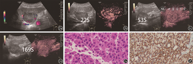
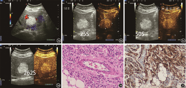
图2 患者男性,69岁,肝细胞癌(甲胎蛋白阴性),慢性肝炎病史30余年。图a:肝S2/3内低回声团,边界尚清,周边可见点状血流信号;图b、c、d:超声造影动脉期高增强,门静脉及延迟期低增强;图e、f:HE及免疫组化[HE×200;Hep(+)(肝细胞抗原阳性),×200] |
讨论
HCA是一种罕见的肝良性肿瘤,常在体检中偶然发现,少数伴腹部不适等症状。雌激素暴露是HCA的主要危险因素,与口服避孕药的使用密切相关,这与国外HCA流行病学相符[6]。本组病例男女比例相近,90%无长期服用避孕药史。随着分子分类和免疫组化标志物的进展,HCA分为4种亚型[2,7]:HNF1A(H-HCA)突变型、β-catenin激活型、炎症型和未分类型肝细胞腺瘤。研究还显示HCA恶性转化与β-catenin激活、肿瘤大(>5 cm)、男性有关[7]。本组HCA病例中有1例74岁男性患者术后7年随访中发现恶变,首次手术肿瘤>5 cm,与既往报道[8, 9]相符,但其对于恶变来源是病灶周边肝组织还是HCA本身还不完全清楚,有待进一步研究。
病理显示HCA由层状或索状肝细胞及少量Kuffer细胞组成,无汇管区及中心静脉,板层间有扩张的血窦,胆管结构较正常肝组织少,可发生脂肪变性、出血及坏死,血流较丰富[10],常发生在无乙肝或肝硬化背景的肝脏中[7,11]。由于HCA复杂的病理变化导致其声像图表现不具有特异性[12],可表现为高回声、低回声或其他回声;CMAL主要是由平滑肌细胞、成熟的脂肪组织及畸形的厚壁血管按不同比例构成,既往研究报道[13]二维超声多表现为高回声;而HCC(AFP-)中细胞成分简单、反射界面少而以低回声为主,该表现与本研究的结果相符。大量文献指出[14, 15],典型的HCA血流信号以周边为主,血管密度小于HCC,但本研究中大部分HCA病灶表现为血流不丰富。可能原因:彩色多普勒对大的或者稍高速的血流敏感,对小的血流敏感度不高,而HCA中以静脉血流及低速低阻动脉为主,所以普通彩色多普勒图上常显示血流信号不丰富。然而,超声造影常显示明显的动脉期快速高增强,这与表2 中的部分结果是相符的。既往研究报道,由于影像学特征存在重叠,HCA常误诊为CAML或HCC等其他肝内占位性病变[2]。在本研究中80% HCA动脉期高增强,门静脉期或延迟期表现为等或低增强,即以“高-等-等”或“高-等-低”增强模式表现常见,甲胎蛋白阴性,不伴乙型肝炎或肝硬化等表现,与其他文献报道[16]是相符的。而既往文献[15, 16]中指出,动脉期向心性高增强是HCA的典型表现,常为均匀性高增强(除外出血的病灶),而在本研究中观察到HCA动脉期向心性高增强所占比例并不高,本研究中没有单独将其列为一个鉴别诊断要点,可能是因为本研究的病例数相对较少。HCC(AFP-)组的超声造影常表现为动脉期高增强,没有明显的向心性或离心性增强,常表现为典型的“高-低-低”增强模式。本研究显示,HCA病灶消退为等增强和低增强的时间均晚于HCC(AFP-)病灶(P均<0.05),HCA病灶常在延迟期才消退为低增强,而HCC(AFP-)病灶造影剂消退较快,可发生在门静脉期早期甚至是动脉期后期消退为低增强,结合肝炎背景,由此可鉴别HCA与HCC(AFP-)。CAML的超声造影表现与HCA病灶相比差异没有统计学意义。CAML动脉期均匀高增强,门静脉及延迟期表现为高或等增强,其消退时间一般较长,常表现为“高-高-等”或“高-高-高”增强模式[4]。但由于CAML中常含有脂肪成分,其二维超声表现以高回声为主。CAML、HCC(AFP-)及HCA的超声造影表现在诊断中具有一定的价值,但日常工作中仍然需要结合临床特点、常规二维超声表现进行鉴别诊断。
本研究仍存在一定的局限性。首先,由于HCA及CAML同属于罕见病,纳入分析的病例数量有限,且未根据免疫组织化学法及基因检测结果将HCA划分为不同亚型,没有单独对不同类型生物学行为和影像学表现进行分析。其次,回顾性设计有其固有的偏倚限制,如由于HCA、CAML和HCC(AFP-)患者的纳入和检查是在不同的时间进行的,超声和超声造影成像方法没有达到100%标准化。
综上所述,HCA常误诊为CAML或HCC,熟悉三者的临床特点、常规二维超声及超声造影特点有助于提高HCA的术前诊断准确率。