Home    中文  
 
  • Search
  • lucene Search
  • Citation
  • Fig/Tab
  • Adv Search
Just Accepted  |  Current Issue  |  Archive  |  Featured Articles  |  Most Read  |  Most Download  |  Most Cited
Superficial Parts Ultrasound

Ultrasound and pathological characteristics of breast metaplastic carcinoma containing squamous cell carcinoma components

  • Yaping Jia ,
  • Shu'e Zeng ,
Expand
  • Ultrasound Department, Hubei Cancer Hospital, Wuhan 430079, China
Corresponding author: Zeng Shu'e, Email:

Received date: 2022-04-26

  Online published: 2023-10-31

Copyright

Copyright by Chinese Medical Association No content published by the journals of Chinese Medical Association may be reproduced or abridged without authorization. Please do not use or copy the layout and design of the journals without permission. All articles published represent the opinions of the authors, and do not reflect the official policy of the Chinese Medical Association or the Editorial Board, unless this is clearly specified.

Abstract

Objective

To explore the ultrasound characteristics of breast metaplastic carcinoma containing squamous cell carcinoma components (MCSC) and compare them with pathological results.

Methods

Nine patients with MCSC confirmed by surgical and pathological examination at Hubei Cancer Hospital were retrospectively selected. Color Doppler ultrasound was used to scan both the breasts and armpits to observe the ultrasound characteristics of the tumors. BI-RADS classification and Adler blood flow grading were performed on the breast tumors. All MCSC patients underwent gross, microscopic, and immunohistochemical examinations after surgery, and the ultrasound features of the tumors were compared with pathological results.

Results

There were 3 cases of squamous cell carcinoma, 1 case of adenosquamous carcinoma, 1 case of metaplastic carcinoma with mesenchymal differentiation, and 4 cases of mixed metaplastic carcinoma. The maximum diameter of the tumors ranged from 1.07-15.00 cm, with a median diameter of 4.48 cm. Five cases showed mixed internal echoes, and 4 showed hypoechogenicity; 9 showed varying degrees of enhancement in posterior echoes; 7 had unclear or unclear boundaries; 8 had an irregular morphology; 3 were accompanied by calcification. Ultrasound examination showed 4 cases of axillary lymph node enlargement. According to Adler blood flow grading, 8 cases were classified as Grade III, and 1 was classified as Grade I. BI-RADS grading suggested category 4b in 1 case and categories 4c-5 in the remaining. Postoperative pathological examination of gross specimens showed cysts of varying sizes in 8 cases, with only 1 case showing no cysts. Immunohistochemistry showed that there were 4 cases (4/9) of triple negative breast cancer.

Conclusion

Mixed metaplastic carcinoma is the most common type in MCSC, and ultrasound imaging shows the appearance of a malignant tumor. The ultrasound features of a larger tumor volume, mixed echoes with enhanced posterior echoes, and abundant blood supply in the tumor parenchyma are meaningful for the diagnosis of MCSC.

Cite this article

Yaping Jia , Shu'e Zeng . Ultrasound and pathological characteristics of breast metaplastic carcinoma containing squamous cell carcinoma components[J]. Chinese Journal of Medical Ultrasound (Electronic Edition), 2023 , 20(08) : 844 -848 . DOI: 10.3877/cma.j.issn.1672-6448.2023.08.010

乳腺化生性癌(metaplastic breast carcinoma,MBC)指乳腺腺上皮不同比例转化为另一种上皮组织或非腺上皮组织和(或)间叶组织1。MBC组织学类型多样,是一种罕见的乳腺恶性肿瘤,在所有乳腺恶性肿瘤中占比仅为0.2%~5.0%2。含有鳞状细胞癌成分的乳腺化生性癌(metaplastic carcinoma with squamous cell component,MCSC)更为少见,本研究中MCSC包括单纯性鳞状细胞癌、腺鳞癌、伴间叶分化的化生性癌和混合型化生性癌。既往关于MBC的报道多为小样本案例报道,对MCSC的研究更为少见,本文通过回顾性分析9例MCSC患者的超声声像图特征并与病理结果进行对比分析,以加深对此罕见肿瘤的认识,为超声诊断MCSC积累一定经验。

资料与方法

一、对象

回顾性分析2014年1月至2020年12月于湖北省肿瘤医院手术切除,经病理检查证实的9例MCSC患者,9例患者均因乳房肿块就诊,均为绝经期或围绝经期女性,年龄44~76岁,平均年龄(51.8±10.9)岁。9例均行乳腺癌改良根治术。所有MCSC患者术后均进行大体、镜下病理和免疫组化检查。

二、仪器与方法

1.超声检查:采用Philips EPIQ5彩色多普勒超声诊断仪,实时高频线阵探头,频率5~12 MHz。患者取仰卧位,按时钟法多切面扫查双侧乳腺及腋窝,观察肿瘤的位置、大小、纵横比,形态、边界、内部回声、后方回声、有无钙化、周边及内部血流情况等,综合以上信息对乳房肿瘤进行乳腺影像报告与数据系统(breast imaging reporting and data system,BI-RADS)分类3,采用彩色多普勒血流成像(color Doppler flow imaging,CDFI)按Adler4半定量法对瘤体内实质部分血流信号丰富程度进行分级。
2.免疫组化观察指标:免疫组化主要观察雌激素受体(estrogen receptor,ER)、孕激素受体(progesterone receptor,PR)和人表皮生长因子受体-2(human epidermal growth factor receptor-2,Her-2)的表达。

结果

一、超声表现

9例MCSC中,1例患者肿瘤为多发性,8例为单发性,其中左乳肿瘤3例,右乳肿瘤6例;外上象限肿瘤7例,乳房内侧肿瘤1例,1例为巨大肿瘤,直径约15 cm;肿瘤最大径1.07~15.00 cm,中位径线为4.48 cm;5例内部呈混合性回声,内均可见大小不等的不规则无回声区,4例内部呈低回声;9例后方回声均可见不同程度增强;7例边界欠清晰、浅分叶状或边缘模糊不清;8例形态不规则;3例伴多发性密集簇状钙化;超声检查有4例患者腋下淋巴结肿大,其中3例经病理证实为淋巴结转移。CDFI显示肿瘤实性部分血流较丰富,Adler血流分级:8例为Ⅲ级,1例为I级。BI-RADS分级:5例5类,3例4c类,1例4b类,超声诊断恶性肿瘤符合率100%(表1)。
表1 9例MCSC的超声表现与病理结果
病例序号 年龄(岁) 肿瘤位置 肿瘤最大径线(cm) 内部回声 Adler血流分级 后方回声增强 钙化 BI-RADS分级 腋下淋巴结肿大 病理分型
1 51 右乳、外上 4.12 实质性 5 混合型化生性癌
2 57 左乳、外上 4.04 囊实性 4c SCC
3 47 右乳、外上 2.78 实质性 I 5 ASC
4 47 右乳内侧 1.07 实质性 4b SCC
5 44 右乳 15.00 囊实性 5 混合型化生性癌
6 45 左乳、外上 2.74 实质性 4c 混合型化生性癌
7 79 右乳、外上 3.54 囊实性 5 混合型化生性癌
8 46 右乳、外上 2.95 实质性 5 SCC
9 51 左乳、外上 4.09 囊实性 4c 伴间叶分化的化生性癌

注:MCSC为含有鳞状细胞癌成分的乳腺化生性癌;SCC为单纯性鳞状细胞癌;ASC为腺鳞癌;BI-RADS为乳腺影像报告与数据系统

二、病理结果

病理诊断乳腺鳞状细胞癌3例,腺鳞癌1例,伴间叶分化的化生性癌1例(癌为高级别浸润性导管癌、导管原位癌及鳞状细胞癌,间叶成分为软骨肉瘤、多形性平滑肌肉瘤),混合型化生性癌4例(1例伴大汗腺分化癌)。
术后病理大体标本示8例(8/9)可见大小不等的囊腔,1例未见囊腔。病理证实腋窝淋巴结转移者3例(3/9),均可见腺癌转移,1例见鳞癌转移。免疫组化显示三阴性乳腺癌者4例(4/9)。本研究4例三阴性乳腺癌中2例为混合型化生性癌,1例为鳞状细胞癌(图1),无腺鳞癌和伴间叶分化的化生性癌(表2)。
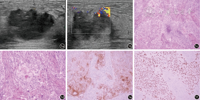
图1 含鳞状细胞癌成分的乳腺化生性癌的超声及病理图像(病例8)。图a为超声图像示肿块呈低回声,边界模糊形态不规则,未显示明显钙化;图b为彩色多普勒图像示肿块血流信号增多;图c为病理图像示肿瘤呈实性片状伴局部坏死,无腺管形成,可见角化珠和细胞间桥(HE ×100);图d为病理图像示肿瘤细胞核深染,核浆比增大,胞浆嗜酸,核分裂象可见(HE ×200);图e为免疫组织化学图像示肿瘤细胞CK5/6胞质阳性(EnVision法×200);图f为免疫组织化学图像示肿瘤细胞P40胞核弥漫强阳性(EnVision法 ×200)
表2 9例MCSC的病理分型与腋窝淋巴结转移情况
病例序号 年龄(岁) 病理分型 腋窝淋巴结转移 免疫组化
1 51 混合型化生性癌 腺癌(4/19枚) ER(-)PR(-)Her-2(浸润性导管癌部分3+,鳞癌部分1+)
2 57 SCC ER(10%,2+)PR(2%,2+)Her-2(-)
3 47 ASC ER(1+,1%)PR(-)Her-2(-)
4 47 SCC ER(-)PR(-)Her-2(2+)
5 44 混合型化生性癌 鳞状细胞癌(1/14枚),腺癌(10/14枚) ER(5%,2+)PR(2%,1+)Her-2(3+)
6 45 混合型化生性癌 腺癌(2/21枚) ER(-)PR(-)Her-2(-)
7 79 混合型化生性癌 ER(-)PR(-)Her-2(-)
8 46 SCC ER(-)PR(-)Her-2(-)
9 51 伴间叶分化的化生性癌 ER(-)PR(-)Her-2(1+)

注:MCSC为含有鳞状细胞癌成分的乳腺化生性癌;SCC为单纯性鳞状细胞癌;ASC为腺鳞癌;ER为雌激素受体;PR为孕激素受体;Her-2为人表皮生长因子受体-2

讨论

MBC与非特殊类型浸润性癌相比具有较高的侵袭性和复发率,预后差。由于乳腺MBC成分构成不同,影像学表现并没有特异性,较难与其他类型的乳腺恶性肿瘤相鉴别,有时甚至表现出良性特征5。大部分MBC是三阴性乳腺癌6,内分泌治疗效果较差,因此对于MBC早发现早治疗至关重要。本研究中9例MCSC患者,发病年龄44~76岁,中位年龄51.8岁,肿块最大径1.07~15.00 cm,中位径线4.48 cm。与既往文献报道所认为的乳腺MBC的发病年龄多为中老年,就诊时乳腺肿瘤体积较大一致7-8。Rakha等9认为东方人乳腺MBC中以鳞状细胞癌最常见,而本研究中MCSC以混合型化生性癌最多(4/9),其次是鳞状细胞癌(3/9)。
本研究9例MCSC中超声检查5例可见大小不等的不规则无回声暗区,超声所见的不规则暗区与大体标本出血坏死囊性区的表现相吻合。镜下囊性区域可见鳞状细胞癌原发灶中心细胞聚集,囊壁可见典型鳞状细胞癌结构,伴坏死及囊腔形成。含有鳞状细胞癌成分的肿瘤生长迅速,内部供血不足导致瘤体内部发生囊性变,鳞状细胞癌由囊壁脱落坏死形成腔隙。本研究观察到9例患者的肿瘤后方回声均呈不同程度的增强,考虑与其内部存在囊性成分有关10;这一表现与多数乳腺浸润性导管癌肿块后方回声衰减明显不同。本研究9例中8例可见大小不等的囊性病变,病理分型中鳞状细胞癌成分所占比例较高(60%~100%),仅1例MCSC的大体标本切开未见囊性病变,该例病理结果为(左乳)混合型化生性癌(浸润性微乳头状癌约60%,浸润性导管癌Ⅲ级成分约40%,局灶呈化生性癌改变,化生性癌成分为鳞状细胞癌)伴间质淋巴细胞浸润。该肿瘤超声表现为实质性肿瘤,可能与鳞状细胞癌含量较少,癌成分较复杂有关。
9例患者的其他超声表现为:边界欠清晰或边缘模糊(7/9)、形态不规则(8/9)、3例均表现为多发性密集簇状钙化(3/9)、CDFI显示病灶实性部分血流较丰富,Adler血流信号分级大多为Ⅲ级(8/9),以上表现均符合乳腺恶性肿瘤的声像图特点。根据乳房肿瘤的以上特征,本研究中BI-RADS分级1例分为4b类,其余均为4c~5类,术前超声将乳腺肿瘤均分类为几乎确定的恶性肿瘤,超声诊断恶性肿瘤与病理结果符合率为100%。
文献报道MBC的腋窝淋巴结转移率低,约8%~40%11,跳过区域淋巴结出现远处转移的发生率高12-13。MCSC的腋窝淋巴结转移较为少见。本研究中,腋窝淋巴结转移共3例(3/9),病理类型均为混合型化生性癌,其中1例患者腋窝淋巴结(11/14枚)可见癌转移,但这转移的11枚淋巴结中只有1枚为鳞状细胞癌转移,另外10枚均为腺癌转移。另外2例患者淋巴结转移均为腺癌转移。3例三阴性乳腺癌中,腋窝淋巴结(2/21枚)可见腺癌转移,鳞状细胞癌均未见淋巴结转移。这可能提示MCSC中鳞状细胞癌的迁移力较腺癌弱。但出现转移往往预示预后更差,因此早期诊断MCSC对改善患者预后意义重大。
本研究免疫组化结果显示9例MCSC中三阴性乳腺癌者仅4例(4/9),病理分型1例为鳞状细胞癌,1例为伴间叶分化的化生性癌,2例为混合型化生性癌。这2例混合型化生性癌中1例鳞状细胞癌成分占比约80%,1例仅局灶呈化生性鳞状细胞癌表现。有研究14-15认为90%的乳腺鳞状细胞癌患者免疫组化为三阴性,本研究3例鳞状细胞癌中仅1例免疫组化为三阴性,另外2例免疫组化表现为ER、PR或Her-2低表达。
综上所述,MCSC的超声表现为恶性肿瘤的声像图特征。肿瘤体积较大、瘤体内部为混合性回声伴后方回声增强、肿瘤实质部分血供丰富的超声表现对诊断MCSC有意义。但本研究病例数少,病理学亚型有限,今后将加大样本量进一步探讨。
1
Zein D, Hughes M, Kumar S, et al. Metaplastic carcinomaof the breast is more aggressive than triple-negative breast cancer: a study from a single institution and review of literature[J]. Clin Breast Cancer, 2017, 17(5): 382-391.

2
Lakhani SR. WHO classification of tumours of the breast[M]. 4th.Lyon: International Agency for Research on Cancer, 2012.

3
Radhakrishna S. Breast diseases: imaging and clinical management[M]. London: Springer, 2015: 61-62.

4
Adler DD, Carson PL, Rubin JM, et al. Doppler ultrasoundcolor flow imaging in the study of breast cancer: preliminary findings[J]. Ultrasound Med Biol, 1990, 16(6): 553-559.

5
Langlands F, Cornford E, Rakha E, et al. Imaging overview of metaplastic carcinomas of the breast: a large study of 71 cases[J]. Br J Radiol, 2016, 89(1064): 20140644.

6
Weigelt B, Eberle C, Cowell CF, et al. Metaplastic breast carcinoma: More than a special type[J]. Nat Rev Cancer, 2014, 14(3): 147-148.

7
Toumi Z, Bullen C, Tang AC, et al. Metaplastic breast carcinoma:A case report and systematic review of the literature[J]. Pathol Int, 2011, 61(10): 582-588.

8
Tzanninis IG, Kotteas EA, Ntanasis-Stathopoulos I, et al. Management and outcomes in metaplastic breast cancer[J]. Clin Breast Cancer, 2016, 16(6): 437-443.

9
Rakha EA, Tanph PH, Varga Z, et al.Prognostic factors in metaplastic carcinoma of the breast: a multi-institutional study[J]. Br J Cancer, 2015, 112(2): 283-289.

10
Salemis NS. Breast abscess as the initial manifestation of primary pure squamous cell carcinoma:A rare presentation and literature review[J]. Breast Dis, 2011, 33(3): 125-131.

11
Mckinnon E, Xiao P. Metaplastic carcinoma of the breast[J]. Arch Pathol Lab Med, 2015, 139(6): 819-822.

12
Pezzi CM, Patel-Parekh L, Cole K, et al. Characteristics and treatment of metaplastic breast cancer: analysis of 892 cases from the National Cancer Data Base[J]. Ann Surg Oncol, 2007, 14(1): 166-173.

13
Park HS, Park S, Kim JH, et al. Clinicopathologic features and outcomes of metaplastic breast carcinoma: comparison with invasive ductal carcinoma of the breast[J]. Yonsei Med J, 2010, 51(6): 864-869.

14
Siegelmann-Danieli N, Murphl TJ, Meschter SC, et al. Primary pure squamous cell carcinoma of the breast[J]. Clin Breast Cancer, 2005, 6(3): 270-272.

15
Liu J, Yu Y, Sun JY, et al. Clinicopathologic characteristics and prognosis of primary squamous cell carcinoma of the breast[J]. Breast Cancer Res Treat, 2015, 149(1): 133-140.

Outlines

/

Copyright © Chinese Journal of Medical Ultrasound (Electronic Edition), All Rights Reserved.
Tel: 010-51322630、2632、2628 Fax: 010-51322630 E-mail: csbjb@cma.org.cn
Powered by Beijing Magtech Co. Ltd