Home    中文  
 
  • Search
  • lucene Search
  • Citation
  • Fig/Tab
  • Adv Search
Just Accepted  |  Current Issue  |  Archive  |  Featured Articles  |  Most Read  |  Most Download  |  Most Cited
Pediatric Ultrasound

Ultrasound assessment of diaphragm function in pediatric patients with Duchenne muscular dystrophy

  • Xuan Zhang ,
  • Yutong Ma ,
  • Yuqian Miao ,
  • Yun Zhang ,
  • Shiwen Wu ,
  • Xiaochu Dang ,
  • Yingying Chen ,
  • Zhaoming Zhong ,
  • Xuejuan Wang ,
  • Miao Hu ,
  • Yanfeng Sun ,
  • Xiuzhu Ma ,
  • Faqin Lyu ,
  • Haiyan Kou ,
Expand
  • Ultrasonic Department, the Third Medical Centre of PLA General Hospital, 100039 Beijing, China
  • Neurology Department, the Third Medical Centre of PLA General Hospital, 100039 Beijing, China
  • Pediatric Department, the Third Medical Centre of PLA General Hospital, 100039 Beijing, China
Corresponding author: Kou Haiyan, Email:

Received date: 2022-08-01

  Online published: 2024-01-08

Copyright

Copyright by Chinese Medical Association No content published by the journals of Chinese Medical Association may be reproduced or abridged without authorization. Please do not use or copy the layout and design of the journals without permission. All articles published represent the opinions of the authors, and do not reflect the official policy of the Chinese Medical Association or the Editorial Board, unless this is clearly specified.

Abstract

Objective

To evaluate the value of ultrasonography in assessing diaphragm function in children with Duchenne muscular dystrophy (DMD).

Methods

A total of 21 children with DMD clinically diagnosed at the Third Medical Center of the PLA General Hospital from May 1, 2021 to March 1, 2022 were collected, and 21 healthy volunteers of the same age group were included as a control group. Diaphragmatic ultrasonography was performed on all subjects, and the main indicators included diaphragmatic thickening rate (DTF) and diaphragmatic mobility (DE). The differences of diaphragm ultrasonography findings, DTF, and DE between the two groups were compared and analyzed.

Results

Compared with normal children, DMD children showed obvious thickening of the diaphragm, enhanced and uneven echo, and weakened respiratory movement. The right anterior axillary line DE in the DMD group was (12.0±5.0) mm, significantly lower than that of the control group (19.0±3.3) mm (t=0.207, P<0.001). The right midclavicular line DE was (11.7±4.1) mm, also significantly lower than that of the control group (20.0±5.3) mm (t=0.603, P<0.001). The DTF [(38.3±19.4)%] in the DMD group was significantly lower than that of the control group [(50.6±10.9)%] (t=0.013, P=0.015). DTF [(32.9±19.0)%] of the right midclavicular line was lower than that of the control group [(49.7±11.5)%] (t=0.079, P=0.002).

Conclusion

Ultrasound can effectively evaluate the diaphragm function in children with DMD, and quantitative measurement of diaphragm DTF and DE is helpful to monitor the condition of children with DMD.

Cite this article

Xuan Zhang , Yutong Ma , Yuqian Miao , Yun Zhang , Shiwen Wu , Xiaochu Dang , Yingying Chen , Zhaoming Zhong , Xuejuan Wang , Miao Hu , Yanfeng Sun , Xiuzhu Ma , Faqin Lyu , Haiyan Kou . Ultrasound assessment of diaphragm function in pediatric patients with Duchenne muscular dystrophy[J]. Chinese Journal of Medical Ultrasound (Electronic Edition), 2023 , 20(10) : 1068 -1073 . DOI: 10.3877/cma.j.issn.1672-6448.2023.10.011

Duchenne肌营养不良(Duchenne muscular dystrophy,DMD)是进行性肌营养不良中最常见的一种类型1,发病率约为1/3500。病因为基因片段缺失导致基因编码抗肌萎缩蛋白缺失2-3,进而发生肌纤维坏死、纤维化及再生能力的丧失,临床上主要表现为进行性骨骼肌退化和心脏、膈肌功能障碍4-5。患者多在20岁前死于呼吸衰竭或心力衰竭6-7。目前DMD患者的康复治疗以控制病情进展为目标,在终生治疗过程中,呼吸功能评估是明确病情进展的重要环节。国外专家共识认为,对于认知受损且无法可靠地进行呼吸功能测试的患儿,应培训使用辅助装置8。文献显示,患者呼吸衰竭主要是因为膈肌受累59。超声可准确评估膈肌移动度(diaphragmatic excursion,DE)、膈肌增厚率(diaphragm thickening fraction,DTF),而DE和DTF与呼吸功能变化呈显著正相关10-11。目前,超声评价DMD患儿膈肌功能的研究鲜见报道,因此,本研究拟通过超声准确测量右锁骨中线、右侧腋前线DE及DTF,从而评估DMD患儿的膈肌运动功能。

资料与方法

一、对象

收集2021年5月1日至2022年3月1日,在解放军总医院第三医学中心接受治疗的DMD患儿21例,均为男性,年龄3~15岁,平均年龄(8.4±3.0)岁。纳入标准:(1)行动困难、双侧腓肠肌假性肥大等临床表型已诊断;(2)已通过分子诊断技术明确诊断。排除标准:(1)临床诊断其他类型肌营养不良;(2)其他原因引起的继发性肢体肥大;(3)中途退出研究及数据不完整。同期纳入同年龄段的健康儿童志愿者21例为对照组,均为男性,平均年龄(8.8±3.2)岁。本研究是一项前瞻性研究,获得解放军总医院伦理委员会的批准(批件号:S2022-165-01),研究取得家属或监护人的知情同意,并签署知情同意书。

二、仪器与方法

1.仪器:使用Philips iE Elite彩色多普勒超声诊断仪,选用C5-1凸阵探头、L11-3线阵探头,频率分别为1~5 MHz、3~11 MHz。
2.膈肌超声检查:患儿取平卧位,于平静呼吸状态下检查。由于以脾脏作为声窗测量左半膈肌运动相对困难,目前超声评估膈肌运动时常以肝脏作为声窗测量右半膈肌。将探头置于右侧锁骨中线第8~10肋间隙,使超声束与膈肌垂直,二维超声图像上显示3层平行的组织结构,包括两条高回声的胸膜层和腹膜层以及中间低回声的肌肉层;膈肌的厚度(diaphragm thickness,Tdi)为胸膜层与腹膜层之间的距离;在二维超声定位膈肌后,使用M型超声观察膈肌运动,膈肌运动幅度至少测量3个呼吸周期,取平均值。
3.观察指标:(1)DE测量:使用凸阵探头测量DE,在M型超声图像上,将采样线垂直于膈肌,以胸膜高回声线为标志,记录波峰与波谷之间的距离,即为DE,DE=膈肌吸气末高度与膈肌呼气末高度的差值(图1)。(2)DTF测量:使用线阵探头测量DTF。在二维超声图像上定位膈肌位置,切换M型模式,使取样线与膈肌垂直,分别测量取样线上吸气末与呼气末Tdi,以获取DTF,DTF=(吸气末Tdi-呼气末Tdi)/呼气末Tdi×100%。同样的方法,将探头置于右侧腋前线肋缘下,获取DE及DTF。
图1 M型超声测量膈肌移动度的图像(箭头所示为膈肌及其运动,双向箭头所示为膈肌移动度)
4.病理组织检查和实验室检查:DMD病例组均进行病理组织检查和实验室检查。(1)单侧肱二头肌病理组织活检,常规石蜡包埋,行苏木精-伊红(hematoxylin eosin,HE)染色,光镜下观察病理变化,并进行基因检测。(2)实验室检查:行血清肌酸激酶(creatine kinase,CK)水平检测。

三、统计学分析

采用SPSS 22.0医学统计软件进行数据分析,符合正态分布的计量资料以表示。采用独立样本t检验比较DMD组与健康儿童组2组数据的组间差异,同一组内吸气末Tdi与呼气末Tdi比较采用配对t检验。以P<0.05为差异有统计学意义

结果

一、一般资料

(一)临床表现

病例组DMD患儿21例,临床主要表现为双侧腓肠肌假性肥大,其中10例(3~8岁)肢体活动能力较同龄人减低,走、跳等行动困难,11例(9~15岁)患儿依靠轮椅出行。同期对照组21例健康儿童志愿者,发育正常,行动灵活无障碍。2组年龄比较,差异无统计学意义(t=0.800,P=0.692)。

(二)病理组织检查和实验室检查结果

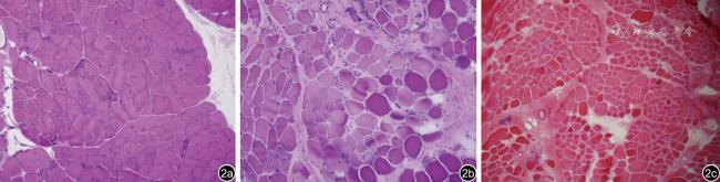
肱二头肌病理组织HE染色见肌纤维大小显著不等,少量坏死纤维及再生肌纤维,肌内膜及肌束膜中、重度增生(图2);基因检测显示外显子缺失或重复。实验室血清CK为(9316.2±5704.9)U/L,明显高于正常值(18~198) U/L。
图2 Duchenne肌营养不良病理图像。图a示肌纤维大小中度不等,偶见坏死纤维,可见较多再生肌纤维,肌内膜及肌束膜轻度增生,血管周围未见炎症细胞浸润(HE ×200);图b示肌纤维大小显著不等,可见少量坏死纤维及再生肌纤维,肌内膜及肌束膜重度增生,血管周围未见炎症细胞浸润(HE ×200);图c示肌纤维大小显著不等,少量坏死肌纤维及再生肌纤维,肌内膜及肌束膜重度增生,血管周围未见炎症细胞浸润(HE ×200)

二、超声表现

(一)二维超声表现

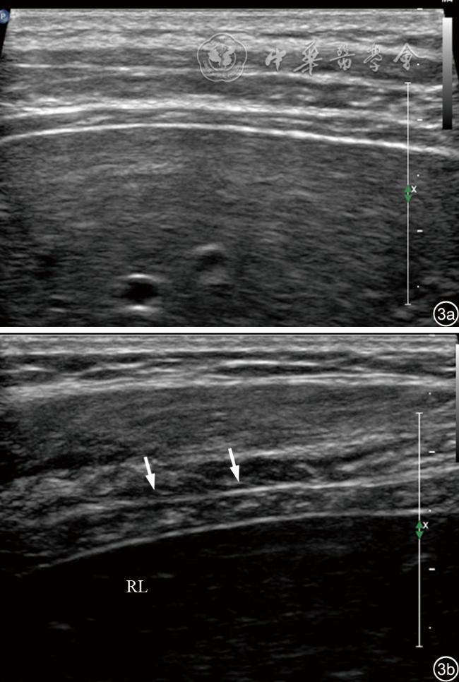
健康儿童膈肌表现为均匀的低回声,中间可见结缔组织形成的条带状高回声线,与胸膜线、腹膜线走行较一致。与正常对照组比较,DMD组右侧腋前线和锁骨中线2个切面呼气末Tdi与吸气末Tdi明显增厚(P均<0.001),膈肌回声增强、不均匀,肌肉间结缔组织形成的条带状高回声线部分消失或减低,正常膈肌结构紊乱或消失(图3)。
图3 正常儿童与Duchenne肌营养不良膈肌二维超声图像。图a为正常儿童膈肌二维超声图像,膈肌表现为均匀的低回声,中间可见结缔组织形成的条带状高回声线,与胸膜线、腹膜线走行较一致;图b为Duchenne肌营养不良患儿膈肌二维超声图像,膈肌较正常儿童增厚,回声增强、不均匀,肌肉间结缔组织形成的条带状高回声线部分紊乱

注:RL为肝右叶

(二)M型超声表现

1. DMD组、正常对照组吸气末Tdi与呼气末Tdi比较:DMD组右侧腋前线吸气末Tdi(4.26±0.60)mm、呼气末Tdi(4.05±0.59)mm,右侧锁骨中线吸气末Tdi(4.62±0.60)mm,呼气末Tdi(4.35±0.59)mm,两切面Tdi随呼吸周期均无明显变化(P=0.593、0.493);而对照组21例健康儿童,右侧腋前线吸气末Tdi(1.72±0.24)mm、呼气末Tdi(0.18±0.21)mm,右侧锁骨中线吸气末Tdi(1.69±0.30)mm、呼气末Tdi(0.99±0.20)mm,两切面Tdi随呼吸周期变化显著(P=0.004、0.006,表1)。
表1 DMD组、正常对照组膈肌吸气末厚度与呼气末厚度比较(mm,
组别 例数 右侧腋前线 t P 右侧锁骨中线 t P
吸气末Tdi 呼气末Tdi 吸气末Tdi 呼气末Tdi
DMD组 21 4.26±0.60 4.05±0.59 0.874 0.593 4.62±0.60 4.35±0.59 0.913 0.493
对照组 21 1.72±0.24 1.18±0.21 0.841 0.004 1.69±0.30 0.99±0.20 0.527 0.006

注:DMD为Duchenne肌营养不良;Tdi为膈肌厚度

2. DMD组与正常对照组DE、DTF比较:DMD组M型超声显示膈肌运动幅度随呼吸变化减低,正常儿童膈肌“大波浪”样表现消失,部分接近平直(图4)。DMD组右侧腋前线和锁骨中线DE分别为(12.0±5.0)、(11.7±4.1)mm,与对照组21例健康儿童的DE(19.0±3.3)、(20.0±5.3)mm比较,差异均有统计学意义(P均<0.001);右侧腋前线和锁骨中线DTF分别为(38.3±19.4)%、(32.9±19.0)%,与对照组21例健康儿童的DTF(50.6±10.9)%、(49.7±11.5)%比较,差异均有统计学意义(P=0.015、0.002,表2)。
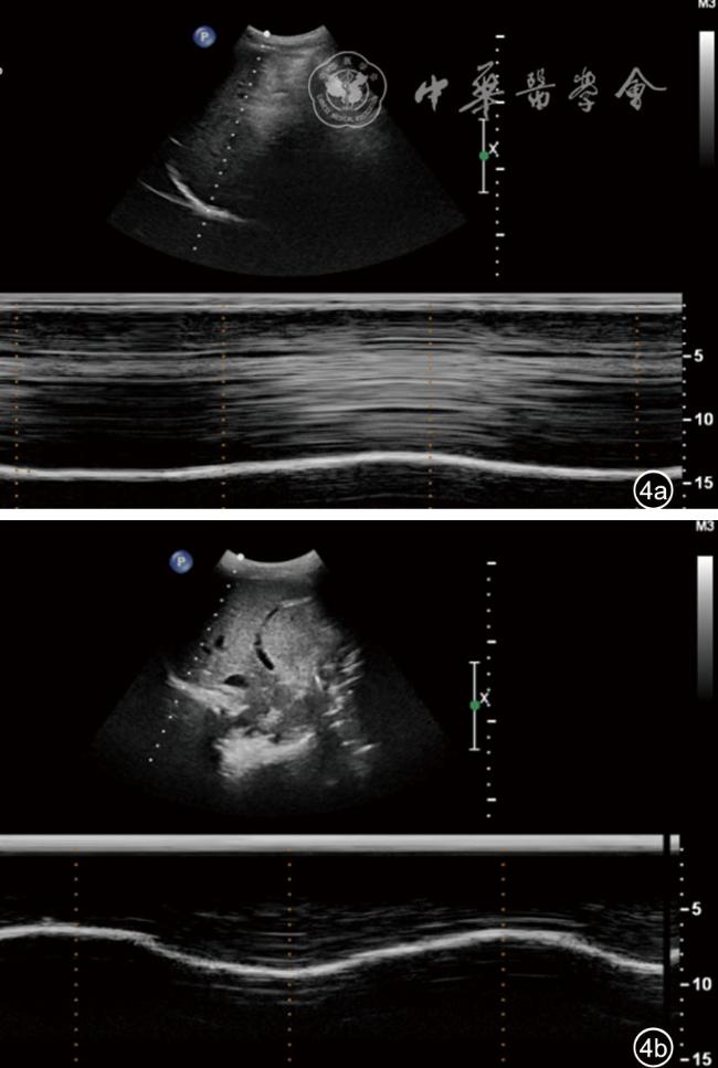
图4 右侧腋前线切面Duchenne肌营养不良患儿与正常儿童膈肌超声图像。图a示Duchenne肌营养不良患儿膈肌运动幅度随呼吸运动变化减弱,接近平直,吸气末与呼气末膈肌的厚度未见明显变化;图b示正常儿童膈肌随呼吸运动变化明显,呈“大波浪”样表现
表2 DMD组与正常对照组DE、DTF对比
组别 例数 右侧锁骨中线DTF(%) 右侧腋前线DTF(%) 右侧锁骨中线DE(mm) 右侧腋前线DE(mm)
DMD组 21 32.9±19.0 38.3±19.4 11.7±4.1 12.0±5.0
对照组 21 49.7±11.5 50.6±10.9 20.0±5.3 19.0±3.3
t 0.079 0.013 0.603 0.207
P 0.002 0.015 <0.001 <0.001

注:DMD为Duchenne肌营养不良;DTF为膈肌增厚率;DE为膈肌移动度

讨论

DMD在疾病进展过程中,膈肌发生纤维化坏死,导致膈肌进行性功能障碍或瘫痪,膈肌的收缩功能减低812,最终引起呼吸衰竭。可靠的肺功能检测和呼吸肌活动测量对于监测DMD疾病进展、识别通气功能不全的早期体征和规划个体呼吸系统管理十分重要13。肺功能检查是DMD肺功能评估的标准814。但对儿童进行肺功能检查,常需进行专业培训及心理健康教育15,因此其应用存在一定局限性。而膈肌作为最主要的吸气肌16,在吸气过程中,承担90%以上的通气,其功能的评估对DMD肺功能的评估具有重要意义17
目前,膈肌功能的影像学评估方法主要包括荧光透视18-19、胸部X线片、CT、MRI和超声检查等。其中X线和CT具有电离辐射20,会导致放射性损伤;MRI是无创检查方法,可以较准确评估膈肌进行性结构的改变以及功能性膈肌损伤21,但此项检查耗时相对较长,仅适合年龄偏大、检查依从性较好的儿童,而多数年龄偏小的患儿无法正常完成MRI检查。超声因具有无创、可实时动态监测、可反复操作等优势,在临床中的应用越来越广泛,尤其对于儿童患者22。但超声对DMD患儿膈肌功能的评估鲜见报道。
有临床研究表明,吸气时膈肌收缩,长度变短,膈肌厚度增加,使用膈肌厚度这一指标评估膈肌功能具有较高的敏感度和特异度23-24。本研究在右侧腋前线、右侧锁骨中线两个切面检查中发现,DMD患儿膈肌增厚,回声增强、杂乱、不均匀,呼气末Tdi和吸气末Tdi较健康儿童组明显增厚,且DMD患儿吸气末Tdi与呼气末Tdi无明显差异,右侧腋前线DTF及右侧锁骨中线DTF均较正常对照组减低,说明DMD患儿呼吸期间膈肌厚度未见明显变化,表明DMD患儿的膈肌收缩功能下降。这也在一定程度证实了既往学者提出的DTF可有效评估膈肌收缩功能25
此外,DMD患儿膈肌运动幅度随着呼吸运动变化明显减弱,甚至部分接近平直,右侧腋前线及右侧锁骨中线DE均较正常对照组减低,与对照组健康儿童膈肌“大波浪”运动的超声表现存在明显差异,这也进一步反映了DMD患儿呼吸期间膈肌收缩能力明显下降。
本研究结果显示,超声可以通过DTF、DE指标评估DMD患儿的膈肌功能,其优势体现在:(1)超声可以较准确地检测DTF、DE变化26;(2)超声技术无创、可实时动态监测18,有利于DMD患儿膈肌功能的动态评估27;(3)膈肌超声可在患者平静呼吸下进行,无需训练和主动配合。
本研究尚存在一些不足之处:纳入样本量偏少,均为男性。原因在于DMD为罕见病,且该病为X连锁隐性遗传性肌肉变性疾病,发病主要为男性患者,女性携带者大多表型正常。因此,期望在后期建立病例库,进行进一步病例积累和研究探讨。
综上所述,超声具有无创、实时、可重复检测的优势,可有效评价DMD患儿膈肌功能,定量测量膈肌DTF、DE有助于DMD患儿病情监测。
1
Siemionow M, Langa P, Harasymczuk M, et al. Human dystrophin expressing chimeric (DEC) cell therapy ameliorates cardiac, respiratory, and skeletal muscle's function in Duchenne muscular dystrophy[J]. Stem Cells Transl Med, 2021,10(10): 1406-1418.

2
Creisméas A, Gazaille C, Bourdon A, et al. TRPC3, but not TRPC1, as a good therapeutic target for standalone or complementary treatment of DMD[J]. J Transl Med, 2021, 19(1): 519.

3
Ebrahimi M, Lad H, Fusto A, et al. De novo revertant fiber formation and therapy testing in a 3D culture model of Duchenne muscular dystrophy skeletal muscle[J]. Acta Biomater, 2021, 132: 227-244.

4
Birnkrant DJ, Bushby K, Bann CM, et al. Diagnosis and management of Duchenne muscular dystrophy, part 1: diagnosis, and neuromuscular, rehabilitation, endocrine, and gastrointestinal and nutritional management[J]. Lancet Neurol, 2018, 17(3): 251-267.

5
Starosta A, Konieczny P. Therapeutic aspects of cell signaling and communication in Duchenne muscular dystrophy[J]. Cell Mol Life Sci, 2021, 78(11): 4867-4891.

6
Writing Group For Practice Guidelines For Diagnosis And Treatment Of Genetic Diseases Medical Genetics Branch Of Chinese Medical Association, Tan H, Liang D, et al. Clinical practice guidelines for Duchenne muscular dystrophy [J]. Zhonghua Yi Xue Yi Chuan Xue Za Zhi, 2020, 37(3): 258-262.

7
谭虎, 梁德生, 邬玲仟. 杜氏进行性肌营养不良的临床实践指南[J]. 中华医学遗传学杂志, 2020, 37(3): 258-262.

8
Birnkrant DJ, Bushby K, Bann CM, et al. Diagnosis and management of Duchenne muscular dystrophy, part 2: respiratory, cardiac, bone health, and orthopaedic management[J]. Lancet Neurol, 2018, 17(4): 347-361.

9
Babbs A, Chatzopoulou M, Edwards B, et al. From diagnosis to therapy in Duchenne muscular dystrophy[J]. Biochem Soc Trans, 2020, 48(3): 813-821.

10
Ahmad A, Esraa S, Amany F, et al. Role of chest ultrasound in diaphragmatic assessment among stable chronic obstructive pulmonary disease patients before and after pulmonary rehabilitation[J]. The Egyptian Journal of Chest Diseases and Tuberculosis, 2021, 70(2): 195-204.

11
Şık N, Çitlenbik H, Öztürk A, et al. Point of care diaphragm ultrasound in acute bronchiolitis: A measurable tool to predict the clinical, sonoimagedata severity of the disease, and outcomes[J]. Pediatr Pulmonol, 2021, 56(5): 1053-1059.

12
McCool FD, Oyieng'o DO, Koo P. The utility of diaphragm ultrasound in reducing time to extubation[J]. Lung, 2020, 198(3): 499-505.

13
Pennati F, LoMauro A, D'Angelo MG, et al. Non-invasive respiratory assessment in Duchenne muscular dystrophy: from clinical research to outcome measures[J]. Life (Basel), 2021, 11(9): 947.

14
Sondekoppam RV, Naik L, Tsui J, et al. Proper use and interpretation of diaphragmatic ultrasonography[J]. Can J Anaesth, 2017, 64(5): 548-549.

15
杨艳红. 肺功能检查对小儿哮喘及咳嗽变异性哮喘的临床应用价值探讨[J].数理医药学杂志, 2021, 34(3): 373-374.

16
王飞飞, 朱晓萍, 马少林. 超声评估膈肌结构和功能[J].中华危重病急救医学, 2017, 29(3): 276-280.

17
van Steveninck AL, Imming LM. Diaphragm dysfunction prior to intubation in a patient with Covid-19 pneumonia; assessment by point of care ultrasound and potential implications for patient monitoring[J]. Respir Med Case Rep, 2020, 31: 101284.

18
Leal BE, Gonçalves MA, Lisboa LG, et al. Validity and reliability of fluoroscopy for digital radiography: a new way to evaluate diaphragmatic mobility[J]. BMC Pulm Med, 2017, 17(1): 62.

19
Weber MD, Lim JKB, Glau C, et al. A narrative review of diaphragmatic ultrasound in pediatric critical care[J]. Pediatr Pulmonol, 2021, 56(8): 2471-2483.

20
Cremonini C, Lewis MR, Jakob D, et al. Diagnosing penetrating diaphragmatic injuries: CT scan is valuable but not reliable[J]. Injury, 2022, 53(1): 116-121.

21
Pennati F, Arrigoni F, LoMauro A, et al. Diaphragm involvement in Duchenne Muscular Dystrophy (DMD): an MRI study[J]. J Magn Reson Imaging, 2020, 51(2): 461-471.

22
王祥, 黄诗倩, 夏祖和, 等. 超声监测膈肌功能在临床中的应用进展[J].中华危重病急救医学, 2021, 33(5): 638-640.

23
Mercurio G, D'Arrigo S, Moroni R, et al. Diaphragm thickening fraction predicts noninvasive ventilation outcome: a preliminary physiological study[J]. Crit Care, 2021, 25(1): 219.

24
Nakanishi N, Okura K, Okamura M, et al. Measuring and monitoring skeletal muscle mass after stroke: a review of current methods and clinical applications[J]. J Stroke Cerebrovasc Dis, 2021, 30(6): 105736.

25
孙强, 山峰, 董海, 等. 超声膈肌增厚率对重症肌无力危象机械通气患者撤机成功的预测价值[J]. 中华危重病急救医学, 2017, 29(7): 619-623.

26
Daniel M, Lang E, Huynh TM, et al. Prevalence and time-course of diaphragmatic dysfunction following lung resection: A repeated ultrasonic assessment[J]. Anaesth Crit Care Pain Med, 2022, 41(2): 101024.

27
Meng P, Ogna A, Fayssoil A. M mode ultrasound and tissue Doppler imaging to assess diaphragm feature in late onset pompe disease[J]. Neurol Int, 2020, 12(3): 55-58.

Outlines

/

Copyright © Chinese Journal of Medical Ultrasound (Electronic Edition), All Rights Reserved.
Tel: 010-51322630、2632、2628 Fax: 010-51322630 E-mail: csbjb@cma.org.cn
Powered by Beijing Magtech Co. Ltd