Home    中文  
 
  • Search
  • lucene Search
  • Citation
  • Fig/Tab
  • Adv Search
Just Accepted  |  Current Issue  |  Archive  |  Featured Articles  |  Most Read  |  Most Download  |  Most Cited
Peripheral Vascular Ultrasound

Hemodynamics changes of the brachial artery to fingertip arterioles on the fistula side upper limb in hemodialysis patients with chronic renal failure

  • Bo Tang 1 ,
  • Jiping Luo 1 ,
  • Tao Zhou 1 ,
  • Duo Huang 1 ,
  • Tingqiong Liu 1 ,
  • Yaping Chen 1 ,
  • Wensheng Yue , 1,
Expand
  • 1.Department of Ultrasound, Affiliated Hospital of North Sichuan Medical College, Nanchong 637000, China
Corresponding author: Yue Wensheng, Email:

Received date: 2022-12-23

  Online published: 2024-03-05

Copyright

Copyright by Chinese Medical Association No content published by the journals of Chinese Medical Association may be reproduced or abridged without authorization. Please do not use or copy the layout and design of the journals without permission. All articles published represent the opinions of the authors, and do not reflect the official policy of the Chinese Medical Association or the Editorial Board, unless this is clearly specified.

Abstract

Objective

To evaluate the hemodynamic changes of the brachial artery to fingertip arterioles in hemodialysis (HD) patients with chronic renal failure by high frequency ultrasound combined with Angio planewave ultrasensitive imaging (Angio PLUS).

Methods

Eighty-one chronic renal failure patients with autogenous arteriovenous fistula (AVF) on long-term regular hemodialysis were prospectively selected at the Affiliated Hospital of North Sichuan Medical College from March 2021 to September 2022, and 103 healthy adults were selected as a control group during the same period. Artery anatomical structures, internal diameter, monophasic waveform, peak systolic velocity (PSV), end diastolic velocity (EDV), mean diastolic velocity (MDV), resistance index (RI), peak systolic blood flow slope (Slope), and blood flow volume (V-Flow) of the brachial artery, ulnar artery, radial artery, ulnar proper palmar digital artery, and radial proper palmar digital artery on the upper limb were acquired by high frequency ultrasound combined with Angio PLUS and compared between and within groups.

Results

The proportion of monophasic waveforms in the flow spectrum of the brachial, ulnar, and radial arteries of the upper limb on the side of the fistula was higher than that of the healthy adult upper limb (χ2=60.106, 8.316, and 11.916, respectively; P<0.05), but there was no statistically significant difference between the ulnar and radial palmar proper digital arteries (P>0.05). The waveforms of all level arterial spectrum on the fistula side upper limb were monophasic; however, the proportion of monophasic waveforms gradually increased from the proximal artery (brachial artery) to the middle artery (ulnar artery and radial artery) to the distal artery (ulnar and radial palmar proper digital artery) on the healthy upper limb. Compared with healthy upper arteries, the internal diameter, PSV, MDV, EDV, and V-Flow of the brachial, ulnar and radial arteries on the fistula side upper limb were increased and RI was decreased (P<0.05), while the internal diameter, MDV, EDV, and V-Flow of the ulnar and radial proper palmar digital artery on the fistula side upper limb were decreased and RI was increased (P<0.05). There was no statistically significant difference in RI between the brachial and ulnar and radial arteries on the fistula side upper limb (P>0.05), but they were all lower than those of the ulnar and radial proper palmar digital artery (P<0.05). The internal diameter, PSV, and V-Flow of arteries on the fistula side upper limb were gradually decreased from the proximal artery (brachial artery) to the middle artery (ulnar artery and radial artery) to the distal artery (ulnar and radial palmar proper digital artery) (P<0.05), which was consistent with the trend of internal diameter, PSV, RI, Slope, and V-Flow of arteries on the healthy upper limb.

Conclusion

By combining high-frequency ultrasound with Angio PLUS technology, the inner diameter and hemodynamic changes of fingertip arterioles can be further quantitatively detected on the basis of observing the brachial, ulnar, and radial arteries on the fistula side in HD patients with chronic renal failure, which could provide a more comprehensive analysis of changes in hemodynamics. The RI of fingertip arterioles is higher than that of the brachial, ulnar, and radial arteries, which has appreciated clinical value for early diagnosis of distal limb hypoperfusion/ischemia on the fistula side in HD patients.

Cite this article

Bo Tang , Jiping Luo , Tao Zhou , Duo Huang , Tingqiong Liu , Yaping Chen , Wensheng Yue . Hemodynamics changes of the brachial artery to fingertip arterioles on the fistula side upper limb in hemodialysis patients with chronic renal failure[J]. Chinese Journal of Medical Ultrasound (Electronic Edition), 2023 , 20(12) : 1276 -1281 . DOI: 10.3877/cma.j.issn.1672-6448.2023.12.010

自体动静脉内瘘(autologous arteriovenous fistula,AVF)是终末期肾病患者进行长期有效血液透析(hemodialysis,HD)的血管通路,其中采用前臂远端桡动脉和头静脉直接吻合是AVF的首选途径,具有可长期使用、并发症少等特点1。但术后部分患者造瘘侧上肢血栓、动脉硬化及肢体远端低灌注缺血综合征等并发症发生率仍较高。常规高频超声和彩色多普勒超声通过监测血管腔内的血流动力学变化,能快速筛查和早期诊断血管并发症,但对微小血管识别的敏感度较低,不能全面评估指端微小血管的灌注情况2。平面波超敏感血流显像(angio plane wave ultrasensitive imaging,Angio PLUS)对微血流显像具有高敏感度及准确性3,可定性定量检测组织或病灶微血管的血流动力学状态4。本研究通过高频超声结合Angio PLUS技术对比分析维持性HD患者造瘘侧上肢与健康成人上肢肱动脉至指端微小动脉血管结构及血流动力学特征和差异,探讨造瘘侧上肢动脉血流动力学变化特点。

资料与方法

一、对象

选取2021年3月至2022年9月因慢性肾衰竭于川北医学院附属医院血液净化中心长期规律HD患者81例,其中男性35例,女性46例,年龄28~91岁,平均年龄(56.38±13.55)岁.所有患者均采用单侧肢体桡动脉-头静脉内瘘,透析时长均大于3个月.同期纳入103例于川北医学院附属医院健康管理中心体检的健康成人作为正常对照组,其中男性45例,女性48例,年龄35~80岁,平均年龄(54.93±10.47)岁.排除标准:(1)Allen实验阳性及先天性血管发育异常;(2)AVF相关血管发现血栓或狭窄;(3)急慢性感染病、严重心血管疾病及风湿血液系统疾病;(4)妊娠期或哺乳期女性.本研究是一项前瞻性研究,且经川北医学院附属医院伦理委员会批准(批件号:2021ER207-2),受试者均签署知情同意书,同意参与本研究。

二、仪器与方法

1. 仪器:采用法国声科Supersonic Aixplorer超声诊断仪,SL15-4高频线阵探头,探头频率4~15 MHz。
2. 肱动脉与尺桡动脉超声参数采集:检查室温23~25 ℃,嘱受试者静息30 min后仰卧,双侧上肢自然伸直与心脏同一水平,掌心向上呈放松休息位,检查时手臂外展约60°。HD患者检查造瘘侧上肢,健康成人检查右侧上肢。探头由大圆肌下缘肱动脉向远端指端微小动脉扫查,观察动脉管壁及腔内血流充盈情况。测量肘横纹上5 cm处肱动脉、腕横纹上2 cm处尺动脉与桡动脉(造瘘侧上肢桡动脉测量处位于瘘口远心端)的血管内径;采用脉冲多普勒超声获取各段动脉血管纵断面管腔内血流频谱图,观察并记录各段动脉收缩期峰值速度(peak systolic velocity,PSV)、舒张末期速度(end diastolic velocity,EDV)、舒张期平均速度(mean diastolic velocity,MDV)、阻力指数(resistance index,RI)、收缩期峰值血流加速度(systolic acceleration,Slope)及每分钟血流容积(blood flow volume,V-Flow)。以上测值均取连续3个心动周期的平均值。
3. 指端微小动脉超声参数采集:嘱受检者掌心向上手指自然伸直,选用Angio PLUS彩色能量成像(color power imaging,CPI)模式分别观察手中指末节指骨区尺侧指掌侧固有动脉与桡侧指掌侧固有动脉。指端微小动脉管径较细,横断面显示困难,于血管纵切面调整血管内血流信号无外溢时测量血管内径。各微小动脉频谱及血流动力学参数测量方法同肱动脉。
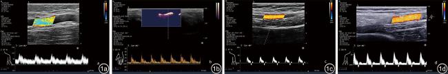
4. 动脉血流频谱形态:根据有无舒张期反向波将频谱分为单相波与非单相波,再进一步根据舒张中晚期频谱波形将非单相波分为典型三相波与非典型三相波(图1)。
图1 动脉血流频谱形态脉冲多普勒超声图像。图a示造瘘侧上肢肱动脉呈单相低阻力型动脉血流频谱;图b示造瘘侧上肢尺侧指掌侧固有动脉血流呈单相高阻力型动脉血流频谱;图c示健康上肢肱动脉呈非典型外周动脉三相波血流频谱(收缩期正向,舒张中晚期负向、正向不连续性低速血流频谱);图d示健康上肢肱动脉呈典型外周动脉三相波血流频谱(收缩期正向,舒张中晚期负向、正向连续性低速血流频谱)

三、统计学分析

采用SPSS 25.0软件对数据进行统计分析,计数资料以例(%)表示,2组间比较采用χ2检验。血流动力学参数等呈正态分布的计量资料以表示,2组间比较采用独立样本t检验;组内各段动脉参数比较采用单因素方差分析,进一步两两比较采用S-N-K检验。以P<0.05为差异有统计学意义。

结果

一、一般资料

HD患者与正常组性别、年龄比较,差异无统计学意义(P均>0.05)。

二、2组间动脉血流频谱形态比较

造瘘侧上肢肱动脉、尺动脉及桡动脉血流频谱单相波占比高于健康上肢(χ2 =60.106、8.316、11.916,P均<0.05),而尺侧及桡侧指掌侧固有动脉单相波占比2组差异无统计学意义(P均>0.05)。造瘘侧上肢各段动脉频谱波形均为单相波,而健康上肢由近段动脉(肱动脉)经中段动脉(尺动脉、桡动脉)至远段动脉(尺侧及桡侧指掌侧固有动脉)血流频谱单相波占比逐渐增高(表1)。
表1 造瘘侧上肢动脉与健康成人上肢动脉血流频谱波形占比[例(%)]
动脉波形 BA UA RA UPPDA RPPDA
造瘘侧上肢(n=81)
单相波 81(100) 81(100) 81(100) 81(100) 81(100)
非单相波
典型三相波 0 0 0 0 0
非典型三相波 0 0 0 0 0
健康上肢(n=103)
单相波 49(47.57) 93(90.30)a 89(86.41)a 103(100)abc 102(99.03)abc
非单相波
典型三相波 34(33.01) 5(4.85) 6(5.82) 0 1(0.97)
非典型三相波 20(19.42) 5(4.85) 8(7.77) 0 0

注:BA为肱动脉;UA为尺动脉;RA为桡动脉;UPPDA为尺侧指掌侧固有动脉;RPPDA为桡侧指掌侧固有动脉;与BA相比,aP<0.05;与UA相比,bP<0.05;与RA相比,cP<0.05

三、2组间动脉超声参数对比

与健康上肢动脉相比,造瘘侧上肢肱动脉、尺动脉及桡动脉内径增大,PSV、MDV、EDV及V-Flow增高,RI降低(P均<0.05);尺侧及桡侧指掌侧固有动脉内径减小,MDV、EDV及V-Flow降低,RI增高(P均<0.05,表2)。
表2 造瘘侧上肢与健康成人上肢动脉血流动力学参数(
动脉 内径(mm) PSV(cm/s) MDV(cm/s) EDV(cm/s) RI Slope(cm/s2 V-Flow(ml/min)
造瘘侧上肢(n=81)
BA 6.24±0.99* 133.29±48.08* 59.18±22.02* 63.33±24.42* 0.53±0.10* 641.91±370.00* 875.69±462.05*
UA 2.52±0.61*a 107.32±42.68*a 47.65±30.61*a 50.51±32.10*a 0.58±0.18* 649.57±315.79* 121.28±101.91*a
RA 2.85±0.86*ab 104.90±63.31*a 54.19±40.94* 56.66±40.63* 0.53±0.23* 411.39±335.73*ab 172.73±158.85*a
UPPDA 0.76±0.23*abc 26.78±14.60abc 7.67±5.57*abc 7.91±5.64*abc 0.74±0.13*abc 200.30±170.49abc 2.67±2.07*abc
RPPDA 0.76±0.29*abc 17.46±9.41*abc 5.27±3.73*abc 5.37±3.81*abc 0.73±0.13*abcd 107.46±87.30*abcd 2.20±1.32*abc
F 685.15 131.17 78.87 89.08 34.40 64.27 214.47
P <0.01 <0.01 <0.01 <0.01 <0.01 <0.01 <0.01
健康上肢(n=103)
BA 3.72±0.65 74.01±17.47 6.95±6.89 7.63±7.10 0.90±0.09 787.20±239.05 68.44±31.85
UA 1.85±0.38a 65.43±15.57a 13.56±9.38a 14.50±9.32a 0.79±0.10a 601.12±150.38a 31.61±26.63a
RA 2.09±0.30ab 58.09±14.02ab 11.90±7.34a 12.64±7.48a 0.79±0.10a 541.57±152.53ab 29.53±18.44a
UPPDA 0.93±0.14abc 30.05±15.95abc 11.17±8.03ab 11.56±6.28ab 0.64±0.11abc 182.31±86.51abc 4.88±3.89abc
RPPDA 0.86±0.14abc 24.99±12.37abcd 8.67±5.96bcd 8.91±6.03bcd 0.66±0.11abc 153.20±86.18abc 3.42±2.76abc
F 991.18 210.59 12.35 13.34 105.07 223.57 172.09
P <0.01 <0.01 <0.01 <0.01 <0.01 <0.01 <0.01

注:PSV为收缩期峰值速度;MDV为舒张期平均速度;EDV为舒张末期速度;RI为阻力指数;Slope为收缩期峰值血流加速度;V-Flow为血流容积;BA为肱动脉;UA为尺动脉;RA为桡动脉;UPPDA为尺侧指掌侧固有动脉;RPPDA为桡侧指掌侧固有动脉;组内比较:与BA比较,aP<0.05;与组内UA比较,bP<0.05;与组内RA比较,cP<0.05;与组内UPPDA比较,dP<0.05;组间比较:与健康成人同名动脉参数比较,*P<0.05

四、2组内动脉超声参数对比

造瘘侧上肢肱动脉、尺动脉及桡动脉之间RI差异无统计学意义(P均>0.05),且均低于尺侧与桡侧指掌侧固有动脉(P均<0.05)。造瘘侧上肢动脉内径、PSV及V-Flow与健康上肢动脉内径、PSV、RI、Slope及V-Flow自近段动脉(肱动脉)经中段动脉(尺动脉、桡动脉)至远段动脉(尺侧及桡侧指掌侧固有动脉)均呈逐渐降低的变化趋势(P<0.05,表2)。

讨论

本研究采用高频超声结合Angio PLUS 技术对造瘘侧上肢肱动脉、尺动脉、桡动脉内径及血流动力学参数进行定性和定量检测,研究结果提示,与健康上肢相比,造瘘侧上肢肱动脉、尺动脉、桡动脉内径增大,血流频谱单相波占比增高,PSV、MDV及EDV流速增快,RI降低,血流量增多,频谱波形均呈现为高速低阻的高流量血流状态,这与既往研究相符5,6,7。AVF成形术后,高压动脉与低压静脉系统连接致心脏回心血流量和每搏输出量增多,血液处于高动力循环状态,动脉内皮细胞所受管壁剪切力及管壁张力增大出现血管重塑使管腔适应性扩张,呈现动脉内径增宽、阻力降低及血流量增多的高流量状态以满足血液透析治疗需求8,9,10
此外,本研究应用Angio PLUS技术检测造瘘侧指端微小动脉,并与健康成人比较,尺侧与桡侧指掌侧固有动脉内径减小,MDV及EDV流速减慢,RI增高,血流量降低,但并无反向血流,血流频谱波形也未发生改变,呈现外周阻力增高的低灌注血流状态,与Bae等11研究结果相符。血液透析患者内瘘成形术前及术后一段时期指端皮肤灌注压改变,指端早期缺血,但Bae等研究中并未直观反映动脉内径及血流动力学变化情况。造瘘侧上肢近心端桡动脉血液大部分经瘘口流入静脉,其余流向桡动脉远心端与尺动脉共同通过掌浅弓、掌深弓等分支供应指端微小动脉12,但部分患者造瘘侧远端肢体仍出现手指末端苍白发凉、麻木疼痛等缺血症状13。目前普遍认为肢体远端缺血为盗血所致,并以出现反向血流为证据,Leon等提出该说法或并不准确,除盗血原因外,动脉管腔狭窄与远端动脉病变引起的无反向血流的低灌注现象也可导致肢体远端缺血14。指端微小动脉内径及血流动力学变化的机制较为复杂,目前尚无法全面解释,可能由于患者自身基础疾病、毒素积聚与血液透析治疗等损伤血管内皮细胞、诱导氧化应激与炎症反应,导致或加快动脉硬化、微小动脉痉挛、管径变细及内环境紊乱等,多种机制共同作用导致造瘘侧指端微小动脉微循环障碍,呈现高外周阻力低灌注缺血状态15,16,17
本研究应用Angio PLUS显像技术检测指端微小动脉,结合肱动脉、尺动脉及桡动脉内径及血流动力学等变化趋势,与健康上肢动脉对比分析发现:造瘘侧上肢近心端动脉(肱动脉、尺动脉及桡动脉)RI无明显差异但均低于远心端动脉(尺侧及桡侧指掌侧固有动脉)RI,与既往研究及本研究中健康上肢近心端至远心端动脉RI逐渐降低的变化趋势18有所不同。AVF成形术后上肢近心端动脉处于高动力循环状态,内径增大腔内血流呈现高速低阻的高流量血流动力学状态,指端微小动脉因微循环障碍等原因导致管径变细,呈现高外周阻力的低灌注缺血状态,上肢近心端至远心端动脉阻力指数变化趋势发生改变,故与健康上肢外周动脉逐渐分支变细阻力指数逐渐降低的变化趋势有所不同。
本研究尚存在一定的局限性:样本量较小,部分动脉血流动力学参数差异并不明显;缺乏AVF成形术前与术后患者自身对比、未考虑术中血管吻合方式、瘘口大小及透析时长对上肢动脉的影响,本研究结果仍需后续临床研究验证。
综上所述,通过高频超声联合Angio PLUS技术可在观察HD患者造瘘侧肱、尺、桡动脉的基础上进一步定量检测指端微小动脉的内径及血流动力学变化,证实其指端微小动脉RI高于肱、尺、桡动脉,对HD患者造瘘侧肢体远端低灌注缺血现象的早期诊断有一定的临床价值。
1
DeVita MV, Khine SK, Shivarov H. Novel approaches to arteriovenous access creation, maturation, suitability, and durability for dialysis [J]. Kidney Int Rep, 2020, 5(6): 769-778.

2
Meola M, Marciello A, Di Salle G, et al. Ultrasound evaluation of access complications: Thrombosis, aneurysms, pseudoaneurysms and infections [J]. J Vasc Access, 2021, 22(1_suppl): 71-83.

3
谭雪艳, 岳文胜, 陈思思, 等. 健康成人上肢肱动脉-指端微循环的血流动力学梯度变化研究 [J/OL]. 中华医学超声杂志(电子版), 2021, 18(2): 193-198.

4
Mohindra N, Jain N, Yadav S, et al. Utility of ultrasound Angio-PLUS imaging for detecting blood flow in breast masses and comparison with color Doppler for differentiating benign from malignant masses [J]. Acta Radiol (Stockholm, Sweden: 1987), 2023, 64(6): 2087-2095.

5
Vallespin J, Meola M, Ibeas J. Upper limb anatomy and preoperative mapping [J]. J Vasc Access, 2021, 22(1_suppl): 9-17.

6
Chytilova E, Jemcov T, Malik J, et al. Role of Doppler ultrasonography in the evaluation of hemodialysis arteriovenous access maturation and influencing factors [J]. J Vasc Access, 2021, 22(1_suppl): 42-55.

7
Zamboli P, Fiorini F, D'Amelio A, et al. Color Doppler ultrasound and arteriovenous fistulas for hemodialysis [J]. J Ultrasound, 2014, 17(4): 253-263.

8
Malik J, Lomonte C, Rotmans J, et al. Hemodialysis vascular access affects heart function and outcomes: Tips for choosing the right access for the individual patient [J]. J Vasc Access, 2021, 22(1_suppl): 32-41.

9
Remuzzi A, Ene-Iordache B. Novel paradigms for dialysis vascular access: upstream hemodynamics and vascular remodeling in dialysis access stenosis [J]. Clin J Am Soc of Nephrol: CJASN, 2013, 8(12): 2186-2193.

10
Tuka V, Malik J, Chytilova E, et al. Long-term arterial adaptation to high blood flow in the feeding artery of vascular access for hemodialysis [J]. J Vasc Access, 2012, 13(3): 305-309.

11
Bae M, Chung SW, Lee CW, et al. Skin perfusion pressure for predicting access-related hand ischemia following arteriovenous fistula surgery based on the brachial artery [J]. J Vasc Access, 2022, 23(3): 383-389.

12
Tan RES, Lahiri A. Vascular anatomy of the hand in relation to flaps [J]. Hand Clin, 2020, 36(1): 1-8.

13
Stolic R. Most important chronic complications of arteriovenous fistulas for hemodialysis [J]. Med Princ Pract, 2013, 22(3): 220-228.

14
Leon C, Asif A. Arteriovenous access and hand pain: the distal hypoperfusion ischemic syndrome [J]. Clin J Am Soc Nephrol, 2007, 2(1): 175-183.

15
Rizzoni D, Agabiti Rosei E. Small artery remodeling in hypertension and diabetes [J]. Curr Hypertens Rep, 2006, 8(1): 90-95.

16
Ahmadmehrabi S, Tang WHW. Hemodialysis-induced cardiovascular disease [J]. Semin Dial, 2018, 31(3): 258-267.

17
Liu WC, Tomino Y, Lu KC. Impacts of indoxyl sulfate and p-cresol sulfate on chronic kidney disease and mitigating effects of AST-120 [J]. Toxins, 2018, 10(9): 367.

18
Badeer HS. Hemodynamics for medical students [J]. Adv in Physiol Educ, 2001, 25(1): 44-52.

Outlines

/

Copyright © Chinese Journal of Medical Ultrasound (Electronic Edition), All Rights Reserved.
Tel: 010-51322630、2632、2628 Fax: 010-51322630 E-mail: csbjb@cma.org.cn
Powered by Beijing Magtech Co. Ltd