
近年来,随着全球范围内肥胖人群的增多,非酒精性脂肪性肝病(non-alcoholic fatty liver disease,NAFLD)的发病率也在逐年上升,NAFLD已成为国内最常见的一种慢性肝病[1],根据病情进展程度可将其分为非酒精性肝脂肪变、非酒精性脂肪性肝炎、肝硬化和肝细胞癌[2]。伴随高比例的代谢并发症,NAFLD显著增加了患者的心血管疾病(cardiovascular disease,CVD)发生风险,因而早期诊断及持续监测肝脏脂肪变性显得尤为重要。虽然肝活检被视为NAFLD诊断的金标准[3],但其为有创操作,可能存在出血或取样不足等问题,因而限制了其广泛应用。因此,一种无创、可量化且能够在一定程度上替代肝活检的检查手段亟待推广。超声引导衰减参数成像(ultrasound-guided attenuation parameter,UGAP)是近年来发展起来的超声技术,它基于衰减系数已知的组织模拟参考模型测量组织声波衰减系数[4],使用常规的超声探头计算超声信号通过肝脏组织的衰减值。超声衰减系数是评价肝脂肪变性程度的重要指标,UGAP作为一种新技术,能提供声衰减的量化数值,可更直观地呈现肝脂肪变性的程度。目前,已有相关研究[5,6]表明UGAP对NAFLD具有良好的诊断性能,但目前尚未见报道针对其在临床环境中的可行性和影响因素进行研究。本研究旨在评估UGAP技术的临床适用性及可重复性,并探讨其相关影响因素,构建Nomogram模型评估NAFLD患者发生CVD的风险,为NAFLD患者心血管疾病的防治提供参考依据。
资料与方法
一、对象
本研究为回顾性横断面研究。选取2022年11月至2023年11月于哈尔滨医科大学第一附属医院就诊的NAFLD患者204例。纳入标准:(1)年龄≥18周岁;(2)符合《非酒精性脂肪性肝病诊疗指南(2010年版)》中列出的NAFLD诊断标准[7];(3)均接受二维超声检查、UGAP技术及实验室检查,如血生化指标检查;(4)未接受过脂肪肝治疗。排除标准:(1)既往行右肝切除术;(2)不能积极配合UGAP检查;(3)妊娠期女性或右上腹有创伤且伤口处于愈合期的患者;(4)服用过影响血脂及肝脏脂肪代谢的药物。纳入符合入排标准的204例NAFLD患者,另随机选取同期73例健康体检者作为对照组。本研究已经哈尔滨医科大学第一附属医院伦理委员会批准(批件号:2023IIT298)。
二、仪器与方法
1.仪器:
使用GE LOGIQ Fortis超声诊断仪,C1-6-D凸阵探头,频率为3.5 MHz。
2.二维超声及UGAP检查:
受试者在检查前需空腹6 h以上。检查由2名具有5年以上超声诊断经验的医师共同完成。按照Hamaguchi评分系统的二维超声分级诊断标准对所有受试者的肝脏脂肪变性程度进行评价,该评分系统基于肝脏回声强度、后方回声衰减、肝肾回声对比、肝内管道显示清晰度,对肝脏的回声信号进行评分[8],分为S0~S3级,其脂肪变性程度依次递增,分别为无、轻度、中度和重度脂肪变性。
受试者随即进行UGAP检查,操作由2名接受UGAP技术专业培训的医师执行。嘱受试者取仰卧位,医师沿腋中线通过肋间隙对肝脏的SⅤ段或SⅥ段进行扫查。在二维超声状态下激活UGAP模式,嘱受试者屏气,在均匀区域设置ROI位置,由于ROI的测量深度固定在4~8 cm,因此操作者只能横向移动ROI位置,以避开大血管和胆管。记录单帧上的8次连续测量结果,将四分位数间距/中位数<0.30的UGAP值定义为有效测量(图1),取中位数作为最终结果。

3.临床资料收集及分组:
记录每位受检者的皮肤-肝包膜距离(skin-to-liver-capsule distance,SLD)、体质量指数(body mass index,BMI)及1周内的血脂及肝功能指标。调查患者相关医疗记录,病历中有确诊NAFLD后被初次诊断为CVD的患者为合并CVD组,临床上检测出NAFLD后未患有CVD者为未合并CVD组。CVD定义为5种心血管结局,包括充血性心力衰竭、冠心病、心绞痛、心肌梗死和卒中[9]。所有CVD的诊断和分类均基于第10版国际疾病分类(International Classification of Diseases,ICD)。
三、统计学分析
采用SPSS 27.0软件对数据进行统计分析,使用R软件制作列线图。符合正态分布的计量资料以 ±s表示,2组间比较采用独立样本t检验。非正态分布的数据,以M(P25,P75)表示,2组间比较采用非参数检验。计数资料以例(%)表示,2组间比较采用χ2检验。应用单因素和多因素线性回归分析各变量与UGAP值之间的关联。将单因素分析中P<0.05的变量纳入多因素回归分析,确定UGAP值的独立影响因素。此外,采用Spearman等级相关来评估UGAP值与肝脂肪变性二维超声诊断分级之间的相关性;采用Kruskal-Wallis检验比较不同程度肝脂肪变性的UGAP值差异。受试者间UGAP值的一致性通过计算组内相关系数(intraclass correlation coefficient,ICC)来评估。采用Logistic回归和Nomogram模型分析NAFLD患者并发CVD的危险因素,并应用ROC曲线及校正曲线评估该模型的临床效能。P<0.05为差异具有统计学意义。
±s表示,2组间比较采用独立样本t检验。非正态分布的数据,以M(P25,P75)表示,2组间比较采用非参数检验。计数资料以例(%)表示,2组间比较采用χ2检验。应用单因素和多因素线性回归分析各变量与UGAP值之间的关联。将单因素分析中P<0.05的变量纳入多因素回归分析,确定UGAP值的独立影响因素。此外,采用Spearman等级相关来评估UGAP值与肝脂肪变性二维超声诊断分级之间的相关性;采用Kruskal-Wallis检验比较不同程度肝脂肪变性的UGAP值差异。受试者间UGAP值的一致性通过计算组内相关系数(intraclass correlation coefficient,ICC)来评估。采用Logistic回归和Nomogram模型分析NAFLD患者并发CVD的危险因素,并应用ROC曲线及校正曲线评估该模型的临床效能。P<0.05为差异具有统计学意义。结果
一、一般资料
本研究共纳入277例受试者,其中NAFLD组204例[男性109例,女性95例,年龄范围23~77岁,平均年龄56(50,63)岁],对照组73例[男性35例,女性38例,年龄范围24~77岁,平均年龄56.5(47,64)岁]。NAFLD组与对照组的BMI、丙氨酸氨基转移酶、门冬氨酸氨基转移酶、血糖、总胆固醇、甘油三酯、高密度脂蛋白、UGAP值差异存在统计学意义(P均<0.05,表1)。
表1 NAFLD组与对照组一般临床资料比较[M(P25,P75)] |
| 临床资料 | 对照组(n=73) | NAFLD组(n=204) | 统计值 | P值 |
|---|---|---|---|---|
| 年龄(岁) | 56.5(47,64) | 56(50,63) | Z=-0.035 | 0.972 |
| BMI(kg/m2) | 22.75(21.85,23.55) | 26.90(25.6,27.7) | Z=-11.490 | <0.001 |
| SLD(cm) | 2.19(1.75,2.42) | 2.13(1.88,2.36) | Z=-0.094 | 0.925 |
| ALT(U/L) | 22.75(18.1,27.15) | 50.50(44.7,60.05) | Z=-11.339 | <0.001 |
| AST(U/L) | 22.95(18.35,26.7) | 53.60(44.15,65.2) | Z=-11.234 | <0.001 |
| GGT(U/L) | 58.55(34.2,86.05) | 63.40(35.9,87.15) | Z=-0.363 | 0.717 |
| Glu(mmol/L) | 5.08(4.67,5.62) | 5.91(5.63,7.06) | Z=-7.595 | <0.001 |
| TC(mmol/L) | 3.75(3.21,4.15) | 5.26(4.98,5.86) | Z=-12.564 | <0.001 |
| TG(mmol/L) | 1.16(0.94,1.35) | 2.28(1.89,2.69) | Z=-12.746 | <0.001 |
| HDL(mmol/L) | 1.21(0.88,1.36) | 1.04(0.92,1.17) | Z=-2.425 | 0.015 |
| LDL(mmol/L) | 3.06(2.32,3.69) | 3.09(2.82,3.63) | Z=-0.492 | 0.623 |
TP(g/L, ±s) ±s) | 67.00±5.75 | 68.02±5.99 | t =-1.264 | 0.207 |
| AKP(U/L) | 73.8(60.2,89.75) | 73.4(64.05,87.8) | Z=-0.788 | 0.431 |
| UGAP-1(dm/cm/MHz) | 0.61(0.59,0.62) | 0.71(0.68,0.74) | Z=-11.498 | <0.001 |
| UGAP-2(dm/cm/MHz) | 0.62(0.6,0.63) | 0.72(0.69,0.76) | Z=-11.462 | <0.001 |
注:BMI为体质量指数;SLD为皮肤-肝包膜距离;ALT为丙氨酸氨基转移酶;AST为门冬氨酸氨基转移酶;GGT为γ-谷氨酰转移酶;Glu为血糖;TC为总胆固醇;TG为甘油三酯;HDL为高密度脂蛋白;LDL为低密度脂蛋白;TP为总蛋白;AKP为碱性磷酸酶;UGAP-1、UGAP-2分别为2位检查医师测量的超声引导衰减参数;NAFLD为非酒精性脂肪性肝病 |
277例受试者中,S0组73例(健康对照组),S1~S3组分别为83、86、35例。S0组UGAP值为0.61(0.59,0.62)dB/cm/MHz,S1组UGAP值为0.68(0.67,0.69)dB/cm/MHz,S2组UGAP值为0.72(0.72,0.74)dB/cm/MHz,S3组UGAP值为0.81(0.79,0.83)dB/cm/MHz,且UGAP值在S0~S3各组间的差异存在统计学意义(H=236.35,P<0.001)。
二、与UGAP相关的影响因素分析
以UGAP值为因变量,年龄、BMI、SLD、丙氨酸氨基转移酶、门冬氨酸氨基转移酶、γ-谷氨酰转移酶、血糖、总胆固醇、甘油三酯、高密度脂蛋白、低密度脂蛋白、总蛋白、碱性磷酸酶、肝脂肪变性程度(赋值:S0=0,S1=1,S2=2,S3=3)为自变量。单因素线性回归分析表明,影响UGAP的因素包括BMI、血糖、丙氨酸氨基转移酶和门冬氨酸氨基转移酶、总胆固醇和甘油三酯以及肝脂肪变性程度(P均<0.05)。将上述指标纳入多因素线性回归分析,剔除方差膨胀因子(variance inflation factor,VIF)>10的变量丙氨酸氨基转移酶,结果显示,UGAP的独立影响因素为BMI、总胆固醇、甘油三酯及肝脂肪变性程度(P均<0.05,表2、表3)。
表2 影响NAFLD患者UGAP值的单因素线性回归分析 |
| 因素 | 非标准化系数 | 标准化系数 | t值 | P值 | R2 | 调整R2 | F(P)值 | |
|---|---|---|---|---|---|---|---|---|
| B | 标准误 | Beta | ||||||
| 年龄 | 0 | 0 | 0.043 | 0.708 | 0.479 | 0.002 | -0.002 | 0.502(0.479) |
| BMI | 0.022 | 0 | 0.936 | 44.099 | <0.001 | 0.876 | 0.876 | 1944.6395(<0.001) |
| SLD | 0.001 | 0.012 | 0.006 | 0.099 | 0.921 | 0 | -0.004 | 0.01(0.921) |
| ALT | 0.004 | 0 | 0.971 | 67.354 | <0.001 | 0.943 | 0.943 | 4536.536(<0.001) |
| AST | 0.002 | 0 | 0.841 | 25.778 | <0.001 | 0.708 | 0.707 | 664.486(<0.001) |
| GGT | 0 | 0 | 0.059 | 0.98 | 0.328 | 0.003 | 0 | 0.96(0.328) |
| Glu | 0.026 | 0.002 | 0.583 | 11.887 | <0.001 | 0.339 | 0.337 | 141.308(<0.001) |
| TC | 0.068 | 0.001 | 0.955 | 53.408 | <0.001 | 0.912 | 0.912 | 2852.434(<0.001) |
| TG | 0.079 | 0.001 | 0.959 | 55.858 | <0.001 | 0.919 | 0.919 | 3120.156(<0.001) |
| HDL | -0.029 | 0.018 | -0.098 | -1.633 | 0.104 | 0.01 | 0.006 | 2.666(0.104) |
| LDL | -0.009 | 0.005 | -0.106 | -1.763 | 0.079 | 0.011 | 0.008 | 3.109(0.079) |
| TP | 0.001 | 0.001 | 0.11 | 1.832 | 0.068 | 0.012 | 0.008 | 3.357(0.068) |
| AKP | 0 | 0 | 0.062 | 1.025 | 0.306 | 0.004 | 0 | 1.051(0.306) |
| 肝脂肪变性程度 | 0.064 | 0.002 | 0.908 | 35.842 | <0.001 | 0.824 | 0.823 | 1284.649(<0.001) |
注:BMI为体质量指数;SLD为皮肤-肝包膜距离;ALT为丙氨酸氨基转移酶;AST为门冬氨酸氨基转移酶;GGT为γ-谷氨酰转移酶;Glu为血糖;TC为总胆固醇;TG为甘油三酯;HDL为高密度脂蛋白;LDL为低密度脂蛋白;TP为总蛋白;AKP为碱性磷酸酶;UGAP为超声引导衰减参数;NAFLD为非酒精性脂肪性肝病 |
表3 影响NAFLD患者UGAP值的多因素线性回归分析 |
| 因素 | 非标准化系数 | 标准化系数 | t值 | P值 | VIF | R2 | 调整R2 | F(P)值 | |
|---|---|---|---|---|---|---|---|---|---|
| B | 标准误 | Beta | |||||||
| 常数 | 0.387 | 0.011 | 35.057 | <0.001 | - | 0.976 | 0.976 | 1841.781(<0.001) | |
| BMI | 4.0×10⁻³ | 0.001 | 0.190 | 7.691 | <0.001 | 6.906 | |||
| AST | 5.75×10⁻⁵ | 0 | 0.024 | 1.330 | 0.185 | 3.652 | |||
| Glu | 0 | 0.001 | -0.006 | -0.465 | 0.643 | 1.600 | |||
| TC | 0.024 | 0.002 | 0.332 | 13.265 | <0.001 | 7.112 | |||
| TG | 0.028 | 0.002 | 0.339 | 11.959 | <0.001 | 9.081 | |||
| 肝脂肪变性程度 | 0.011 | 0.001 | 0.153 | 7.463 | <0.001 | 4.774 | |||
注:BMI为体质量指数;AST为门冬氨酸氨基转移酶;Glu为血糖;TC为总胆固醇;TG为甘油三酯;UGAP为超声引导衰减参数;NAFLD为非酒精性脂肪性肝病 |
三、UGAP技术评价肝脂肪变性的可行性分析
UGAP值和肝脂肪变性程度之间存在显著的正相关关系(r=0.923,P<0.001)。一致性分析结果表明,UGAP技术操作者间的总体一致性很好(ICC=0.977,P<0.001),S0~S3各分组检查结果的一致性也较好(ICC均>0.800,P均<0.001,表4)。
表4 UGAP值的操作者间一致性分析 |
| 参数 | 组内相关系数 | 95%CI | P值 |
|---|---|---|---|
| 总体 | 0.977 | 0.972~0.982 | <0.001 |
| 肝脂肪变性诊断分级 | |||
| S0 | 0.885 | 0.823~0.926 | <0.001 |
| S1 | 0.851 | 0.779~0.901 | <0.001 |
| S2 | 0.887 | 0.832~0.925 | <0.001 |
| S3 | 0.909 | 0.827~0.953 | <0.001 |
注:UGAP为超声引导衰减参数 |
四、NAFLD合并CVD组与未合并CVD组患者的基线资料比较
NAFLD合并CVD组与未合并CVD组比较,年龄、BMI、收缩压、UGAP值、血糖、吸烟组间差异有统计学意义(P均<0.05,表5)。
表5 NAFLD合并CVD组与未合并CVD组患者的基线资料比较[M(P25,P75)] |
| 资料 | NAFLD合并CVD组(n=83) | NAFLD未合并CVD组(n=121) | 统计值 | P值 |
|---|---|---|---|---|
| 年龄(岁) | 62(57,66) | 52(46,58) | Z=-7.269 | <0.001 |
| BMI(kg/m2) | 27.6(26.7,29.6) | 26.3(25.1,27.2) | Z=-6.223 | <0.001 |
| 收缩压(mmHg) | 130(130,140) | 125(120,140) | Z=-3.846 | <0.001 |
| 舒张压(mmHg) | 85(80,90) | 83(80,90) | Z=-0.364 | 0.715 |
| UGAP(dm/cm/MHz) | 0.76(0.71,0.81) | 0.69(0.68,0.72) | Z=-7.483 | <0.001 |
| TC(mmol/L) | 4.95(4.15,5.51) | 5.14(4.78,5.63) | Z=-1.903 | 0.057 |
| TG(mmol/L) | 1.94(1.53,2.44) | 2.01(1.48,2.53) | Z=-0.464 | 0.643 |
HDL(mmol/L, ±s) ±s) | 1.05±0.20 | 1.06±0.21 | t =0.242 | 0.809 |
| LDL(mmol/L) | 3.13(2.32,3.66) | 3.07(2.89,3.58) | Z=-0.744 | 0.457 |
| Glu(mmol/L) | 6.39(5.86,7.99) | 5.78(5.43,6.09) | Z=-6.00 | <0.001 |
| 性别[例(%)] | χ2=0.915 | 0.339 | ||
| 男性 | 41(37.6) | 68(62.4) | ||
| 女性 | 42(44.2) | 53(55.8) | ||
| 吸烟[例(%)] | χ2=17.043 | <0.001 | ||
| 否 | 32(28.1) | 82(71.9) | ||
| 是 | 51(56.7) | 39(43.3) |
注:1 mmHg=0.133 kPa;BMI为体质量指数;UGAP为超声引导衰减参数;TC为总胆固醇;TG为甘油三酯;HDL为高密度脂蛋白;LDL为低密度脂蛋白;Glu为血糖;NAFLD为非酒精性脂肪性肝病;CVD为心血管疾病 |
五、NAFLD合并CVD的多因素逐步Logistic影响因素分析
将上述差异有统计学意义(P<0.05)的变量作为自变量纳入多因素逐步Logistic回归方程分析中,结果显示,年龄、BMI和UGAP值是NAFLD合并CVD的独立危险因素(P均<0.05,表6)。
表6 NAFLD合并CVD的多因素逐步Logistic回归分析 |
| 因素 | B | 标准误差 | Wald χ2值 | P值 | OR值 | 95%CI |
|---|---|---|---|---|---|---|
| 年龄 | 0.29 | 0.047 | 38.2 | <0.001 | 1.337 | 1.219~1.466 |
| BMI | -0.473 | 0.205 | 5.321 | 0.021 | 0.623 | 0.417~0.931 |
| UGAP | 69.919 | 13.449 | 27.027 | <0.001 | 2.32×1030 | 8.27×10¹⁸~6.51×1041 |
| 常量 | -54.662 | 8.212 | 44.31 | <0.001 | 0 |
注:BMI身体质量指数;UGAP为超声引导衰减参数 |
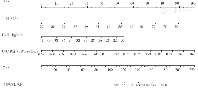
六、NAFLD并发CVD风险预测列线图模型的构建
讨论
受控衰减参数(controlled attenuation parameters,CAP)技术是一种检测声衰减的手段,已有诸多研究证实其在量化肝脏脂肪含量方面的有效性[12,13],但CAP技术在肥胖患者中的检测成功率和准确性表现不佳,且在区分中度和重度肝脂肪变性方面的能力也相对较弱[14]。较CAP技术更为新颖的UGAP技术则是基于已知衰减系数的组织模拟参考模型测量组织衰减系数[4]。通过固定深度(4 cm)、固定频率(3.5 MHz)和已知体模作为参照,来补偿肝脏组织在不同深度、频率和散射特性下的声波传播差异。在UGAP模式下,声波发射和接收条件与标准体模中的条件保持一致,并通过对照标准体模数据,对目标组织的声波回波进行调整[15],调整后的声波便能更准确地反映由于组织衰减引起的变化。本研究中NAFLD组与对照组的UGAP值存在显著差异,在S0~S3各组间的UGAP值差异也存在统计学意义(P<0.001),且随着肝脂肪变性程度的加重,UGAP值也呈升高趋势,与Kuroda等[16]发现UGAP值与肝脂肪变性程度呈显著正相关的研究结果一致。通过计算ICC,进一步分析UGAP技术的可重复性,结果显示UGAP的操作者间测量一致性很好,其有望成为临床常规定量诊断脂肪肝的一种有前景的工具。
鉴于肝脏是体内重要的代谢器官,参与机体的血糖和血脂调节[17]。本研究还探讨了UGAP技术的相关影响因素,单因素分析显示BMI、血糖、丙氨酸氨基转移酶和门冬氨酸氨基转移酶、总胆固醇和甘油三酯及肝脂肪变性程度是UGAP值的重要影响因素(P<0.05)。多因素线性回归分析结果显示,BMI、总胆固醇、甘油三酯和肝脂肪变性程度是UGAP的独立影响因素(P<0.05),这反映了UGAP与血脂水平及肝脂肪变性的严重程度存在紧密联系。肝脏是机体脂质代谢的关键部位,体内总胆固醇、甘油三酯均可通过载脂蛋白运输到肝脏[18],当NAFLD患者肝脏内甘油三酯聚集增多,受损的肝细胞释放信号促进肝细胞再生,继而诱导肝细胞脂肪变性,这一内在机制提示临床医师可结合超声检测结果及患者的肝功能、血脂水平来全面评估脂肪肝的严重程度。
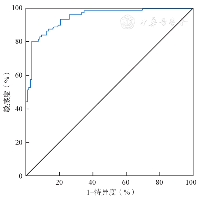
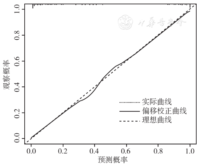
NAFLD与CVD具有多种共同的心脏代谢风险要素,且二者之间相互影响关联。早期监测NAFLD并发CVD的危险因素对预防突发的心脏病等危险状况、延缓甚至逆转疾病的发展非常重要。本研究表明,年龄、BMI、UGAP值是NAFLD患者并发CVD的独立危险因素。North等[19]对衰老与CVD的内在联系做出综述,认为年龄是与CVD相关的主要危险因素之一。Yang等[20]运用轨迹分析的方法,分析BMI轨迹对CVD终身风险的影响,发现CVD终身风险随着BMI轨迹水平的升高而增加,与本研究结果相吻合。基于上述危险因素构建了NAFLD患者并发CVD的风险预测Nomogram模型,该模型预测的ROC曲线下面积达到0.948,Youden指数:0.774;校准曲线显示观测值与预测值之间具有良好的一致性,提示该模型的区分度及有效性均较好,能够作为临床早期预测NAFLD患者合并CVD风险的有效工具。此外,由本研究列线图还可发现BMI低的患者,UGAP值越高,合并CVD的风险越高;UGAP值相同的情况下,与BMI较高的脂肪肝患者相比,BMI低的人更易并发CVD,与Kim等[21]发现瘦型NAFLD患者具有更高的CVD发病风险的研究结果一致。其机制可能与内脏脂肪累积有关,体型偏瘦的人皮下脂肪含量虽不高,但内脏脂肪含量很高,内脏脂肪比皮下脂肪更易影响血脂和CVD的发生风险[22]。内脏脂肪含量增加不仅与代谢器官的异位脂质沉积密切相关,还会通过代谢平衡失调、炎症反应等方式进一步影响心血管健康。因此,BMI低的人群患有脂肪肝发生CVD的风险更高。随着对NAFLD认识的加深,以及更可靠的NAFLD诊断工具在临床中的应用,期望未来NAFLD和进展期肝硬化的增长趋势能够得到有效控制,不良的CVD结局能够得到预防。
本研究尚存在以下局限性。首先,由于样本量较小,有必要通过更大规模的队列研究来验证本研究得出的初步结论。其次,这是一项单中心研究,结果可能存在偏差。此外,本研究构建的Nomogram模型尚未在外部数据集进行验证。因此,未来研究需纳入其他中心的样本以强化外部验证的可靠性。
综上所述,UGAP作为一项新技术,表现出良好的操作者间可重复性,且UGAP值与BMI、血脂水平及肝脂肪变性程度独立相关。因此,UGAP技术具有良好的可行性和临床适用性。此外,本研究构建的Nomogram模型具有实际的临床应用价值,为NAFLD患者早期预测并发CVD的风险提供了可靠的依据。