
肌少症已被定义为一种渐进性、全身性的骨骼肌疾病,表现为肌肉质量和功能的加速丧失[1],与多种疾病的加速发展显著相关,如癌症[2,3]、心血管疾病[4,5]和慢性阻塞性肺疾病[6],同时也与老年人不良结局的风险增加有关[1,4]。根据2019年亚洲肌少症工作组(Asian Working Group for Sarcopenia,AWGS)的诊断标准,肌少症可通过生物电阻抗分析(bioelectrical impedance analysis,BIA)或双能X 射线吸收测定法(dual-energy X-ray absorptiometry,DXA)评估肌肉质量,并结合包括握力测试、5 次起坐时间测试以及简易体能状况评估量表(short physical performance battery,SPPB)等一系列临床测试综合评估肌肉力量和身体表现进行明确诊断[7]。根据该诊断标准进行肌少症的诊断流程相对复杂,其中应用的2 种肌肉质量评估方法也具有一定局限性,DXA 具有辐射性且缺乏便携性,而BIA 结果会受机体水合状态的影响[8]。因此,有必要进一步探索更安全、便捷、有效的肌少症评估方法。
资料与方法
一、对象
二、仪器与方法
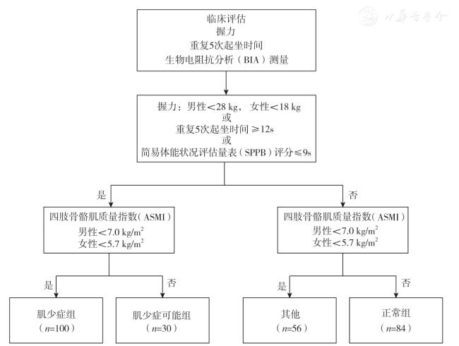
1.临床评估:在人体测量方面,测量所有参与者的身高、体质量,并计算体质量指数(body mass index,BMI)。在肌肉质量评估方面,采用BIA 测量所有参与者的躯干肌肉质量、肢体肌肉质量,并据此计算四肢骨骼肌质量指数(appendicular skeletal muscle mass index,ASMI)。在肌肉力量评估方面,测量以下指标:握力、5 次起坐时间和SPPB 评分。在握力测量中,参与者站立位,双臂自然下垂,双手交替用力握住握力计,每侧测量2 次,并记录最大值。在5 次起坐时间测试中,参与者坐在一张46 cm 高、无扶手的椅子上,双手交叉放在肩上,不使用上身力量的情况下尽可能快地连续完成5 次起立和坐下,在参与者最后1 次坐下时记录时间(使用压力感应垫记录时间,以最大程度地减少评估者偏差)。依据临床各项测试的结果通过上述分组流程对研究参与者进行诊断分组。
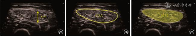
2.肌肉超声检查:使用彩色多普勒超声诊断仪进行检查(型号:迈瑞RESONA7),应用高分辨线阵L9-3U 探头,中心频率为5.8 MHz。团队前期的Meta 分析发现股直肌的厚度(muscle thickness,MT)和横截面积(cross-sectional area,CSA)是预测肌少症患者肌肉质量的最佳参数[11],选择股直肌作为本研究的目标检测肌肉。参与者取平卧位,肌肉处于放松状态,检查医师以软尺测量髂前上棘与髌骨连线中点(股直肌中点)并作标记,探头以尽量轻的压力、垂直于大腿长轴方向放置于标记处,采集B 模式图像并测量MT、CSA 和肌肉回声强度(muscle echogenicity intensity,MEI)等指标(图2)。
3.影像组学分析:所采集的超声图像被导入ITK-SNAP(版本3.8.0)软件进行手动图像分割,评估的感兴趣区域(region of interest,ROI)为超声图像成像区域内的股直肌。ROI 的勾画由1 位具有2 年肌骨超声检查经验的超声医师完成,并由1位具有10 年肌骨超声检查经验的超声医师进行复核确认。
从ITK-SNAP 软件获得的图像和ROI 被导入Pyradiomics 库进行影像组学特征提取。提取的特征包括一阶特征(直方图和形态学特征)以及二阶参数。二阶参数主要涉及灰度共生矩阵、灰度游程长度矩阵、灰度大小区域矩阵、邻域灰度差矩阵和灰度依赖矩阵。
在数据集上构建特征标签时,采用最小绝对收缩和选择算子(least absolute shrinkage and selection operator,LASSO)回归模型。LASSO 模型将所有回归系数压缩至零,并基于正则化权重λ 值精确地将众多不相关特征的系数设置为零。为了确定最优λ 值,采用5 折交叉验证并设定了最小标准,最终得到的λ 值对应的交叉验证误差最低。具有非零系数的剩余特征被用于回归模型拟合,并组合起来形成一个独特的影像组学特征标签。
为了区分肌少症组和正常组,本研究基于从超声图像中所提取的影像组学特征开发了一个影像组学模型,原始数据被随机等分为5 个子集,在模型训练和测试过程中按4 ∶1 比例将4 个子集作为训练集,1 个子集作为测试集,使用受试者操作特征(receiver operating characteristic,ROC)曲线、校准曲线、决策曲线分别评估模型的区分度、校准度及临床收益。
三、统计学分析
所有数据均采用R 软件(版本4.0.2)进行统计分析。性别为计数资料,以频数和百分比描述,采用χ2 检验比较肌少症组与正常组间差异;年龄、身高、体质量、BMI 和超声测量指标为满足正态分布的计量资料,以 ±s 描述,组间比较采用t 检验;四肢肌肉量、ASMI、握力、SPPB 评分和5 次起坐时间为不满足正态分布的计量资料,以M(QR)描述,组间差异比较采用Wilcoxon 秩和检验。采用Spearman 相关性检验分析超声测量指标与临床指标的相关性。采用ROC 曲线下面积(area under curve,AUC)值分别评估各项超声测量指标对肌少症的诊断效能,并计算敏感度和特异度。由于男性和女性肌少症的诊断标准存在差异,本研究分别计算了各项指标在男性和女性人群中的诊断AUC 值及截断值。正常组和肌少症组参与者的分组标准包括握力、ASMI、5 次起坐时间、SPPB 评分等临床指标,将未参与该分组诊断流程的基础临床指标(性别、年龄、BMI)、超声测量指标(MT、CSA、MEI)和影像组学模型评价结果纳入Logistic 回归分析,筛选老年人肌少症的独立影响因素,并建立联合诊断模型。
±s 描述,组间比较采用t 检验;四肢肌肉量、ASMI、握力、SPPB 评分和5 次起坐时间为不满足正态分布的计量资料,以M(QR)描述,组间差异比较采用Wilcoxon 秩和检验。采用Spearman 相关性检验分析超声测量指标与临床指标的相关性。采用ROC 曲线下面积(area under curve,AUC)值分别评估各项超声测量指标对肌少症的诊断效能,并计算敏感度和特异度。由于男性和女性肌少症的诊断标准存在差异,本研究分别计算了各项指标在男性和女性人群中的诊断AUC 值及截断值。正常组和肌少症组参与者的分组标准包括握力、ASMI、5 次起坐时间、SPPB 评分等临床指标,将未参与该分组诊断流程的基础临床指标(性别、年龄、BMI)、超声测量指标(MT、CSA、MEI)和影像组学模型评价结果纳入Logistic 回归分析,筛选老年人肌少症的独立影响因素,并建立联合诊断模型。在进行影像组学分析时,通过Pearson 相关性检验并设定0.90 的相关性阈值来降低特征空间的维度,从而去除共线性特征。在选定轮廓稳定的特征并降低特征空间维度后,使用Wilcoxon 检验对训练集进行单变量分析,针对二元结局设定显著性水平为0.05。随后,在测试集上执行相同的检验,以检查训练数据中发现的相关性是否一致。当双侧P<0.05 时认为差异具有统计学意义。
结 果
一、2 组临床和超声特征比较
表1 2 组老年研究对象临床资料和超声测量指标比较 |
| 项目 | 肌少症组(n=100) | 正常组(n=84) | 统计值 | P 值 |
|---|---|---|---|---|
| 性别[ 人数(%)] | χ 2=0.06 | 0.800 | ||
| 女性 | 59(59.0) | 48(57.1) | ||
| 男性 | 41(41.0) | 36(42.9) | ||
| 年龄(岁,±s) | 74.3±8.2 | 66.4±6.2 | t=7.36 | < 0.001 |
| 身高(m,±s) | 1.61±0.08 | 1.67±0.08 | t=-5.05 | < 0.001 |
| 体质量(kg,±s) | 49.26±9.19 | 58.03±9.53 | t=-6.34 | < 0.001 |
| BMI(kg/m2,±s) | 18.99±3.02 | 20.89±2.96 | t=-4.30 | < 0.001 |
| 四肢肌肉量[kg,M(QR)] | 13.71(12.37,16.91) | 18.78(16.07,21.28) | Z=-7.56 | < 0.001 |
| ASMI[kg/m2,M(QR)] | 5.40(5.00,6.23) | 6.60(6.18,7.30) | Z=-8.35 | < 0.001 |
| 握力[kg,M(QR)] | 19.75(16.25,24.20) | 27.30(23.95,38.05) | Z=-7.80 | < 0.001 |
| SPPB 评分[ 分,M(QR)] | 10(8,11) | 12(12,12) | Z=-8.68 | < 0.001 |
| 5 次起坐时间[s,M(QR)] | 14.04(10.99,18.32) | 8.99(7.81,10.24) | Z=-8.54 | < 0.001 |
| MT(cm,±s) | 1.27±0.25 | 1.53±0.25 | t=-6.93 | < 0.001 |
| CSA(cm2,±s) | 5.31±1.28 | 6.75±1.28 | t=-7.60 | < 0.001 |
| MEI(±s) | 30 663.67±1783.66 | 29 285.24±2045.77 | t=4.87 | < 0.001 |
注:BMI 为体质量指数,ASMI 为四肢骨骼肌质量指数,SPPB 为简易体能状况评估量表,MT 为肌肉厚度,CSA 为肌肉横截面积,MEI 为肌肉回声强度 |
超声指标和临床指标的Spearman 相关性分析结果表明,在总人群中,MT 与身高(r=0.323,P<0.001)、 体质量(r=0.570,P<0.001)、BMI(r=0.482,P<0.001)、 四肢肌肉量(r=0.526,P <0.001)、ASMI(r=0.587,P <0.001)、 握力(r=0.452,P<0.001)、SPPB 评 分(r=0.313,P<0.001) 均呈正相关; 类似的,CSA 与身高(r=0.342,P=0.001)、 体 质 量(r=0.649,P<0.001)、BMI(r=0.585,P<0.001)、 四 肢肌肉量(r=0.570,P<0.001)、ASMI(r=0.640,P<0.001)、 握 力(r=0.512,P<0.001)、SPPB评分(r=0.313,P<0.001)也均呈正相关,且相关性高于MT(图3a)。MT 和CSA 还分别与年龄(r=-0.245,P<0.001;r=-0.233,P<0.001)、5 次起坐时间(r=-0.271,P<0.001;r=-0.308,P<0.001)呈负相关。而MEI 与身高(r=-0.294,P<0.001)、体质量(r=-0.261,P<0.001)、四肢肌肉量(r=-0.358,P<0.001)、ASMI(r=-0.358,P<0.001)、 握力(r=-0.366,P<0.001)、SPPB评分(r=-0.202,P=0.006)呈负相关,与年龄(r=0.178,P=0.015)、5 次起坐时间(r=0.217,P=0.003)呈正相关(图3a)。类似的趋势在男性人群以及女性人群中也可分别观察到(图3b、3c)。
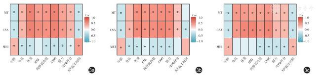
表2 各项超声指标对老年人肌少症的诊断效能 |
| 指标 | AUC | 截断值 | 敏感度(%) | 特异度(%) |
|---|---|---|---|---|
| 总人群 | ||||
| MT(cm) | 0.761 | 1.38 | 66 | 76 |
| CSA(cm2) | 0.795 | 6.27 | 82 | 64 |
| MEI | 0.696 | 28 950.19 | 83 | 60 |
| 男性 | ||||
| MT(cm) | 0.773 | 1.53 | 73 | 75 |
| CSA(cm2) | 0.804 | 6.41 | 76 | 78 |
| MEI | 0.672 | 28 803.12 | 73 | 58 |
| 女性 | ||||
| MT(cm) | 0.758 | 1.37 | 75 | 69 |
| CSA(cm2) | 0.800 | 5.17 | 89 | 61 |
| MEI | 0.712 | 28 928.05 | 85 | 65 |
注:MT 为超声测量的股直肌肌肉厚度,CSA 为超声测量的股直肌横截面积,MEI 为超声测量的肌肉回声强度,AUC 为曲线下面积 |
二、模型建立及诊断效能评估
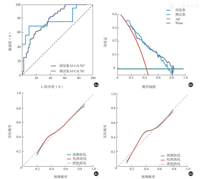
表3 老年人肌少症影响因素的单因素及多因素Logistic 回归分析结果 |
| 指标 | 单因素分析 | 多因素分 | ||
|---|---|---|---|---|
| OR 值(95% 可信区间) | P 值 | OR 值(95% 可信区间) | P 值 | |
| 女性 | 1.079(0.600 ~ 1.943) | 0.799 | ||
| 年龄 | 1.157(1.101 ~ 1.216) | < 0.001 | 1.157(1.085 ~ 1.233) | <0.001 |
| BMI | 0.809(0.728 ~ 0.899) | < 0.001 | 0.819(0.687 ~ 0.975) | 0.025 |
| MT | 0.017(0.004 ~ 0.070) | < 0.001 | 0.243(0.023 ~ 2.575) | 0.240 |
| CSA | 0.415(0.310 ~ 0.556) | < 0.001 | 0.688(0.420 ~ 1.127) | 0.138 |
| MEI | 1.000(1.000 ~ 1.001) | < 0.001 | 1.000(1.000 ~ 1.000) | 0.266 |
| 影像组学模型 | 2.649(1.916 ~ 3.663) | < 0.001 | 2.043(1.347 ~ 3.100) | <0.001 |
注:BMI 为体质量指数,MT 为超声测量的股直肌肌肉厚度,CSA 为超声测量的股直肌横截面积,MEI 为超声测量的肌肉回声强度 |
讨 论
肌少症的临床表现主要包括骨骼肌肌肉质量减少及功能损失,一项针对肌少症全球患病率的研究报道,根据不同的诊断临界值和评估工具,肌少症在老年人中的患病率为10%~27%[20],且与老年人发生不良结局的风险增加以及多种慢性病的不良预后有关[1,2,3,4]。因此,早期诊断肌少症并及时干预,对于改善老年人生存质量和疾病预后至关重要。然而,目前肌少症的诊断流程需要通过一系列的临床评估完成[7],过程相对复杂,在实际应用中受到一定限制,亟需进一步探索一种安全、便捷、有效的肌少症评估方法。超声是一种常用于肌肉疾病检查的影像学方式,可对肌少症患者的肌肉质量进行定量评估[21]。此外,影像组学可通过高通量提取图像特征并建立与生物学信息之间的关联,从而提高疾病的诊断、预后预测的准确性[22]。基于此,本研究建立了基于超声图像的影像组学模型,并分别评估了超声测量指标、影像组学模型对老年人肌少症的诊断价值,建立了一种包含影像组学评价结果的联合诊断模型,可为肌少症诊断提供一个便捷、有效的方法。
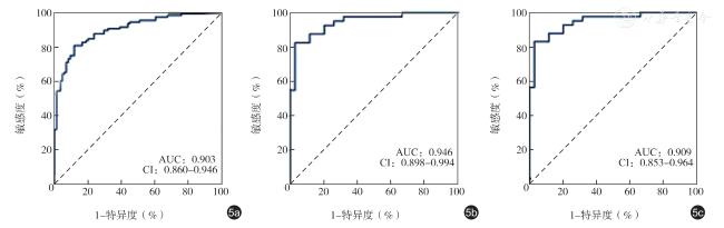
在本研究中,通过超声测量获得的股直肌肌肉大小相关指标MT 和CSA 以及肌肉回声指标MEI与四肢肌肉量、ASMI、握力等临床肌肉评估指标相关性良好,并且这些超声指标对肌少症具有中等的独立诊断价值。这与现有研究的结果类似:Chen 等[23]通过超声测量股直肌MT 和CSA 诊断肌少症的AUC 值分别为0.73 和0.81,刘菲等[24]通过结合超声与人工智能技术分析股直肌MT 诊断肌少症的AUC 值为0.735。此外,本研究基于股直肌超声图像所建立的影像组学模型,对肌少症具有良好的诊断价值,在训练集和测试集中的诊断AUC 值分别为0.787 和0.781,且模型具有较高真实性和可靠性。包含该影像组学模型评价结果、年龄与BMI 所建立的联合诊断模型对肌少症诊断具有高度准确性,在总人群中诊断AUC 值达0.903,在男性中的诊断AUC 值达0.946,在女性中的诊断AUC 值达0.909。本研究结果表明,通过联合股直肌超声图像影像组学模型评价结果和2 个简单的基础临床指标,即年龄和BMI,可实现对肌少症的高准确度诊断。
目前关于肌少症的影像组学研究多基于CT 图像开展,主要通过影像组学对癌症患者的腹部CT图像中第三腰椎层面的骨骼肌或腰大肌区域进行分析,从而预测患者的不良预后风险[25,26]。已有研究证实超声评估肌肉质量与CT 或MRI 的结果相关性较高,Souza 等[27]在慢性肾病患者中对比了超声和CT 测量的平均股直肌CSA,证实2 种方法具有良好的一致性,Mendis 等[11]的研究证实通过超声测量的髂腰肌、缝匠肌和股直肌的面积与MRI测量的结果差异无统计学意义。本研究基于超声成像方式,在正常以及患肌少症的老年人群中开展研究,建立了基于超声图像影像组学模型,通过该影像组学模型可实现高准确度的肌少症诊断,相比CT 与MRI,超声具有无辐射、禁忌证少等优势,有利于开展大规模的老年人肌少症筛查。
本研究也存在一定的局限性,包括样本量有限,且所建立的模型尚未通过大规模临床数据和多中心数据进行验证。此外,在建立图像组学模型的过程中,所有ROI 均由医师手动勾画,而这是一项耗时的工作,未来可深入探索的方面包括纳入自动肌肉分割模型,并将本研究中建立的模型应用于大规模、多中心临床数据进行验证。
综上所述,本研究所建立的基于股直肌超声图像的影像组学模型对肌少症具有良好的独立诊断价值,将该影像组学模型评价结果与年龄、BMI 结合可实现对肌少症的高准确度诊断,从而避免一系列临床测量的复杂诊断流程,有望推动开展肌少症的大规模社区筛查,有助于实现肌少症的早期诊断。