Home    中文  
 
  • Search
  • lucene Search
  • Citation
  • Fig/Tab
  • Adv Search
Just Accepted  |  Current Issue  |  Archive  |  Featured Articles  |  Most Read  |  Most Download  |  Most Cited
Pediatric Ultrasound

Ultrasonic manifestations and diagnostic value of torsion of the pedicle of old ovarian cysts in infants and young children

  • Liling Lu 1 ,
  • Xiuzhen Yang 1 ,
  • Bin Xu 1 ,
  • Lei Zhao 1 ,
  • Jingjing Qian 1 ,
  • Xiaoying Li 1 ,
  • Biao Wang 1 ,
  • Jingjing Ye , 1, 2,
Expand
  • 1 Department of Ultrasonography,Children's Hospital of Zhejiang University School of Medicine, Hangzhou 310052, China
  • 2 Heart Center, Children's Hospital of Zhejiang University School of Medicine, Hangzhou 310052, China

Corresponding author:

Ye Jingjing, Email:

Received date: 2025-03-24

  Online published: 2025-09-29

Copyright

Copyright by Chinese Medical Association No content published by the journals of Chinese Medical Association may be reproduced or abridged without authorization. Please do not use or copy the layout and design of the journals without permission. All articles published represent the opinions of the authors, and do not reflect the official policy of the Chinese Medical Association or the Editorial Board, unless this is clearly specified.

Abstract

Objective

To explore the ultrasound imaging features of torsion of the pedicle of old ovarian cysts in infants and young children.

Methods

A retrospective study was conducted on 60 infants or young children under the age of 1 year who were diagnosed with ovarian cysts and admitted to the Children's Hospital Affiliated to Zhejiang University School of Medicine from January 2021 to July 2024. The ultrasound features and pathological manifestations were analyzed, and the sensitivity, specificity, positive predictive value, negative predictive value, and accuracy of ultrasound features for diagnosing torsion of the pedicle of old ovarian cysts were calculated to evaluate their diagnostic efficacy.

Results

Out of 60 cases of ovarian cysts in infants and young children, 34 cases were old ovarian cyst torsion. The main ultrasound features and their diagnostic efficacy were: calcification (sensitivity: 67.6%; specificity: 80.8%; positive predictive value: 82.1%; negative predictive value: 65.6%); intracystic layering (sensitivity: 67.6%; specificity: 73.1%; positive predictive value: 76.7%; negative predictive value: 63.3%); and vortex sign-like pedicle structure (sensitivity: 47.1%; specificity: 100%; positive predictive value: 100%; negative predictive value: 59.1%). The diagnostic sensitivity, specificity, positive predictive value, and negative predictive value were 26.5%, 100%, 100%, and 51.0%, respectively, when the three characteristics were present simultaneously. The accuracy of ultrasound diagnosis of torsion of old ovarian cyst pedicle was 88.2% (30/34).

Conclusion

The torsion of the pedicle of old ovarian cysts in infants and young children has characteristic ultrasound manifestations, including calcification, intracystic layering, and vortex sign-like pedicle structures, which have good diagnostic value.

Cite this article

Liling Lu , Xiuzhen Yang , Bin Xu , Lei Zhao , Jingjing Qian , Xiaoying Li , Biao Wang , Jingjing Ye . Ultrasonic manifestations and diagnostic value of torsion of the pedicle of old ovarian cysts in infants and young children[J]. Chinese Journal of Medical Ultrasound (Electronic Edition), 2025 , 22(08) : 748 -753 . DOI: 10.3877/cma.j.issn.1672-6448.2025.08.009

新生儿卵巢囊肿发病率约为1%,一般在妊娠晚期通过产前超声检查发现,是产前超声检查中最常见的一种腹腔囊肿,其性质多为良性,部分可自行消退1,2,3。目前新生儿卵巢囊肿的发病机制尚不明确,多数学者认为与胎儿卵巢对胎盘或母体血液中发现的某些促性腺激素和雌激素的过度反应有关4。大部分新生儿卵巢囊肿在出生时无明显症状5,6,7,需要引起注意的是,新生儿卵巢囊肿最常见的并发症是卵巢扭转,其发生率为38%~55%8-9,且卵巢囊肿宫内陈旧性扭转发生率较高。解剖学研究表明,新生儿骨盆容积有限且卵巢支持韧带相对松弛,这一特殊解剖特征使卵巢囊肿容易进入腹腔,活动空间增大,继而增加了卵巢囊肿蒂扭转的风险10。鉴于卵巢囊肿蒂扭转可能导致不可逆的生殖功能损害,建立准确的早期诊断标准具有重要临床意义。因此,本研究对浙江大学医学院附属儿童医院60例婴幼儿卵巢囊肿进行分析,总结其超声表现特征,旨在为临床实践提供客观、可靠的诊断依据。

资料与方法

一、对象

回顾性选取2021年1月至2024年7月浙江大学医学院附属儿童医院收治的60例确诊为婴幼儿卵巢囊肿的病例。年龄范围:0~12个月。纳入标准:(1)年龄≤12月龄;(2)具有完整超声影像资料;(3)经手术病理检查或长期随访确诊。本研究经浙江大学医学院附属儿童医院伦理委员会审批通过:(批件号:2025-IRB-0112-P-01)。

二、方法

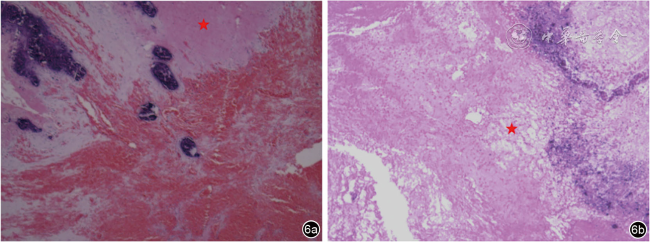
采用回顾性分析方法对60例研究对象的超声图像进行分析。超声特征及其病理表现的定义为:(1)钙化:将点状强回声称为钙化,可伴或不伴声影,一般在卵巢囊肿的囊壁可探及;钙化在病理切片中表现为特征性的深蓝色团块状沉积。(2)囊内分层:超声图像主要表现为液体分层征和回缩的团状中等回声,提示出血的发生;在病理切片中,出血表现为红细胞在血管腔外的聚集或含铁血黄素的沉着。(3)漩涡征样蒂结构:超声检查中漩涡征表现为特征性的螺旋状同心圆结构,部分病例可清晰显示扭转蒂部的连续性;其病理切片主要表现为胞质嗜酸性和细胞核的碎裂消失,伴或不伴局部出血钙化,提示组织存在缺血性坏死。计算超声特征的诊断敏感度、特异度、阳性预测值、阴性预测值、准确性,依据上述指标评估超声特征的诊断效能。

结果

一、病理及随访结果

60例确诊为卵巢囊肿的病例,根据病理结果及随访结果分为以下两类:(1)单纯性卵巢囊肿26例;(2)陈旧性卵巢囊肿蒂扭转34例,其中28例通过手术病理结果确诊,6例因随访发现自体截肢现象确诊。

二、婴幼儿陈旧性卵巢囊肿蒂扭转的超声特征及诊断效能

1.钙化:本研究共有28例病例的超声图像中出现钙化(图1图2),其中陈旧性卵巢囊肿蒂扭转中有23例存在钙化。以钙化作为陈旧性卵巢囊肿蒂扭转的诊断标准,其诊断敏感度为67.6%(23/34),特异度为80.8%(21/26),阳性预测值为82.1%(23/28),阴性预测值为65.6%(21/32)。
图1 婴幼儿陈旧性卵巢囊肿蒂扭转超声图像表现为钙化。图a为女性婴幼儿出生1个月,超声可见右侧腹囊性包块,内见液体分层征,囊壁可见散在环状钙化点(箭头所示),超声诊断右侧卵巢及囊肿陈旧性扭转坏死;图b为女性新生儿出生5 d,超声可见左侧腹囊实性包块,内见液体分层征,囊壁可见多发钙化斑(箭头所示),超声诊断左侧卵巢及囊肿陈旧性扭转坏死
图2 婴幼儿陈旧性卵巢囊肿蒂扭转病理图片中钙化表现。图a为女性婴幼儿出生1个月,超声诊断左侧卵巢囊肿陈旧性蒂扭转,病理提示广泛坏死组织伴钙化改变,箭头所示可见蓝色团块状沉积(HE ×40);图b为女性新生儿出生14 d,超声诊断盆腔囊性肿块,卵巢来源首先考虑,病理提示卵巢囊肿蒂扭转伴坏死钙化,箭头所示可见深蓝色团块状沉积(HE ×40)
2.囊内分层:本研究共有30例病例的超声图像出现囊内分层,病理切片提示出血(图3图4),其中23例确诊为陈旧性卵巢囊肿蒂扭转。以囊内分层作为陈旧性卵巢囊肿蒂扭转的诊断标准,其诊断敏感度为67.6%(23/34),特异度为73.1%(19/26),阳性预测值为76.7%(23/30),阴性预测值为63.3%(19/30)。
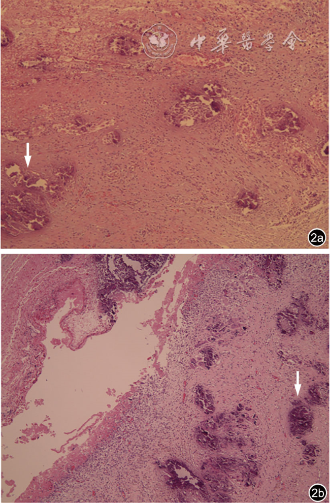
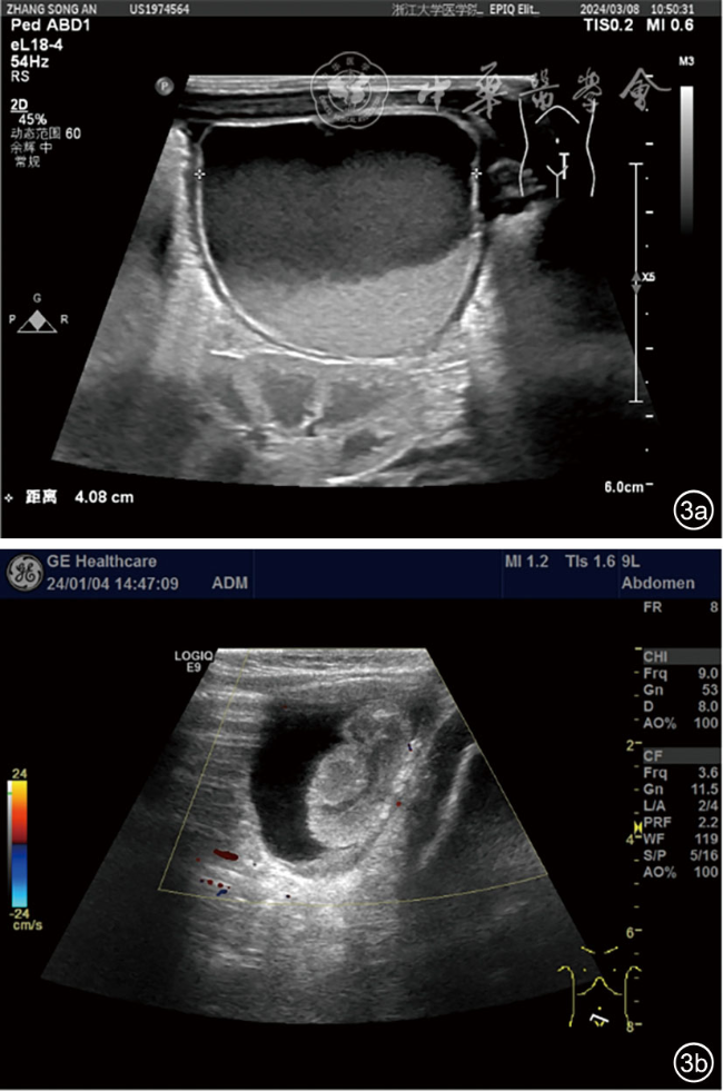
图3 婴幼儿陈旧性卵巢囊肿囊内分层超声图像。图a为女性婴幼儿,出生1个月,超声可见左下腹囊性包块,内见液体分层征,超声诊断左侧卵巢囊肿伴扭转;图b为女性婴幼儿,出生2个月,超声可见盆腔左侧囊实性包块,内见回缩样团块回声,囊壁可见斑片状钙化,超声诊断左侧卵巢来源囊肿及左侧卵巢陈旧性扭转坏死
图4 婴幼儿陈旧性卵巢囊肿病理图片中出血表现。图a为女性新生儿,出生1 d,超声诊断右侧卵巢来源占位伴出血可能,病理提示右侧卵巢黄素化囊肿伴广泛出血坏死,箭头所示可见红细胞聚集(HE ×100);图b为女性婴幼儿,出生2个月,超声诊断左侧卵巢囊肿伴扭转,病理提示左侧卵巢囊肿伴广泛出血水肿坏死及钙化,箭头所示可见红细胞聚集(HE ×40)
3.漩涡征样蒂结构:本研究有16例存在漩涡征样蒂结构,并且最终均确诊为陈旧性卵巢囊肿蒂扭转(图5图6)。以漩涡征样蒂结构作为陈旧性卵巢囊肿蒂扭转的诊断标准,其诊断敏感度为47.1%(16/34)),特异度为100%(26/26),阳性预测值为100%(16/16),阴性预测值为59.1%(26/44)。
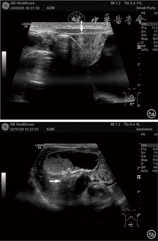
图5 婴幼儿陈旧性卵巢囊肿漩涡征样蒂结构超声图像。图a为女性新生儿,出生1 d,超声可见左中腹囊性包块,包块左下缘见螺旋样同心圆结构(箭头所示),扭转约360°,超声诊断左中腹囊性包块,左侧卵巢来源可能性大合并扭转考虑;图b为女性新生儿,出生8 d,超声可见右下腹囊性包块,内见液体分层征,包块后下缘低回声团,呈蒂样结构(箭头所示),超声诊断右侧卵巢来源囊肿及右侧卵巢陈旧性扭转坏死可能性大
图6 婴幼儿陈旧性卵巢囊肿缺血性坏死病理图像。图a为女性婴幼儿,出生4个月,超声诊断右侧卵巢囊肿陈旧性扭转坏死,病理提示卵巢囊肿蒂扭转伴坏死,红色星号处可见细胞核碎裂消失(HE ×40);图b为女性婴幼儿,出生1个月,超声诊断右侧卵巢来源囊肿及卵巢陈旧性扭转坏死,病理提示卵巢囊肿蒂扭转伴坏死,红色星号处可见细胞核碎裂消失(HE ×40)
4.钙化或囊内分层的诊断效能:本研究中存在钙化或囊内分层特征的病例共38例,其中31例确诊为陈旧性卵巢囊肿蒂扭转,其诊断敏感度为91.2%(31/34),特异度为73.1%(19/26),阳性预测值为81.6%(31/38),阴性预测值为86.4%(19/22)。
5.同时具备以上3种特征的诊断效能:本研究中同时存在钙化、囊内分层、漩涡征样蒂结构的病例共9例,全部确诊为陈旧性卵巢囊肿蒂扭转。3种特征同时具备的诊断敏感度为26.5%(9/34),特异度为100%(26/26),阳性预测值为100%(9/9),阴性预测值为51.0%(26/51)。

三、陈旧性卵巢囊肿蒂扭转的超声诊断准确性

在34例陈旧性卵巢囊肿蒂扭转的病例中,4例超声诊断错误,其中3例诊断为畸胎瘤(图7a~7c),1例诊断为肠重复畸形(图7d),但最终病理结果均提示为卵巢囊肿蒂扭转坏死(图7e~7h)。超声诊断陈旧性卵巢囊肿蒂扭转的准确性为88.2%(30/34)。
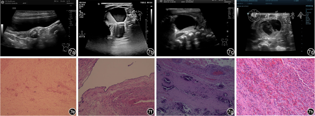
图7 陈旧性卵巢囊肿蒂扭转超声误诊病例超声及病理表现。图a为女性婴幼儿,出生12个月,超声可见盆腔内不均质团块回声,内见斑片状强回声伴声影,超声诊断左侧卵巢来源畸胎瘤;图b为女性婴幼儿,出生1个月,超声可见右侧附件区囊性包块,内见液体分层征,超声诊断右附件畸胎瘤;图c为女性新生儿,出生4 d,超声可见盆腔内囊实性包块,内见液体分层征,超声诊断盆腔囊实性包块,畸胎瘤待排;图d为女性新生儿,出生3 d,超声可见右侧腹囊性包块,内透声差,见分隔,超声诊断右侧腹囊性包块,肠重复畸形可能;图e(与图a为同一病例)病理提示左侧卵巢组织广泛坏死伴钙化改变,可见少许深蓝色团块状沉积和细胞核的碎裂消失(HE ×40);图f(与图b为同一病例)病理提示卵巢单纯性囊肿伴坏死钙化,可见散在细胞核的碎裂消失(HE ×100);图g(与图c为同一病例)病理提示卵巢囊肿蒂扭转伴坏死,可见深蓝色团块状沉积和红细胞在血管外聚集(HE ×200);图h(与图d为同一病例)病理提示卵巢囊肿蒂扭转伴出血坏死钙化,可见红细胞散在分布(HE ×200)

讨论

本研究通过对60例婴幼儿卵巢囊肿病例的系统分析,发现34例为陈旧性卵巢囊肿蒂扭转,26例为单纯性卵巢囊肿,表明陈旧性卵巢囊肿蒂扭转在婴幼儿中的发病率并不低。为了更好地诊断婴幼儿陈旧性卵巢囊肿蒂扭转,本研究发现了陈旧性卵巢囊肿蒂扭转的3个关键诊断特征:(1)钙化(诊断特异度80.8%);(2)囊内分层(诊断特异度73.1%);(3)漩涡征样蒂结构(诊断特异度100%)。因此,如果发现婴幼儿卵巢囊肿的超声图像中出现这些特征,则要高度警惕其为陈旧性卵巢囊肿蒂扭转的可能性。值得注意的是,虽然漩涡征具有高度特异性(100%),但其敏感度相对较低(47.1%),这一现象可能归因于:(1)新生儿肠道气体干扰导致图像质量受限;(2)新生儿卵巢囊肿早期发生宫内扭转导致的蒂部纤维化改变难以在超声中辨别11,12,13,14
此外,在诊断准确性评估中,有4例存在误诊,3例误诊为畸胎瘤,1例误诊为肠重复畸形。误诊为畸胎瘤的原因可能是因为卵巢畸胎瘤在儿童和青少年中的发病率较高15-16,并且有些卵巢畸胎瘤的超声图像中也会出现液体分层征。但是实际上卵巢畸胎瘤在婴幼儿中是相对罕见的,多数研究统计的卵巢畸胎瘤病例平均发生年龄在11岁左右17-18。另外,卵巢畸胎瘤所呈现的液体分层征的超声特征通常在青春期后的患者中较为常见。最重要的是畸胎瘤特征性的超声表现其实是囊腔内结节样高回声伴声影(Rokitansky结节)19。所以当看到婴幼儿附件区的包块合并有钙化或囊内分层的超声表现时,应首先考虑为陈旧性卵巢囊肿蒂扭转而非畸胎瘤。
目前,对于卵巢囊肿的治疗措施包括连续超声随访监测、产前超声引导下穿刺抽吸减压、囊肿剥除术或患侧卵巢和(或)输卵管切除术11。其中手术治疗的效果是毋庸置疑的,但也是极具风险的,并且如切除患侧卵巢,会对患者的后续生活带来不可逆转的影响20。本研究在分析陈旧性卵巢囊肿蒂扭转的病例中发现,有6例超声诊断为陈旧性卵巢囊肿蒂扭转的患者在治疗上选择了随访,并且在随访过程中出现了肿物的缩小及脱落现象,即自体截肢现象,这也提示并不是所有的卵巢囊肿蒂扭转都需要紧急手术治疗。
综上所述,在婴幼儿陈旧性卵巢囊肿蒂扭转的超声诊断中,钙化、囊内分层以及漩涡征样蒂结构的特征具有一定的应用价值。除漩涡征样蒂结构之外,还可以根据是否存在钙化、囊内分层来进行评估,而不一定局限于寻找漩涡征样蒂结构,这在一定程度解决了在实际超声检查中因漩涡征样蒂结构难以辨别而遗漏陈旧性卵巢囊肿蒂扭转的难题。
1
Mudholkar VG, Acharya AS, Kulkarni AM, et al. Antenatally diagnosed neonatal ovarian cyst with torsion [J]. Indian J Pathol Microbiol, 2011, 54(1): 228-229.

2
Erol O, Erol MB, Isenlik BS, et al. Prenatal diagnosis of fetal ovarian cyst: case report and review of the literature [J]. J Turk Ger Gynecol Assoc, 2013, 14(2): 119-122.

3
Kwak DW, Sohn YS, Kim SK, et al. Clinical experiences of fetal ovarian cyst: diagnosis and consequence [J]. J Korean Med Sci, 2006, 21(4): 690-694.

4
Tyraskis A, Bakalis S, Scala C, et al. A retrospective multicenter study of the natural history of fetal ovarian cysts [J]. J Pediatr Surg, 2018, 53(10): 2019-2022.

5
Bagolan P, Giorlandino C, Nahom A, et al. The management of fetal ovarian cysts [J]. J Pediatr Surg, 2002, 37(1): 25-30.

6
Shapiro EY, Kaye JD, Palmer LS. Laparoscopic ovarian cystectomy in children [J]. Urology, 2008, 73(3): 526-528.

7
Foley PT, Ford WD, McEwing R, et al. Is conservative management of prenatal and neonatal ovarian cysts justifiable? [J]. Fetal Diagn Ther, 2005, 20(5): 454-458.

8
Bryant AE, Laufer MR. Fetal ovarian cysts: incidence, diagnosis and management [J]. J Reprod Med, 2004, 49(5): 329-337.

9
Monnery-Noché ME, Auber F, Jouannic JM, et al. Fetal and neonatal ovarian cysts: is surgery indicated? [J]. Prenat Diagn, 2008, 28(1): 15-20.

10
崔晏春, 贾立群, 王晓曼. 新生儿及婴儿期卵巢囊肿蒂扭转的超声表现 [J/CD].中华医学超声杂志(电子版), 2019, 16(3): 198-201.

11
Akın MA, Akın L, Özbek S, et al. Fetal-neonatal ovarian cysts--their monitoring and management: retrospective evaluation of 20 cases and review of the literature [J]. J Clin Res Pediatr Endocrinol, 2010, 2(1): 28-33.

12
Ogul H, Havan N, Pirimoglu B, et al. Prenatal and postnatal ultrasonographic findings of the torsioned ovarian cyst: a case report and brief literature review [J]. Int Surg, 2015, 100(3): 514-517.

13
Dimitraki M, Koutlaki N, Nikas I, et al. Fetal ovarian cysts. Our clinical experience over 16 cases and review of the literature [J]. J Matern Fetal Neonatal Med, 2012, 25(3): 222-225.

14
Luzzatto C, Midrio P, Toffolutti T, et al. Neonatal ovarian cysts: management and follow-up [J]. Pediatr Surg Int, 2000, 16(1-2): 56-59.

15
Sisler CL, Siegel MJ. Ovarian teratomas: a comparison of the sonographic appearance in prepubertal and postpubertal girls [J]. AJR Am J Roentgenol, 1990, 154(1): 139-141.

16
Surratt JT, Siegel MJ. Imaging of pediatric ovarian masses [J]. Radiographics, 1991, 11(4): 533-548.

17
Gkrozou F, Tsonis O, Vatopoulou A, et al. Ovarian teratomas in children and adolescents: Our own experience and review of literature [J]. Children (Basel), 2022, 18, 9(10): 1571.

18
Özcan R, Kuruoğlu S, Dervişoğlu S, et al. Ovary-sparing surgery for teratomas in children [J]. Pediatr Surg Int, 2013, 29(3): 233-237.

19
Sisler CL, Siegel MJ. Ovarian teratomas: a comparison of the sonographic appearance in prepubertal and postpubertal girls [J]. AJR Am J Roentgenol, 1990, 154(1): 139-141.

20
Morray JP, Geiduschek JM, Ramamoorthy C, et al. Anesthesia-related cardiac arrest in children: initial findings of the Pediatric Perioperative Cardiac Arrest (POCA) Registry [J]. Anesthesiology, 2000, 93(1): 6-14.

Outlines

/

Copyright © Chinese Journal of Medical Ultrasound (Electronic Edition), All Rights Reserved.
Tel: 010-51322630、2632、2628 Fax: 010-51322630 E-mail: csbjb@cma.org.cn
Powered by Beijing Magtech Co. Ltd