
2022 , Vol. 19 >Issue 01: 8 - 16
DOI: https://doi.org/10.3877/cma.j.issn.1672-6448.2022.01.003
胎儿单侧小脑发育不全的产前超声诊断
Copy editor: 安京媛
收稿日期: 2020-04-11
网络出版日期: 2022-02-23
基金资助
国家自然科学基金(81771598)
国家重点研发计划(2018YFC1002202)
深圳市科技计划项目(JCYJ20170307091013214)
版权
Prenatal diagnosis of fetal unilateral cerebellar hypoplasia by ultrasound and review of the literature
Received date: 2020-04-11
Online published: 2022-02-23
Copyright
探讨胎儿单侧小脑发育不全(UCH)产前超声声像图特征。
总结2018年1月至2019年12月于南方医科大学附属深圳妇幼保健院,在胎儿系统超声检查中诊断为UCH的6例胎儿的产前超声声像图表现,与MRI、引产后超声及病理解剖结果进行对比,并结合产前诊断该疾病的相关文献对UCH胎儿产前超声诊断特点及临床预后进行分析。
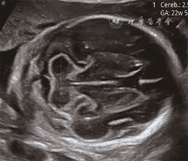
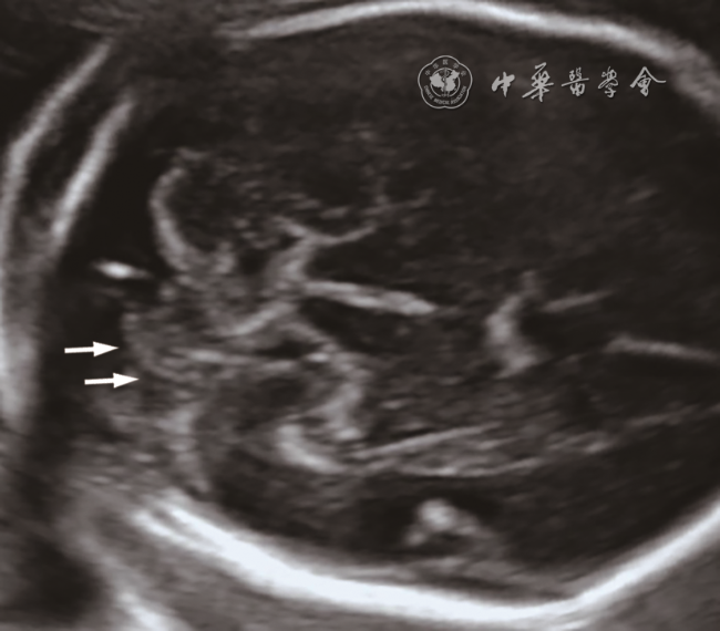
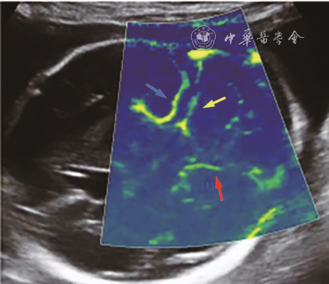
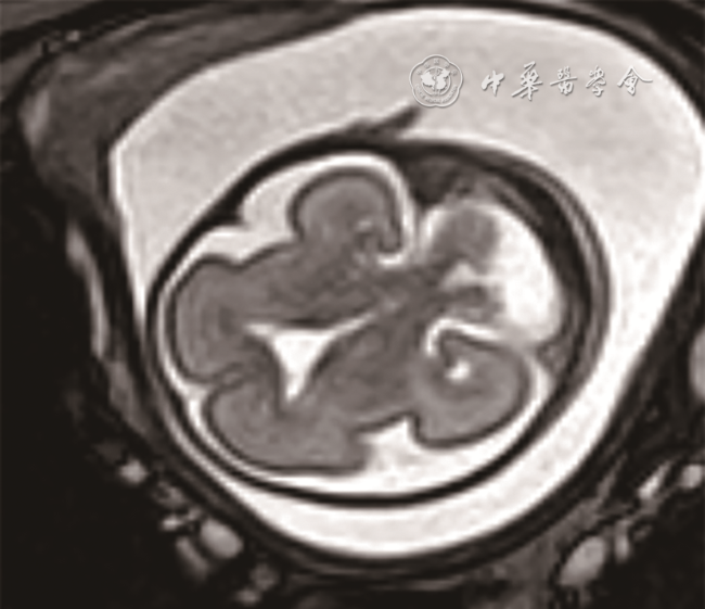
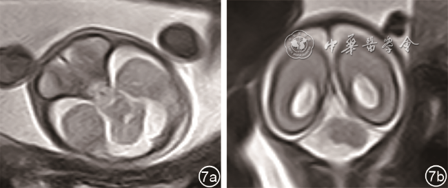
6例UCH胎儿产前超声声像图典型表现:两侧小脑半球不对称和(或)小脑横径减小,伴或不伴蚓部异常,发育不全侧小脑减小,形态失常,其中3例受累侧小脑半球边界不规则,对侧小脑形态及大小均正常。临床预后:6例胎儿中1例活产,随访至1岁,未出现神经症状。结合本研究6例UCH胎儿及文献报道的36例胎儿产前和产后检查结果分析均显示:单纯UCH胎儿、不合并颅内异常的UCH胎儿以及无产前生长受限、早产等不良孕产史的UCH胎儿短期预后均好于合并颅内异常的UCH胎儿。
UCH有特征性产前超声表现,产前超声发现小脑半球不对称,一侧小脑减小、形态失常时可作出诊断,诊断不明确时可结合MRI进行鉴别诊断。
丁妍 , 文华轩 , 廖伊梅 , 曾晴 , 彭桂艳 , 李胜利 . 胎儿单侧小脑发育不全的产前超声诊断[J]. 中华医学超声杂志(电子版), 2022 , 19(01) : 8 -16 . DOI: 10.3877/cma.j.issn.1672-6448.2022.01.003
To explore the prenatal ultrasound features of fetal unilateral cerebellar hypoplasia.
The prenatal ultrasonograms of six fetuses at Shenzhen Maternity and Child Healthcare Hospital Affiliated to Nanfang Medical University, who were diagnosed as having unilateral cerebellar hypoplasia were retrospectively reviewed. The prenatal ultrasound characteristics were summarized, and compared with the features of magnetic resonance imaging (MRI), postnatal ultrasound, and autopsy examinations. A literature review of prenatal diagnosis of such abnormality was performed.
The prenatal ultrasonograms of the six cases of unilateral cerebellar hypoplasia were characterized as asymmetry of the cerebellar hemispheres and/or a decrease in the transverse cerebellar diameter, with or without deformity of the cerebellar vermis. The cerebellum volume of the affected side decreased, along with an abnormal morphology, while the contralateral side was normal. Three cases had irregular borders in the affected side. After birth, one of the six fetuses was followed to one year old without neurological symptoms. Based on the prenatal and postnatal examination results of the six cases in the present study and those of 36 cases reported in the literature, it was found that cases with isolated unilateral cerebellar hypoplasia and those without intracranial abnormalities or other adverse pregnancy history like prenatal growth restriction or premature birth had better short-term prognosis than unilateral cerebellar hypoplasia fetuses with intracranial abnormalities.
Unilateral cerebellar hypoplasia can be prenatally diagnosed based on the characteristic ultrasonic features and MRI can be used for differential diagnosis.
Key words: Ultrasonography, prenatal; Fetus; Cerebellar hypoplasia
表1 6例UCH胎儿产前影像检查结果及围产期结局 |
| 检查年份 | 检出异常(胎儿/孕周) | 胎儿颅内表现a | 可能的病因 | 围产期结局与预后 | ||
|---|---|---|---|---|---|---|
| 产前超声表现 | 产前MRI检查(4例) | 小脑蚓部情况 | ||||
| 2018 | 例1/26 | 小脑半球形态失常,左右不对称,右侧小脑半球形态不规则,边界尚规则,小脑半球间可见一囊肿与第四脑室及后颅窝池相通 | 未检查 | 蚓部发育不全 | 不详 | 不详 |
| 2018 | 例2/24 | 左侧小脑体积减小,边界尚规则,后颅窝池扩大,与第四脑室相通;神经元移行障碍、胼胝体发育不良,超声提示:LCH | 未检查 | 蚓部发育不良 | 不详 | 引产 |
| 2019 | 例3/26 | 左侧小脑体积减小,边界不规则,蛛网膜囊肿;彩色多普勒:左侧小脑上动脉未显示;超声提示:LCH | 后颅窝池囊肿;诊断 LCH | 蚓部发育不良 | 小脑供血动脉异常 | 引产 |
| 2019 | 例4/23 | 左侧小脑体积减小,边界尚规则;超声提示:LCH | 诊断:LCH | 蚓部正常 | 不详 | 引产 |
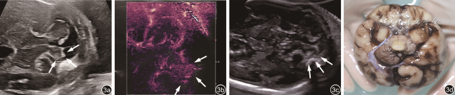
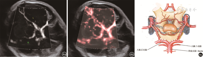
| 2019 | 例5/25 | 左侧小脑体积减小,边界不规则,左侧小脑半球囊性无回声区,后颅窝池与第四脑室相通;彩色多普勒:左侧小脑上动脉未显示;超声提示:LCH | 左侧小脑半球片状异常信号,考虑液化囊变;诊断:LCH | 蚓部显示不清 | 小脑供血动脉异常 | 引产后尸检 |
| 2019 | 例6/22 | 右侧小脑体积减小,边界不规则;超声提示:RCH | 诊断:RCH | 蚓部正常 | 不详 | 活产,随访至1岁,生长发育正常,未出现神经症状 |
注:a6例均未合并颅外异常和相关综合征;UCH为单侧小脑发育不全;LCH为左侧小脑发育不全;RCH为右侧小脑发育不全 |
表2 36例单侧小脑发育不全胎儿的相关文献报道 |
| 文献 | 分类(例数) | 产前颅内主要影像学表现及诊断 | 妊娠结局及产后随访检查情况 |
|---|---|---|---|
| [1] | 单纯UCH(8例) | 小脑半球不对称7例(此处出现高回声3例;T2WI病灶呈低信号2例);合并蚓部原裂未显示1例;孕21~35周诊断LCH 4例,RCH 4例 | 活产8例。出生后2周~7个月MRI均证实为单纯UCH;可能继发于小脑出血2例,病因不明6例;随访14个月~9年患儿发育正常7例,出现异常1例 |
| 合并异常UCH(15例) | 小脑半球不对称,出现边界不规则及小脑叶裂异常等改变(小脑半球处出现高回声2例,MRI信号改变5例);合并蚓部异常4例,脑干不对称1例,严重脑室扩张1例,后颅窝囊性变4例;合并FGR 1例,心脏畸形1例,肠道回声增强1例;孕25~33周诊断LCH 11例,RCH 4例 | 活产13例(早产低体重儿1例),引产或胎死宫内2例;产后病理及45 d~5个月MRI均证实为UCH,此外蚓部异常4例,脑干异常4例,椎动脉发育不良1例,幕上多发异常1例;胎盘病理示胎儿血栓性血管病变2例;可能与小脑出血或缺血性损害相关7例,CMV感染1例,病因不明7例;产后诊断PHACE综合征 4例;随访2个月~4年患儿发育正常8例,随访5个月~5年出现异常7例 | |
| [10] | 合并异常UCH(1例) | 孕31周MRI示左侧脑干发育不全,诊断:LCH | 活产。MRI及MRA诊断:LCH、一侧脑干发育不全、左侧PICA发育不良、基底动脉瘤;可能与小脑缺血性损害相关。出生后3个月出现右臂轻微无力,17个月时出现急性可逆性凝视异常,右侧偏瘫,肌张力增加 |
| [13] | 合并异常UCH(1例) | 孕21周超声诊断:LCH | 活产。产后MRI诊断:LCH,左侧后颅窝池小,一侧脑桥发育不良;病因不明;出生几天后患儿肌张力减退,2岁3个月有智力偏低、肌张力减退、左侧肢体轻度共济失调 |
| [18] | 单纯UCH(1例) | 孕24周声像图示右侧小脑半球处显示2 cm高回声肿块,孕29周同部位显示5 mm囊性肿块,孕33周诊断:RCH | 活产。产后超声证实为RCH;可能继发于小脑出血;随访18个月患儿生长发育正常 |
| [22] | 合并异常UCH(1例) | 孕24周超声诊断:RCH,合并眼-耳-椎骨发育不良、FGR | 引产。尸检证实为RCH并诊断为Goldenhar综合征,病因不明 |
| [23] | 合并异常UCH(1例) | 孕24周超声诊断:RCH,合并蚓部发育不全 | 活产。产后MRI诊断:RCH,蚓部发育不全;诊断为PHACE综合征;病因不明;随访5个月发育正常 |
| [25] | 单纯UCH(1例) | 孕21周声像图示左侧小脑半球高回声区,孕23周同部位低回声伴边缘不规则高回声区,孕25周超声显示两侧小脑半球轻微不对称;MRI T2WI示左侧小脑半球低信号肿块,孕31周诊断:LCH | 孕33周时胎死宫内。尸检证实为LCH,胎盘病理示胎儿血栓性血管病变;可能继发于小脑出血 |
| [26] | 单纯UCH(1例) | 孕21周声像图示右侧小脑半球处显示高回声肿块,孕23周肿块直径缩小,孕24周诊断:RCH | 引产。尸检证实RCH合并小脑海绵状血管瘤致出血性脑梗死 |
| [27] | 单纯UCH(1例) | 孕17周超声诊断:RCH | 引产。尸检证实为RCH;可能因药物引产失败导致小脑血管破裂 |
| [28] | 合并异常UCH(1例) | 孕17 周声像图示后颅窝高回声肿块,孕19周同部位混合回声肿块,孕27周肿块消失,诊断:LCH | 活产。产后3个月声像图示左侧小脑囊性结构,产后28个月MRI诊断:LCH,脑干、一侧小脑脚发育不全;可能与小脑出血或缺血性损害相关;3岁时患儿出现轻微震颤,智力和语言发育正常 |
| [29] | 合并异常UCH(1例) | 孕22周声像图示左侧小脑半球高回声,4 d后高回声体积缩小,第四脑室增大,孕30周诊断 LCH合并蚓部完全缺失,轻度肠扩张、FGR | 引产。尸检证实为LCH,蚓部完全缺失;可能继发于小脑出血 |
| [30] | 合并异常UCH(1例) | 孕18周超声怀疑Dandy-Walker畸形、轻度脑积水,孕21周MRI诊断为LCH,孕22周怀疑Dandy-Walker变异;合并蚓部发育不全 | 引产。尸检证实为LCH,蚓部发育不全,并考虑神经元移行障碍;病因不明 |
| [31] | 合并异常UCH(1例) | 孕31周声像图示左侧侧脑室增宽,孕32周双侧侧脑室不对称增宽,孕34周MRI诊断:RCH,脑室内出血;合并蚓部发育不全 | 活产。产后超声及MRI诊断:RCH,脑干、蚓部发育不全,左侧脑白质轻度萎缩伴囊肿,出血性脑室不对称扩大。可能与脑室出血引起小脑传入神经通路中断相关;出生后异常局促运动和抖动,出生后6~9个月患儿时出现偏瘫,15个月时右侧轻微偏瘫,右侧注意力减少,斜视、右眼视野缩小 |
| [32] | 合并异常UCH(1例) | 孕29周声像图示后颅窝囊性病变向右侧小脑延伸,MRI诊断RCH且右侧小脑半球处囊性病变;合并蚓部发育不全 | 引产。尸检证实为RCH,蚓部发育不全,病理检查示囊性病变为小脑实质坏死后液化;可能与缺血性脑出血相关 |
注:UCH为单侧小脑发育不全;RCH为右侧小脑发育不全;LCH为左侧小脑发育不全;MRI为磁共振成像;MRA为磁共振血管成像;PICA为小脑下后动脉;CMV为巨细胞病毒;FGR为宫内发育迟缓 |
| 1 |
Massoud M, Cagneaux M, Garel C, et al. Prenatal unilateral cerebellar hypoplasia in a series of 26 cases: significance and implications for prenatal diagnosis [J]. Ultrasound Obstet Gynecol, 2014, 44(4): 447-454.
|
| 2 |
Boltshauser E. Cerebellum-small brain but large confusion: a review of selected cerebellar malformations and disruptions [J]. Am J Med Genet A, 2004, 126A(4): 376-385.
|
| 3 |
Benbir G, Kara S, Yalcinkaya BC, et al. Unilateral cerebellar hypoplasia with different clinical features [J]. Cerebellum, 2011, 10(1): 49-60.
|
| 4 |
李胜利, 罗国阳. 胎儿畸形产前超声诊断学 [M]. 2版. 北京: 科学出版社, 2017: 11-23.
|
| 5 |
Boltshauser E, Steinlin M, Martin E, et al. Unilateral cerebellar aplasia [J]. Neuropediatrics, 1996, 27(1): 50-53.
|
| 6 |
Granados-Alzamora V, Pascual-Pascual SI, Pascual-Castroviejo I. Unilateral cerebellar hypoplasia: an alteration of vascular origin? [J]. Rev Neurol, 2003, 36(9): 841-845.
|
| 7 |
赵继宗. 血管神经外科学 [M]. 北京: 人民卫生出版社, 2013: 97-98; 102-105.
|
| 8 |
Hess CP, Fullerton HJ, Metry DW, et al. Cervical and intracranial arterial anomalies in 70 patients with PHACE syndrome [J]. AJNR Am J Neuroradiol, 2010, 31(10): 1980-1986.
|
| 9 |
Harbord MG, Finn JP, Hall-Craggs MA, et al. Moebius' syndrome with unilateral cerebellar hypoplasia [J]. J Med Genet, 1989, 26(9): 579-582.
|
| 10 |
Akkas-Yazici S, Benbir G, Kocer N, et al. Unilateral cerebellar and brain stem hypoplasia in a child with a postnatal diagnosis of dissecting aneurysm in basilar artery [J]. Neuropediatrics, 2014, 45(6): 392-395.
|
| 11 |
Mendelsohn DB, Hertzanu Y, Glass RB, et al. Unilateral cerebellar hypoplasia [J]. J Comput Assist Tomogr, 1983, 7(6): 1077-1078.
|
| 12 |
Gupta P, Hedgire S, Kalyanpur T, et al. Isolated unilateral cerebellar hypoplasia. A case report [J]. Neuroradiol J, 2006, 19(5): 606-608.
|
| 13 |
Poretti A, Limperopoulos C, Roulet-Perez E, et al. Outcome of severe unilateral cerebellar hypoplasia [J]. Dev Med Child Neurol, 2010, 52(8): 718-724.
|
| 14 |
Poretti A, Boltshauser E, Huisman M, et al. Prenatal cerebellar disruptions: neuroimaging spectrum of findings in correlation with likely mechanisms and etiologies of injury [J]. Neuroimaging Clin N Am, 2016, 26(3): 359-372.
|
| 15 |
Poretti A, Leventer RJ, Cowan FM, et al. Cerebellar cleft: a form of prenatal cerebellar disruption [J]. Neuropediatrics, 2008, 39(2): 106-112.
|
| 16 |
Leibovitz Z, Guibaud L, Garel C, et al. The cerebellar "tilted telephone receiver sign" enables prenatal diagnosis of PHACES syndrome [J]. Eur J Paediatr Neurol, 2018, 22(6): 900-909.
|
| 17 |
Malinger G, Lev D, Lerman-Sagie T. The fetal cerebellum. Pitfalls in diagnosis and management [J]. Prenat Diagn, 2009, 29(4): 372-380.
|
| 18 |
Robins JB, Mason GC, Watters J, et al. Case report: cerebellar hemi-hypoplasia [J]. Prenat Diagn, 1998, 18(2): 173-177.
|
| 19 |
韩红, 丁红, 季正标, 等. 微血流成像技术在肝肿瘤血流检测中的应用价值及与彩超的比较研究 [J]. 中国超声医学杂志, 2019,35(4): 331-334.
|
| 20 |
吕伯实, 夏晖, 孙丰刚, 等. 人小脑上动脉的应用解剖学研究 [J]. 解剖科学进展, 2007, 13(4): 303-305.
|
| 21 |
Boltshauser E. Cerebellar imaging--an important signpost in paediatric neurology [J]. Childs Nerv Syst, 2001, 17(4-5): 211-216.
|
| 22 |
Martinelli P, Maruotti GM, Agangi A, et al. Prenatal diagnosis of hemifacial microsomia and ipsilateral cerebellar hypoplasia in a fetus with oculoauriculovertebral spectrum [J]. Ultrasound Obstet Gynecol, 2004, 24(2): 199-201.
|
| 23 |
Erturk O, Uygunoglu U, Celkan T, et al. Prenatal unilateral cerebellar hypoplasia diagnosed as PHACE syndrome [J]. Childs Nerv Syst, 2016, 32(4): 587-588.
|
| 24 |
Steiner JE, McCoy GN, Hess CP, et al. Structural malformations of the brain, eye, and pituitary gland in PHACE syndrome [J]. Am J Med Genet A, 2018, 176(1): 48-55.
|
| 25 |
Malinger G, Zahalka N, Kidron D, et al. Fatal outcome following foetal cerebellar haemorrhage associated with placental thrombosis [J]. Eur J Paediatr Neurol, 2006, 10(2): 93-96.
|
| 26 |
Lerner A, Gilboa Y, Gerad L, et al. Sonographic detection of fetal cerebellar cavernous hemangioma with in-utero hemorrhage leading to cerebellar hemihypoplasia [J]. Ultrasound Obstet Gynecol, 2006, 28(7): 968-971.
|
| 27 |
Afadapa FK, Elsapagh K. Isolated one-sided cerebellar agenesis following an attempted medical termination of pregnancy [J]. J Obstet Gynaecol, 2006, 26(6): 581-582.
|
| 28 |
Mancini J, Lethel V, Hugonenq C, et al. Brain injuries in early foetal life: consequences for brain development [J]. Dev Med Child Neurol, 2001, 43(1): 52-55.
|
| 29 |
Yüksel A, Batukan C. Fetal cerebellar hemorrhage in a severely growth-restricted fetus: natural history and differential diagnosis from Dandy-Walker malformation [J]. Ultrasound Obstet Gynecol, 2003, 22(2): 178-181.
|
| 30 |
Sharma G, Heier L, Kalish RB, et al. Use of fetal magnetic resonance imaging in patients electing termination of pregnancy by dilation and evacuation [J]. Am J Obstet Gynecol, 2003, 189(4): 990-993.
|
| 31 |
Gallini F, Luciano R, Pane M, et al. Crossed cerebellar atrophy of prenatal onset [J]. Childs Nerv Syst, 2006, 22(7): 734-736.
|
| 32 |
Abergel A, Lacalm A, Massoud M, et al. Expanding Porencephalic Cysts: Prenatal Imaging and Differential Diagnosis [J]. Fetal Diagn Ther, 2017, 41(3): 226-233.
|
/
| 〈 |
|
〉 |