
2022 , Vol. 19 >Issue 05: 459 - 466
DOI: https://doi.org/10.3877/cma.j.issn.1672-6448.2022.05.012
pH响应型纳米探针用于超声/光声成像引导下的乳腺癌光热治疗
Copy editor: 吴春凤
收稿日期: 2021-09-25
网络出版日期: 2022-06-16
基金资助
国家自然科学基金面上项目(81971628)
重庆市教委科学技术研究项目(KJZD-K201900401)
重庆市卫健委中青年医学高端人才项目(2019GDRC006)
版权
pH-responsive nanoprobes for ultrasonic/photoacoustic imaging guided photothermal therapy of breast cancer in vitro
Received date: 2021-09-25
Online published: 2022-06-16
Copyright
评估负载聚多巴胺(PDA)的pH响应诊疗一体化碳酸钙(CaCO3)纳米探针的体外超声/光声双模态成像效果及光热治疗疗效。
通过一锅气相扩散法及两步法制备聚乙二醇(PEG)修饰的搭载PDA的CaCO3纳米探针(CaCO3-PDA-PEG),观察其宏观及微观形貌、粒径、红外光谱、稳定性等表征;监测该纳米探针体外超声/光声双模态成像效果和pH响应超声造影显像能力,评估纳米探针的光热转换性能和细胞毒性并通过共聚焦显微镜观察其肿瘤细胞靶向能力;最后,通过细胞增殖实验和活死细胞染色评估纳米探针的体外光热杀瘤效率。
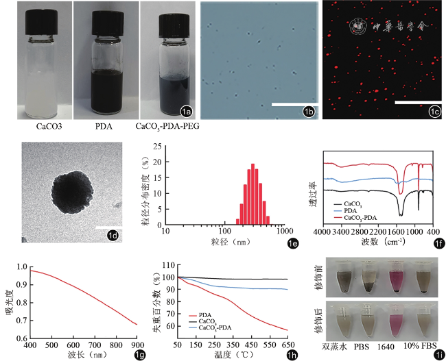
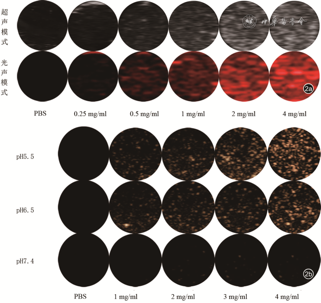
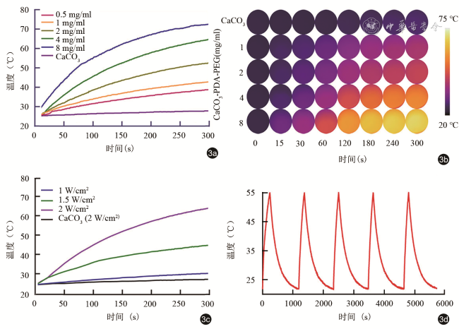
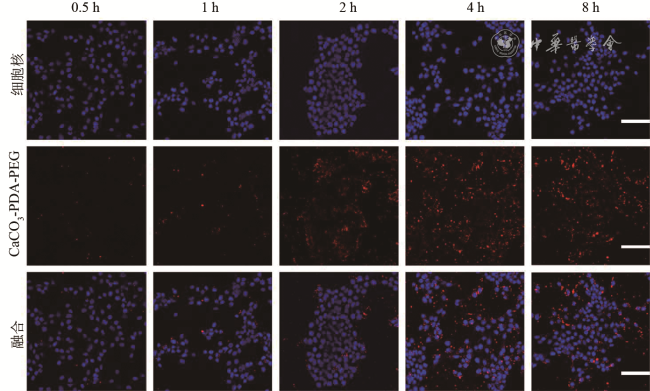
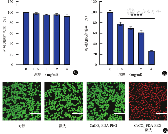
成功制备CaCO3-PDA-PEG纳米探针,呈球形,大小均一,平均粒径约为258 nm,表面电位约为-21 mV;该纳米探针可明显增强光声显像效果,光声信号随纳米探针浓度升高而增强;在肿瘤酸性微环境中,CaCO3-PDA-PEG遇H+可产生二氧化碳气泡进而增强超声造影成像;激光共聚焦显微镜下观察,纳米探针可被动靶向4T1细胞;CaCO3-PDA-PEG(4 mg/ml)与4T1乳腺癌细胞共同孵育,无激光辐照时,细胞存活率高达92.31%;联合808 nm激光辐照后,对肿瘤细胞具有明显的光热杀伤效应,细胞存活率下降至26.61%,同时激光共聚焦显微镜下见大量红色荧光(死细胞)。
所制备的CaCO3-PDA-PEG纳米探针具有良好的pH响应能力,可显著增强体外超声/光声双模态显像效果和光热治疗疗效,为进一步开发可视化肿瘤精准光热治疗方式奠定了基础。
万莉 , 唐芮 , 操雨婷 , 程晨 , 林晓红 , 蒋琴琴 , 胡中倩 , 李攀 . pH响应型纳米探针用于超声/光声成像引导下的乳腺癌光热治疗[J]. 中华医学超声杂志(电子版), 2022 , 19(05) : 459 -466 . DOI: 10.3877/cma.j.issn.1672-6448.2022.05.012
To prepare CaCO3 nanoprobes loaded with polydopamine (PDA) of pH-responsive diagnosis and treatment, and evaluate their effects in ultrasonic/photoacoustic dual-mode imaging and photothermal therapy of breast cancer in vitro.
PEG-modified CaCO3 nanoprobes (CaCO3-PDA-PEG) were prepared by one-pot gas phase diffusion method and two-step method. The macroscopic and microscopic morphology, particle size, infrared spectrum, stability, and other characteristics of CaCO3-PDA-PEG nanoprobes were evaluated. The ultrasonic/photoacoustic dual-mode imaging effect and pH-responsive CEUS imaging ability of the nanoprobes were detected. Then, we evaluated the photothermal conversion efficiency and cytotoxicity of the nanoprobes. Also, the tumor cell targeting ability was assessed by confocal microscopy. Finally, cell proliferation assay and live and dead cell staining were used to evaluate the tumor killing efficiency of the nanoprobes in vitro.
We prepared the CaCO3-PDA-PEG nanoprobes successfully. These spherical nanoprobes were uniform in size, with an average particle size of ~258 nm and a surface potential of ~-21 mV. Besides, the nanoprobes could obviously enhance the photoacoustic imaging signal with the increase of the concentration. Furthermore, CaCO3-PDA-PEG nanoprobes could produce CO2 bubbles when exposed to H+ in the acidic tumor microenvironment (pH 6.5 and pH 5.5), which enhanced CEUS imaging signal. Confocal laser scanning microscopy showed that the nanoprobes could target tumor cells efficiently in a passive way. When incubated with 4T1 cells, these nanoprobes (4 mg/ml) showed a negligible cytotoxicity as the cell survival rate was as high as 92.31%. Yet, once irradiated by an 808 nm laser, CaCO3-PDA-PEG nanoprobes had an obvious photothermal killing effect. The tumor cell survival rate decreased to 26.61% and there was a large amount of red fluorescence (dead cells) under confocal laser scanning microscope.
The prepared pH-responsive CaCO3-PDA-PEG nanoprobes could significantly enhance the ultrasonic/photoacoustic dual-mode imaging signal and photothermal therapy efficacy for tumors in vitro, laying a foundation for the further development of accurate visualized photothermal therapy for tumors.
Key words: pH-responsive; Photothermal therapy; Breast cancer; Nanoprobes; Dual mode imaging
| 1 |
|
| 2 |
|
| 3 |
|
| 4 |
|
| 5 |
|
| 6 |
|
| 7 |
|
| 8 |
何雨蓓, 郝兰, 李倩茹, 等. 基于酞菁铁的靶向乳腺癌纳米粒多模态成像和光热效应的实验研究 [J]. 第三军医大学学报, 2020, 42(8): 772-782.
|
| 9 |
|
| 10 |
|
| 11 |
|
| 12 |
|
| 13 |
|
| 14 |
|
| 15 |
|
/
| 〈 |
|
〉 |
