
2023 , Vol. 20 >Issue 07: 755 - 760
DOI: https://doi.org/10.3877/cma.j.issn.1672-6448.2023.07.015
超声造影在脑胶质瘤切除术术中的应用价值
通信作者:
杨秀华,Email:yangxiuhua@hrbmu.edu.cnCopy editor: 吴春凤
收稿日期: 2022-01-01
网络出版日期: 2023-07-05
版权
Application value of intraoperative contrast-enhanced ultrasound in surgical resection of glioma
Corresponding author:
Yang Xiuhua, Email: yangxiuhua@hrbmu.edu.cnReceived date: 2022-01-01
Online published: 2023-07-05
Copyright
评估超声造影在脑胶质瘤切除术术中的应用价值。
选取2020年9月至2021年7月于哈尔滨医科大学附属第一医院神经外科行脑胶质瘤切除术的27例患者(术前MRI诊断为脑胶质瘤),所有患者术中均行二维超声及超声造影检查,以术后病理作为诊断金标准,明确脑胶质瘤病理类型。采用随机区组设计方差分析比较高级别脑胶质瘤的肿瘤组织、正常脑组织和供血动脉超声造影参数[开始强化时间(TTS)、达峰时间(TTP)、峰值强度(API)]的差异,组间多重比较采用Tukey HSD检验;采用配对t检验比较低级别脑胶质瘤的肿瘤组织和正常脑组织超声造影参数的差异。
27例脑胶质瘤患者中,根据术后病理回报,其中高级别脑胶质瘤16例,低级别脑胶质瘤11例。在高级别脑胶质瘤中,肿瘤组织的TTS为(30.94±2.11)s、TTP为(45.04±2.74)s、API为(79.56±7.35)dB;主要供血动脉的TTS为(28.88±1.54)s、TTP为(43.04±2.85)s、API为(134.27±14.57)dB;正常脑组织的TTS为(35.88±2.16)s、TTP为(46.41±2.75)s、API为(48.02±4.79)dB;高级别脑胶质瘤肿瘤组织的TTS、TTP低于正常脑组织、高于主要供血动脉,API高于正常脑组织、低于主要供血动脉,差异均具有统计学意义(P均<0.001)。在低级别脑胶质瘤中,肿瘤组织的TTS为(23.62±1.90)s、TTP为(35.66±2.38)s、API为(103.67±7.32)dB;正常脑组织的TTS为(27.97±2.23)s、TTP为(44.11±4.68)s、API为(67.27±11.59)dB;低级别脑胶质瘤肿瘤组织的TTS、TTP低于正常脑组织,API高于正常脑组织,差异均具有统计学意义(t=-14.80、-7.41、8.67,P均<0.001)。
术中超声造影可为脑胶质瘤定位及切除范围的确定提供帮助,可最大限度提高肿瘤切除范围。
王晗宇 , 张司可 , 张羽 , 万欣 , 贺秋霞 , 李明明 , 杨秀华 . 超声造影在脑胶质瘤切除术术中的应用价值[J]. 中华医学超声杂志(电子版), 2023 , 20(07) : 755 -760 . DOI: 10.3877/cma.j.issn.1672-6448.2023.07.015
To investigate the application value of intraoperative contrast-enhanced ultrasound (CEUS) in surgical resection of glioma.
A total of 27 patients who underwent glioma resection at Neurosurgery Department of the First Affiliated Hospital of Harbin Medical University from September 2020 to July 2021 (diagnosed as having glioma by MRI before surgery) were selected. All patients underwent intraoperative two-dimensional ultrasound and CEUS. Postoperative pathology was used as the diagnostic gold standard to define the pathological type of glioma. Random block design analysis of variance was used to compare CEUS parameters between tumor tissue, normal brain tissue, and the blood supplying arteries of high-grade glioma. Tukey HSD test was used for multiple comparisons among groups. Paired t test was used for comparison of CEUS parameters between the normal brain tissue and tumor tissue of low-grade glioma.
Among the 27 patients with glioma, 16 had high-grade glioma and 11 had low-grade glioma according to postoperative pathology. In high-grade glioma, the time to start (TTS), time to peak (TTP), and absolute peak intensity (API) were (30.94±2.11) s, (45.04±2.74) s, and (79.56±7.35) dB, respectively. The TTS, TTP, and API of the main supplying arteries were (28.88±1.54) s, (43.04±2.85) s, and (134.27±14.57) dB, respectively. The TTS, TTP, and API of normal brain tissue were (35.88±2.16) s, (46.41±2.75) s, and (48.02±4.79) dB, respectively. The TTS and TTP of high-grade glioma tumor tissue were significantly lower than those of normal brain tissue and higher than those of the main supplying arteries, and the API was significantly higher than that of normal brain tissue and lower than that of the main supplying arteries (P<0.001). In low-grade glioma, the TTS, TTP, and API were (23.62±1.90) s, (35.66±2.38) s, and (103.67±7.32) dB, respectively. The TTS, TTP, and API of normal brain tissue were (27.97±2.23) s, (44.11±4.68) s, and (67.27±11.59) dB, respectively. The TTS and TTP of low-grade glioma tumor tissue were significantly lower than those of normal brain tissue, while the API of low-grade glioma tumor tissue was significantly higher than that of normal brain tissue (t=-14.80, -7.41, and 8.67, respectively, P<0.001 for all).
Intraoperative CEUS can contribute to the localization of glioma and the determination of the extent of resection (EOR), thus providing valuable information for surgeons, maximizing the EOR of the tumor, and prolonging the survival of patients.
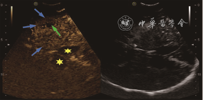
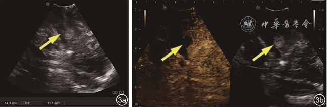
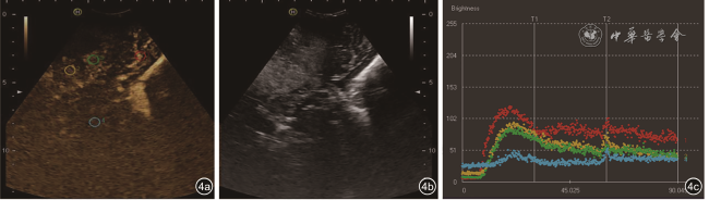
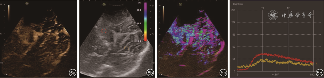
图4 高级别脑胶质瘤术中超声及超声造影表现。图a为高级别脑胶质瘤术中超声造影图像,感兴趣区域(ROI)1(红色圆圈)为供血动脉,ROI 2(黄色圆圈)、ROI 3(绿色圆圈)为肿瘤组织,ROI 4(蓝色圆圈)为周围正常脑组织;图b为高级别脑胶质瘤术中二维灰阶超声图像,肿瘤组织与周围正常脑组织分界模糊;图c为高级别脑胶质瘤超声造影时间-强度曲线图像,可见供血动脉显著快进,且呈明显高增强,且2处肿瘤组织ROI的开始强化时间、达峰时间均低于正常脑组织,峰值强度均高于正常脑组织 |
表1 高级别脑胶质瘤及其供血动脉与正常脑组织间的超声造影时间-强度曲线指标比较( |
| 组别 | TTS(s) | TTP(s) | API(dB) |
|---|---|---|---|
| 供血动脉 | 28.88±1.54 | 43.04±2.85 | 134.27±14.57 |
| 肿瘤组织 | 30.94±2.11a | 45.04±2.74a | 79.56±7.35a |
| 正常脑组织 | 35.88±2.16bc | 46.41±2.75bc | 48.02±4.79bc |
| F值 | 116.87 | 66.74 | 678.08 |
| P值 | <0.001 | <0.001 | <0.001 |
注:TTS为开始强化时间,TTP为达峰时间,API为峰值强度;a与供血动脉比较,b与供血动脉比较,c与a比较,差异均具有统计学意义(P均<0.001) |
表2 低级别脑胶质瘤与正常脑组织间的超声造影时间-强度曲线指标比较( |
| 组别 | TTS(s) | TTP(s) | API(dB) |
|---|---|---|---|
| 肿瘤组织 | 23.62±1.90 | 35.66±2.38 | 103.67±7.32 |
| 正常脑组织 | 27.97±2.23 | 44.11±4.68 | 67.27±11.59 |
| t值 | -14.80 | -7.41 | 8.67 |
| P值 | <0.001 | <0.001 | <0.001 |
注:TTS为开始强化时间,TTP为达峰时间,API为峰值强度 |
| 1 |
|
| 2 |
|
| 3 |
|
| 4 |
|
| 5 |
|
| 6 |
|
| 7 |
|
| 8 |
|
| 9 |
|
| 10 |
|
| 11 |
|
| 12 |
|
| 13 |
|
| 14 |
|
| 15 |
|
| 16 |
|
| 17 |
|
| 18 |
|
| 19 |
|
| 20 |
|
| 21 |
|
| 22 |
|
| 23 |
|
| 24 |
|
| 25 |
葛亚娟, 杨磊, 高军喜, 等. 术中超声造影定量分析在诊断不同级别胶质瘤瘤体及瘤周水肿的临床价值 [J]. 中国超声医学杂志, 2015, 31(3): 193-196.
|
| 26 |
吴意赟, 张心怡, 蔡婷, 等. 超声造影在颅内病变手术中的应用 [J]. 肿瘤影像学, 2020, 29(6): 536-540.
|
| 27 |
|
/
| 〈 |
|
〉 |

