
2023 , Vol. 20 >Issue 06: 642 - 646
DOI: https://doi.org/10.3877/cma.j.issn.1672-6448.2023.06.012
孤立型主动脉缩窄的超声心动图诊断及术后随访研究
Copy editor: 汪荣
收稿日期: 2022-02-25
网络出版日期: 2023-10-31
版权
Echocardiographic diagnosis and postoperative follow-up study of isolated aortic coarctation
Received date: 2022-02-25
Online published: 2023-10-31
Copyright
探讨孤立型主动脉缩窄(COA)的超声心动图特点以及超声心动图在COA诊断治疗及随访中的价值。
纳入2009年3月至2019年10月确诊的121例孤立型COA患儿为研究对象,根据主动脉狭窄程度将患儿分为轻度狭窄(37例)、中度狭窄(34例)、重度狭窄(50例)3组。回顾性分析其超声心动图特点,包括左心室舒张末期内径(LVEDd)、左心室射血分数(LVEF)、室间隔及左心室壁厚度、冠状动脉内径等,并记录进行手术治疗且术后超声随访时间超过1年的20例患儿的超声心动图参数,与术前进行对比分析。
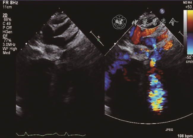
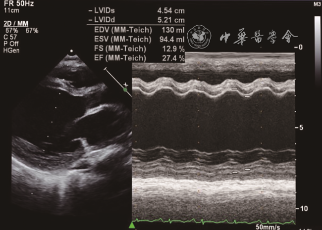
121例患儿中,超声心动图检查首诊误诊3例,误诊率为2.5%(3/121)。部分患儿存在LVEDd增大(67/121,55.4%)、LVEF减低(40/121,33.1%)、左心室心内膜回声增粗增厚(58/121,47.9%)、室间隔和(或)左心室壁增厚(41/121,33.9%)、左冠状动脉内径增宽(12/121,9.9%)等心内继发病变。其中LVEDd增大、左心室心内膜回声增粗增厚、LVEF减低占比在不同狭窄程度组间差异有统计学意义(P均<0.05)。20例随访患儿,与术前比较,手术后1 d、6个月、1年修复处管腔内径增宽、跨狭窄处压差下降、LVEDd减小,差异有统计学意义(P均<0.05);术后1 d的LVEF较术前无显著变化(P>0.05),术后6个月、1年LVEF逐渐恢复,与术前及术后1 d相比差异均有统计学意义(P均<0.05)。20例随访患儿中,2例分别于术后第2年、第8年随访中出现主动脉术后再缩窄。
超声心动图诊断孤立型COA的准确性较高,掌握其超声特点可减少误诊的发生。该疾病手术治疗近中期预后较好,超声心动图在术后定期随访中可发挥重要作用。
吴群 , 张鑫 , 李培 , 王芳韵 , 郑淋 , 卫海燕 , 马宁 . 孤立型主动脉缩窄的超声心动图诊断及术后随访研究[J]. 中华医学超声杂志(电子版), 2023 , 20(06) : 642 -646 . DOI: 10.3877/cma.j.issn.1672-6448.2023.06.012
To explore the echocardiographic characteristics of isolated coarctation of the aorta (COA) and evaluate the value of echocardiography in its diagnosis and follow-up.
A total of 121 patients who were diagnosed with isolated COA from March 2009 to October 2019 were analyzed retrospectively to identify the echocardiographic characteristics of this disease. The echocardiographic parameters included left ventricular end-diastolic diameter (LVEDd), left ventricular ejection fraction (LVEF), ventricular septum and left ventricular wall thickness, coronary artery diameter, etc. According to the degree of COA, the patients were divided into three groups: mild stenosis (37 cases), moderate stenosis (34 cases), and severe stenosis (50 cases). Among them, 20 patients who underwent surgical repair and were followed for more than 1 year after surgery were analyzed to evaluate the effect of operation by comparing the preoperative and postoperative ultrasound data.
All 121 cases were diagnosed by echocardiography while 3 cases of them were misdiagnosed initially. Other ultrasound findings in our group included increased LVEDd (67/121, 55.4%), decreased LVEF (40/121, 33.1%), left ventricular endocardial thickening (58/121, 47.9%), ventricular septum and/or left ventricular wall thickening (41/121, 33.9%), and left coronary artery diameter widening (12/121, 9.9%). Among them, LVEDd increase, left ventricular endocardial thickening, and LVEF decrease had significant differences between the groups that were divided by stenosis degree (P<0.05). Twenty patients were followed by echocardiography at 1 day, 6 months, and 1 year after the operation, and compared with preoperative data, the diameter of the repaired lumen was widened, the pressure difference across the stenosis decreased, and the LVEDd decreased at 1 day, 6 months, and 1 year after surgery, with statistically significant differences (P<0.05). There was no significant change in LVEF 1 day after surgery compared to that before surgery (P>0.05), but LVEF gradually recovered 6 months and 1 year after surgery, with statistically significant differences compared to that before and 1 day after surgery (P<0.05). Two patients had recoarctation in the 2nd and 8th years after surgery, respectively.
The accuracy of echocardiography in the diagnosis of isolated COA is high. Understanding the echocardiographic characteristics can reduce the occurrence of misdiagnosis. The prognosis of isolated COA is satisfactory after surgical treatment. Echocardiography plays an important role in regular postoperative follow-up of patients with this disease.
Key words: Aortic constriction; Echocardiography; Congenital heart disease
表1 孤立型COA不同狭窄程度组间的超声心动图表现比较[例(%)] |
| 组别 | 例数 | LVEDd增大 | 左心室心内膜增粗增厚 | 左心室壁增厚 | LVEF减低 | 左冠状动脉内径增宽 |
|---|---|---|---|---|---|---|
| 轻度狭窄 | 37 | 6(16.2) | 6(16.2) | 8(21.6) | 2(5.4) | 1(2.7) |
| 中度狭窄 | 34 | 20(58.8) | 15(44.1) | 11(32.4) | 6(17.6) | 5(14.7) |
| 重度狭窄 | 50 | 41(82.0) | 37(74.0) | 22(44.0) | 32(64.0) | 6(12.0) |
| χ2值 | 37.466 | 28.725 | 4.803 | 38.066 | 3.217 | |
| P值 | <0.001 | <0.001 | 0.091 | <0.001 | 0.195 |
注:COA为主动脉缩窄;LVEDd为左心室舒张末期内径;LVEF为左心室射血分数 |
表2 20例孤立型COA患儿手术前后超声心动图参数比较( |
| 时间 | D(n=20,mm) | PG(n=20,mmHg) | LVEDd(n=17,mm) | LVEF(n=10,%) | 室间隔和(或)室壁增厚(n=11,mm) |
|---|---|---|---|---|---|
| 术前 | 2.93±0.88 | 64.85±15.41 | 38.52±5.68 | 44.60±5.54 | 4.52±0.37 |
| 术后1 d | 6.31±1.60a | 27.55±8.70a | 34.62±6.92a | 48.50±7.43 | 4.16±0.35 |
| 术后6个月 | 6.45±1.72a | 27.05±8.80a | 32.99±5.29a | 60.40±14.69ab | 3.95±0.62 |
| 术后1年 | 6.94±1.44a | 29.64±11.28a | 33.48±3.89a | 69.60±5.95abc | 4.04±0.52 |
| F值 | 32.165 | 52.961 | 3.575 | 16.968 | 1.479 |
| P值 | <0.001 | <0.001 | 0.019 | <0.001 | 0.235 |
注:1 mmHg=0.133 kPa;COA为主动脉缩窄;D为缩窄处内径;PG为跨狭窄处压差;LVEDd为左心室舒张末期内径;LVEF为左心室射血分数;与术前相比,aP<0.05;与术后1 d相比,bP<0.05;与术后6个月相比,cP<0.05 |
| [1] |
|
| [2] |
|
| [3] |
|
| [4] |
张鑫, 马桂琴, 金兰中, 等. 超声心动图对主动脉缩窄伴左心室收缩功能降低患者的诊断及术后随访价值 [J/CD]. 中华医学超声杂志(电子版), 2014, 11(8): 625-629.
|
| [5] |
北京地区超声心动图协作组. 超声心动图规范化检测心脏功能与正常值 [M]. 北京: 科学技术文献出版社, 2006.
|
| [6] |
中华医学会儿科学分会心血管学组, 中国医师协会心血管内科医师分会儿童心血管专业委员会, 中华儿科杂志编辑委员会. 儿童心力衰竭诊断和治疗建议(2020年修订版) [J]. 中华儿科杂志, 2021, 59(2): 84-94.
|
| [7] |
郑淋, 杜忠东, 金兰中, 等. 超声心动图评价儿童冠状动脉内径正常参考值范围及其临床意义 [J]. 中华儿科杂志, 2013, 51(5): 371-376.
|
| [8] |
|
| [9] |
|
| [10] |
|
| [11] |
|
| [12] |
王迎超, 赵菁, 张玲, 等. 不同程度缩窄主动脉弓致小鼠心肌肥厚及心力衰竭的评价与比较 [J]. 中华急诊医学杂志, 2016, 25(8): 1027-1030.
|
| [13] |
|
| [14] |
|
| [15] |
|
/
| 〈 |
|
〉 |

