
2023 , Vol. 20 >Issue 08: 849 - 853
DOI: https://doi.org/10.3877/cma.j.issn.1672-6448.2023.08.011
术前经皮超声造影对乳腺癌腋窝前哨淋巴结转移及负荷的诊断价值
Copy editor: 汪荣
收稿日期: 2022-04-26
网络出版日期: 2023-10-31
版权
Value of preoperative percutaneous contrast-enhanced ultrasound in diagnosis of axillary sentinel lymph node metastasis and burden in breast cancer
Received date: 2022-04-26
Online published: 2023-10-31
Copyright
探讨术前经皮超声造影对乳腺癌腋窝前哨淋巴结(SLN)转移以及腋窝淋巴结负荷的诊断价值。
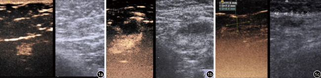
收集2020年7月至2021年12月于哈尔滨医科大学附属肿瘤医院乳腺外科就诊的75例乳腺癌患者,术前经皮超声造影确定SLN并记录其增强模式(分为Ⅰ型均匀增强、Ⅱ型不均匀增强、Ⅲ型无增强)。以病理诊断作为金标准,0~2个SLN转移为有限淋巴结负荷,≥3个SLN转移为高淋巴结负荷,应用四格表分析增强模式诊断腋窝SLN转移及负荷的效能。
超声造影对75例乳腺癌患者SLN的检出率为97.3%(73/75),共检出88个SLN。其中Ⅰ型淋巴结40个,Ⅱ型淋巴结42个,Ⅲ型淋巴结6个。超声造影增强模式诊断SLN转移的敏感度为100%,特异度为62.5%,阳性预测值为50%,阴性预测值为100%,诊断符合率为72.7%。71例T1、T2期乳腺癌患者中24例有SLN转移,其中6例为高淋巴结负荷,18例为低淋巴结负荷。超声造影增强模式诊断SLN负荷状态的敏感度为100%,特异度为56.9%,准确性为60.6%。阳性预测值为17.6%,阴性预测值为100%。
经皮超声造影可有效识别腋窝SLN,超声造影增强模式有助于预测腋窝SLN转移及评估负荷状态。
邵华 , 那子悦 , 荆慧 , 李博 , 王秋程 , 程文 . 术前经皮超声造影对乳腺癌腋窝前哨淋巴结转移及负荷的诊断价值[J]. 中华医学超声杂志(电子版), 2023 , 20(08) : 849 -853 . DOI: 10.3877/cma.j.issn.1672-6448.2023.08.011
To assess the value of preoperative percutaneous contrast-enhanced ultrasound (CEUS) in the diagnosis of axillary sentinel lymph node (SLN) metastasis in breast cancer and the value of enhancement mode in diagnosing axillary lymph node burden.
Seventy-five breast cancer patients at the Breast Surgery Department of Harbin Medical University Cancer Hospital were enrolled from July 2020 to December 2021. Preoperative percutaneous CEUS was used to identify SLNs and record their enhancement pattern (divided into type Ⅰ uniform enhancement, type Ⅱ non-uniform enhancement, and type Ⅲ no enhancement). Pathological diagnosis was used as the gold standard. The value of enhancement pattern analysis in diagnosing axillary SLN metastasis and burden was then assessed by fourfold table.
The detection rate of CEUS for SLN in 75 breast cancer patients was 97.3% (73/75), and a total of 88 SLNs were detected. Among them, there are 40 type Ⅰlymph nodes, 42 type Ⅱ lymph nodes, and 6 type Ⅲ lymph nodes. The sensitivity, specificity, positive predictive value, negative predictive value, and accuracy of CEUS enhancement patterns in predicting SLN metastasis were 100%, 62.5%, 50%, 100%, and 72.7%, respectively. Twenty-four of 71 patients with stage T1/T2 breast cancer had sentinel lymph node metastasis, of whom 6 had high lymph node load and 18 had low lymph node load. The sensitivity, specificity, accuracy, positive predictive value, and negative predictive value of the enhancement patterns of CEUS in diagnosing SLN burden status were 100%, 56.9%, 60.6%, 17.6%, and 100%, respectively.
Percutaneous CEUS is effective in identifying axillary SLNs, and CEUS enhancement patterns can help predict axillary SLN metastasis and burden.
表1 75例患者基本特征[例(%)] |
| 基本特征 | 占比(n=75) |
|---|---|
| 年龄(岁) | |
| ≤35 | 9(12.0) |
| 36~50 | 45(60.0) |
| >50 | 21(28.0) |
| 肿瘤直径 | |
| <2 cm | 13(17.3) |
| 2~5 cm | 58(77.3) |
| >5 cm | 4(5.4) |
| 病理类型 | |
| 浸润性导管癌 | 63(84.0) |
| 浸润性小叶癌 | 2(2.7) |
| 黏液癌 | 3(4.0) |
| 导管内癌 | 7(9.3) |
| 分子亚型 | |
| Luminal A型 | 4(5.3) |
| Luminal B型 | 56(74.7) |
| HER-2过表达 | 11(14.7) |
| 基底型 | 4(5.3) |
| 淋巴结转移情况(个) | |
| 无转移 | 58(65.9) |
| 有转移 | 30(34.1) |
表3 T1、T2期乳腺癌SLN超声造影增强模式与病理结果对照(例) |
| 超声造影增强模式 | 病理结果 | 总计 | |
|---|---|---|---|
| 阳性(高淋巴结负荷) | 阴性(有限淋巴结负荷) | ||
| Ⅰ型(有限淋巴结负荷) | 0 | 37 | 37 |
| Ⅱ型(高淋巴结负荷) | 2 | 28 | 30 |
| Ⅲ 型(高淋巴结负荷) | 4 | 0 | 4 |
| 总计 | 6 | 65 | 71 |
注:SLN为前哨淋巴结 |
| 1 |
|
| 2 |
左梦, 张海宇, 巴黎, 等. 超声造影联合声触诊组织成像定量技术对乳腺癌前哨淋巴结转移的评估[J/OL]. 中华医学超声杂志(电子版), 2021, 18(2): 171-176.
|
| 3 |
|
| 4 |
|
| 5 |
|
| 6 |
|
| 7 |
|
| 8 |
|
| 9 |
|
| 10 |
郭晓霞, 刘昱含, 李潜. 乳腺癌前哨淋巴结超声造影不均匀增强模式的表现分析[J]. 中华实用诊断与治疗杂志, 2019, 33(4): 386-388.
|
| 11 |
|
| 12 |
|
| 13 |
|
| 14 |
|
| 15 |
胥桐, 郭丽苹, 方红, 等. 经皮超声造影对乳腺癌前哨淋巴结的定性评估[J]. 中国医学影像学杂志, 2020, 28(2): 86-89.
|
| 16 |
|
| 17 |
|
/
| 〈 |
|
〉 |