
2023 , Vol. 20 >Issue 10: 1074 - 1080
DOI: https://doi.org/10.3877/cma.j.issn.1672-6448.2023.10.012
先天性原发隔异位型肺静脉异位引流的超声心动图诊断
Copy editor: 汪荣
收稿日期: 2022-09-26
网络出版日期: 2024-01-08
基金资助
浙江省公益技术应用研究项目(LGF22H180002)
版权
Echocardioimagedata diagnosis of anomalous pulmonary venous connection caused by congenital malposition of the septum primum
Received date: 2022-09-26
Online published: 2024-01-08
Copyright
分析原发隔异位(MSP)型肺静脉异位引流(APVC)的超声心动图诊断特点,以期为临床手术提供更多的信息。
回顾性收集2015年1月至2022年4月在浙江大学医学院附属儿童医院经心脏外科手术或CTA证实的21例MSP型APVC患儿的临床资料及超声心动图表现,分析纳入患儿的一般资料、合并畸形、超声心动图特征、误诊情况及外科手术和随访情况。
21例MSP型APVC患儿中完全性肺静脉异位引流(TAPVC)8例、部分性肺静脉异位引流(PAPVC)13例;1例合并多脾综合征。超声心动图表现为肺静脉均开口于解剖左心房的后壁,继发隔发育不良或缺失,原发隔不同程度的偏移导致部分或全部的肺静脉血液引流至解剖右心房。超声首诊正确诊断18例(18/21,85.7%),误诊3例,分别为:1例MSP型TAPVC误诊为三房心合并PAPVC,1例MSP型PAPVC误诊为TAPVC合并继发孔房间隔缺损(ASD),1例MSP型PAPVC误诊为继发孔ASD。20例患儿经心脏外科手术治疗,术后随访4~69个月(平均34个月),所有患儿均预后良好。
超声心动图能正确诊断MSP型APVC及其合并的心内畸形,有助于手术方式的合理选择,提高手术治疗成功率。
张宝富 , 俞劲 , 叶菁菁 , 俞建根 , 马晓辉 , 刘喜旺 . 先天性原发隔异位型肺静脉异位引流的超声心动图诊断[J]. 中华医学超声杂志(电子版), 2023 , 20(10) : 1074 -1080 . DOI: 10.3877/cma.j.issn.1672-6448.2023.10.012
To analyze the echocardioimagedata features of anomalous pulmonary venous connection (APVC) caused by malposition of the septum primum (MSP) in order to provide more information for clinical surgical treatment of this disease.
The clinical data and echocardioimagedata manifestations of 21 patients with APVC caused by MSP confirmed by cardiac surgery or computed tomography angiography at the Children's Hospital of Zhejiang University School of Medicine between January 2015 and April 2022 were retrospectively collected, and the general information, comorbidities, echocardioimagedata features, misdiagnosis, surgical procedures, and follow-up data of the included children were analyzed.
Among the 21 patients included, 8 had total anomalous and 13 had partial APVC directly to the right atrium; 1 case was complicated with polysplenia syndrome. The absent of the superior limbic band of the septum secundum was revealed by echocardiography. The four pulmonary veins were connected with the posterior wall of the anatomical left atrium, which became totally or partially incorporated into the right atrium because of leftward displacement of the septum primum. Eighteen cases (18/21, 85.7%) were correctly diagnosed by echocardiography. Three cases were misdiagnosed as cor triatriatum combined with partial APVC, total APVC combined with ostium secundum atrial septal defects (ASD), and ostium secundum ASD, respectively. Twenty patients were treated by cardiac surgery. The follow-up period ranged from 4~69 months (mean, 34 months) after the operation, and all patients were alive and doing well.
Echocardiography can correctly diagnose APVC caused by MSP and other intracardiac malformations, which is helpful for the rational selection of surgical methods and the improvement of the success rate of surgical treatment.
表1 21例原发隔异位型肺静脉异位引流患儿临床资料、超声心动图、CTA及手术结果 |
| 病例序号 | 年龄 | 性别 | 超声心动图表现 | CTA表现 | 手术结果 |
|---|---|---|---|---|---|
| 1 | 12天 | 男 | MSP,TAPVC,SPMD,PLSVC,TR,PH | - | MSP,TAPVC,SPMD |
| 2 | 7个月23天 | 女 | MSP,PAPVC,SPMD,TR,PH | MSP,PAPVC,SPMD,PH | MSP,PAPVC,SPMD |
| 3 | 1岁3个月 | 男 | CT,PAPVC,ASD(Ⅱ),TR,PH | MSP,TAPVC,SPMD,PH | MSP,TAPVC,SPMD |
| 4 | 6天 | 女 | MSP,TAPVC,SPMD,PDA,TR,PH | MSP,TAPVC,SPMD,PDA,ARSA | MSP,TAPVC,SPMD,PDA |
| 5 | 1个月13天 | 女 | MSP,TAPVC,SPMD,TR,PH | MSP,TAPVC,SPMD,PH | MSP,TAPVC,SPMD |
| 6 | 2个月2天 | 男 | MSP,TAPVC,SPMD,TR,PH | MSP,TAPVC,SPMD,PH | MSP,TAPVC,SPMD |
| 7 | 4个月10天 | 女 | MSP,TAPVC,SPMD,COA,TR,PH | MSP,TAPVC,SPMD,COA,PH | MSP,TAPVC,SPMD,COA |
| 8 | 6个月28天 | 女 | MSP,TAPVC,SPMD,TR,PH | MSP,TAPVC,SPMD,PH | MSP,TAPVC,SPMD |
| 9 | 3个月22天 | 女 | TAPVC,ASD(Ⅱ),PDA,PLSVC,TR,PH | MSP,PAPVC,SPMD,PDA,PH | MSP,PAPVC,SPMD,PDA |
| 10 | 7岁1个月 | 女 | MSP,PAPVC,SPMD,TR,PH | - | MSP,PAPVC,SPMD |
| 11 | 1岁1个月 | 男 | MSP,PAPVC,SPMD,TR,PLSVC | - | MSP,PAPVC,SPMD |
| 12 | 3岁11个月 | 女 | MSP,PAPVC,SPMD,TR,PH | MSP,PAPVC,SPMD,PH | MSP,PAPVC,SPMD |
| 13 | 4个月28天 | 女 | MSP,PAPVC,SPMD,TR | MSP,PAPVC,SPMD | MSP,PAPVC,SPMD |
| 14 | 1岁3个月 | 男 | MSP,PAPVC,SPMD,TR,PH | - | MSP,PAPVC,SPMD |
| 15 | 3岁3个月 | 女 | MSP,PAPVC,SPMD,TR,PH | - | MSP,PAPVC,SPMD |
| 16 | 10个月18天 | 男 | MSP,PAPVC,SPMD,TR,PH | MSP,PAPVC,SPMD,PH | MSP,PAPVC,SPMD |
| 17 | 2个月3天 | 男 | MSP,TAPVC,SPMD,PH | MSP,TAPVC,SPMD,PH | MSP,TAPVC,SPMD |
| 18 | 6个月19天 | 女 | MSP,PAPVC,RSPVS,SPMD,TR,PH | MSP,PAPVC,SPMD,PH | MSP,PAPVC,RSPVS,SPMD |
| 19 | 4岁11个月 | 男 | ASD(Ⅱ),TR | - | MSP,PAPVC,SPMD |
| 20 | 7天 | 男 | MSP,PAPVC,PS(LAI,AVSD,PDA,COA,ⅡVC,AzVD PLSVC,TR,PH) | MSP,PAPVC,PS(LAI,AVSD,PDA,COA,ⅡVC,AzVD PLSVC,PH) | - |
| 21 | 1个月23天 | 男 | MSP,PAPVC,SPMD,TR | MSP,PAPVC,SPMD | MSP,PAPVC,SPMD |
注:ARSA为迷走的右锁骨下动脉;ASD(Ⅱ)为继发孔房间隔缺损;AVSD为房室共同通道;AzVD为奇静脉扩张;COA为主动脉缩窄;CT为三房心;ⅡCV为下腔静脉离断;LAI为左心房异构;MSP为原发隔异位;PAPVC为部分性肺静脉异位引流;PDA为动脉导管未闭;PH为肺动脉高压;PLSVC为永存左上腔静脉;PS为多脾综合征;RSPVS为右上肺静脉狭窄;SPMD为原发隔异位型缺损;TAPVC为完全性肺静脉异位引流;TR为三尖瓣反流;-表示无相关数据;CTA为CT血管造影 |
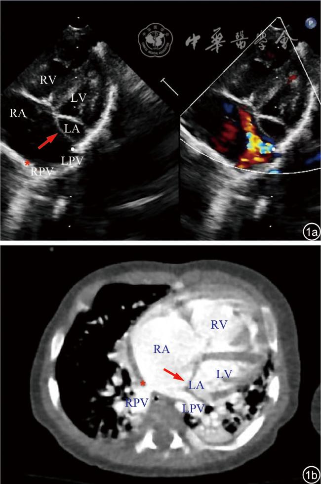
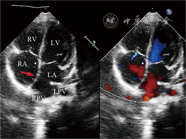
图1 原发隔异位型完全性肺静脉异位引流CT血管造影(CTA)及超声图像。图a为超声心动图胸骨旁四腔心切面显示原发隔明显左移(红色箭头所示),继发隔上边缘带缺如(红色星号所示),导致左、右肺静脉全部回流入右心房;图b为CTA图像显示继发隔上边缘带缺如(红色星号所示),原发隔明显左移(红色箭头所示),所有肺静脉回流入右心房注:LA为左心房;LPV为左肺静脉;LV为左心室;RA为右心房;RPV为右肺静脉;RV为右心室 |
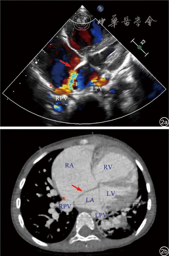
图2 原发隔异位型右侧肺静脉异位引流CT血管造影(CTA)及超声图像。图a为超声心动图胸骨旁四腔心切面显示原发隔左移(红色箭头所示),继发隔上边缘带缺如(红色星号所示),右侧肺静脉回流入右心房;图b为CTA图像显示继发隔上边缘带缺如(红色星号所示),原发隔左移(红色箭头所示)附着于左、右肺静脉间,右侧肺静脉回流入右心房注:LA为左心房;LPV为左肺静脉;LV为左心室;RA为右心房;RPV为右肺静脉;RV为右心室 |
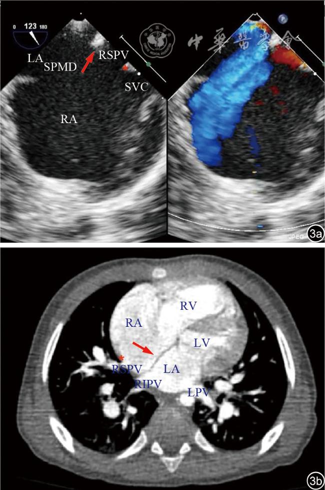
图3 原发隔异位型右上肺静脉异位引流CT血管造影(CTA)及超声图像。图a为经食管超声心动图显示原发隔左移(红色箭头所示),继发隔上边缘带缺如(红色星号所示),导致右上肺静脉回流入右心房;图b为CTA图像显示继发隔上边缘带缺如(红色星号所示),原发隔左移(红色箭头所示)附着于右上、右下肺静脉间,右上肺静脉回流入右心房注:LA为左心房;LPV为左肺静脉;LV为左心室;RA为右心房;RIPV为右下肺静脉;RSPV为右上肺静脉;RV为右心室;SPMD为原发隔异位型缺损;SVC为上腔静脉 |
| 1 |
|
| 2 |
吴明君, 刘畅, 付秀婷. 彩色多普勒超声心动图对先天性肺静脉狭窄的诊断价值[J/CD]. 中华医学超声杂志(电子版), 2012, 9(7): 620-622.
|
| 3 |
陈树宝. 儿童肺动脉高压超声心动图评估[J]. 中国实用儿科杂志, 2015, 30(6): 410-416.
|
| 4 |
|
| 5 |
|
| 6 |
|
| 7 |
|
| 8 |
|
| 9 |
吴力军, 洪雯静, 张玉奇, 等. 先天性原发隔异位的多普勒超声心动图诊断[J].中华超声影像学杂志, 2020, 29(6): 494-498.
|
| 10 |
|
| 11 |
|
| 12 |
|
/
| 〈 |
|
〉 |
