
2023 , Vol. 20 >Issue 11: 1114 - 1118
DOI: https://doi.org/10.3877/cma.j.issn.1672-6448.2023.11.002
高帧频超声造影对小肾透明细胞癌假包膜的评价价值
Copy editor: 吴春凤
收稿日期: 2023-09-29
网络出版日期: 2024-01-15
版权
Value of high-frame-rate contrast-enhanced ultrasound in evaluation of pseudocapsule sign in small clear cell renal carcinoma
Received date: 2023-09-29
Online published: 2024-01-15
Copyright
探讨高帧频超声造影(H-CEUS)评价小肾透明细胞癌(S-CCRCC)假包膜的价值。
回顾性分析2022年9月至2023年4月在解放军总医院第一医学中心经手术病理证实为S-CCRCC的患者80例,按照肿瘤大小分为2组:最大径≤2.0 cm组(30例)和最大径2.1~4.0 cm 组(50例)。对研究对象分别行常规超声造影及H-CEUS检查并留存图像资料,再由2名观察者分别对超声造影和H-CEUS图像进行分析,按造影过程中假包膜的显示程度给出评价结果:完全可见、部分可见、完全不可见。对超声造影和H-CEUS图像特征和结果进行统计分析,采用Kappa检验进行2名观察者间的一致性检验。
2名观察者对2组S-CCRCC假包膜的评价结果:H-CEUS的显示均优于常规超声造影,应用H-CEUS评价S-CCRCC周边假包膜完全可见的比例较常规超声造影有所提高;常规超声造影模式下,2名观察者对S-CCRCC周边假包膜的评价一致性一般:在≤2.0 cm组,Kappa系数为0.605[95%可信区间(CI)为0.396~0.814];在2.1~4.0 cm 组,Kappa系数为0.745(95%CI为0.568~0.922);H-CEUS模式下,2名观察者对S-CCRCC周边假包膜的评价一致性较好:在≤2.0 cm组,Kappa系数为0.813(95%CI为0.646~0.980);在2.1~4.0 cm 组,Kappa系数为0.862(95%CI为0.709~1.000)。
H-CEUS可以使S-CCRCC的假包膜显示得更加清楚,而且不同观察者间评价结果的一致性上有明显提高。
李秋洋 , 赵萍 , 李静波 , 宋禄达 , 朱嘉宁 , 姜波 , 罗渝昆 . 高帧频超声造影对小肾透明细胞癌假包膜的评价价值[J]. 中华医学超声杂志(电子版), 2023 , 20(11) : 1114 -1118 . DOI: 10.3877/cma.j.issn.1672-6448.2023.11.002
To evaluate the value of high-frame-rate contrast-enhanced ultrasound (H-CEUS) in evaluating the pseudocapsule sign in small clear cell renal carcinoma (S-CCRCC).
A retrospective analysis was performed on 80 cases of S-CCRCC confirmed by surgery and pathology at the First Medical Center of PLA General Hospital from September 2022 to April 2023, and they were divided into two groups according to tumor size: maximum diameter ≤ 2.0 cm group (n=30) and maximum diameter 2.1-4.0 cm group (n=50). Conventional CEUS and H-CEUS examinations were performed on the study subjects and the image data were recorded. Then, the conventional CEUS and H-CEUS images were analyzed by two observers respectively, and the evaluation results were given according to the display degree of the pseudocapsule sign during conventional CEUS and H-CEUS: completely visible, partially visible, and completely invisible. The features and results of CEUS and H-CEUS images were statistically analyzed, and Kappa test was used to assess the consistency in the evaluation results of the two observers.
In the evaluation results of the two groups of S-CCRCC, the display of the pseudocapsule sign by H-CEUS was better than that by CEUS, and the proportion of cases with completely visible pseudocapsule sign of S-CCRCC was improved. In the CEUS mode, the Kappa coefficient for the pairwise comparison between the two observers was 0.605 in the ≤2.0 cm group [95% confidence interval (CI): 0.396-0.814], and it was 0.745 (95%CI: 0.568-0.922) in the 2.1-4.0 cm group. In the H-CEUS model, the Kappa coefficient in the ≤2.0 cm group was 0.813 (95%CI: 0.646-0.980), and it was 0.862 (95%CI: 0.709-1.000) in the 2.1-4.0 cm group.
H-CEUS can make the pseudocapsule sign of S-CCRCC more clearly displayed, and the consistency of evaluation results between different observers is significantly improved.
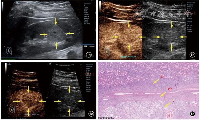
图1 36岁男性患者,右肾中部高回声结节,接受机器人辅助肾部分切除术,术后病理证实为肾透明细胞癌。图a为灰阶超声图像,显示右肾中部高回声结节(结节最大径为2.8 cm);图b超声造影(帧频为10帧/s)显示皮质期(23 s)结节周边假包膜部分可见,呈稍高增强(箭头所示);图c高帧频超声造影(帧频为41帧/s)显示皮质期(22 s)结节周边假包膜完全可见,呈明显高增强(箭头所示);图d病理图片(HE×4)可见完整结构依次为肾实质(K)、假包膜(PC)、肿瘤组织(T) |
表1 不同观察者间应用常规超声造影及高帧频超声造影评价肿瘤最大径≤2.0 cm的小肾透明细胞癌假包膜征象的一致性分析[例(%)] |
| 观察者B | 观察者A | 合计 | ||
|---|---|---|---|---|
| 完全可见 | 部分可见 | 完全不可见 | ||
| 常规超声造影 | ||||
| 完全可见 | 15(50.0) | 1(3.3) | 0(0) | 16(53.3) |
| 部分可见 | 3(10.0) | 6(20.0) | 3(10.0) | 12(40.0) |
| 完全不可见 | 0(0) | 1(3.3) | 1(3.3) | 2(6.7) |
| 合计 | 18(60.0) | 8(26.7) | 4(13.3) | 30(100) |
| 高帧频超声造影 | ||||
| 完全可见 | 17(56.7) | 1(3.3) | 0(0) | 18(60.0) |
| 部分可见 | 2(6.6) | 6(20.0) | 1(3.3) | 9(30.0) |
| 完全不可见 | 0(0) | 0(0) | 3(10.0) | 3(10.0) |
| 合计 | 19(63.3) | 7(23.3) | 4(13.3) | 30(100) |
表2 不同观察者间应用常规超声造影及高帧频超声造影评价肿瘤最大径2.1~4.0 cm的小肾透明细胞癌假包膜征象的一致性分析[例(%)] |
| 观察者B | 观察者A | 合计 | ||
|---|---|---|---|---|
| 完全可见 | 部分可见 | 完全不可见 | ||
| 常规超声造影 | ||||
| 完全可见 | 35(70.0) | 0(0) | 1(2.0) | 36(72.0) |
| 部分可见 | 3(6.0) | 3(6.0) | 2(4.0) | 8(16.0) |
| 完全不可见 | 0(0) | 1(2.0) | 5(10.0) | 6(12.0) |
| 合计 | 38(76.0) | 4(8.0) | 8(16.0) | 50(100) |
| 高帧频超声造影 | ||||
| 完全可见 | 38(76.0) | 0(0) | 0(0) | 38(76.0) |
| 部分可见 | 0(0) | 4(8.0) | 2(4.0) | 6(12.0) |
| 完全不可见 | 1(2.0) | 0(0) | 5(10.0) | 6(12.0) |
| 合计 | 39(78.0) | 4(8.0) | 7(14.0) | 50(100) |
| 1 |
|
| 2 |
|
| 3 |
|
| 4 |
|
| 5 |
|
| 6 |
|
| 7 |
|
| 8 |
朱嘉宁, 李静波, 罗渝昆, 等. 常规超声及超声造影在肾上腺肿瘤诊断中的应用 [J]. 中国研究型医院, 2022, 9(3): 57-60.
|
| 9 |
费翔, 罗渝昆, 李楠, 等. 高帧频超声造影在肝富血供占位性病变动脉期中的成像优势与临床价值 [J/OL]. 中华医学超声杂志(电子版), 2020, 17(9): 827-833.
|
| 10 |
韩鹏, 费翔, 罗渝昆, 等. 高帧频超声造影在鉴别诊断胆囊腺瘤性息肉与胆固醇性息肉中的临床应用 [J/OL]. 中华医学超声杂志(电子版), 2020, 17(9): 815-820.
|
| 11 |
梁舒媛, 罗渝昆, 费翔, 等. 高帧频超声造影在鉴别浅表淋巴结性质中的应用 [J/OL]. 中华医学超声杂志(电子版), 2020, 17(9): 841-847.
|
| 12 |
李楠, 梁舒媛, 费翔, 等. 高帧频超声造影对颈动脉粥样硬化斑块内新生血管的评价价值 [J/OL]. 中华医学超声杂志(电子版), 2020, 17(9): 854-859.
|
| 13 |
|
| 14 |
华琳, 阎岩, 张建. 关于对诊断一致性Kappa系统的探讨 [J]. 数理医药学杂志, 2006, 19(5): 518-520.
|
| 15 |
|
| 16 |
|
| 17 |
|
| 18 |
|
| 19 |
|
| 20 |
|
| 21 |
|
| 22 |
|
/
| 〈 |
|
〉 |