
2023 , Vol. 20 >Issue 11: 1119 - 1124
DOI: https://doi.org/10.3877/cma.j.issn.1672-6448.2023.11.003
高帧频超声造影对慢性肝病患者≤3 cm肝局灶性病变定性诊断的应用价值
Copy editor: 吴春凤
收稿日期: 2023-07-21
网络出版日期: 2024-01-15
版权
Value of high-frame-rate contrast-enhanced ultrasound in diagnosis of small focal liver lesions in patients with chronic liver disease
Received date: 2023-07-21
Online published: 2024-01-15
Copyright
探讨高帧频超声造影(H-CEUS)在慢性肝病患者≤3 cm肝局灶性病变(FLL)定性诊断方面的临床应用价值。
选取2018年1月至2023年4月至瑞金医院及瑞金无锡分院超声科进行肝常规超声造影(CEUS)和H-CEUS检查的153例慢性肝病患者(共176个病灶,病灶最大径均≤3 cm)的CEUS资料进行回顾性分析。所有入组病灶最终由穿刺活检、手术病理、CT增强或MRI增强等其他影像学诊断经随访证实,并将入组病例分为良性组和恶性组。所有入组病例均进行常规CEUS、H-CEUS检查并分别记录静态和动态图像。由2位医师分别对以上检查进行独立分析并记录结果,采用χ2检验比较良、恶性组内这2种CEUS模式特征(开始增强时间、增强程度、增强均匀性、增强方向和血管构型)的差异。采用χ2检验统计分析常规CEUS与H-CEUS在慢肝患者≤3 cm FLL定性诊断的诊断效能,并计算相应的诊断敏感度、特异度、准确性等。
良、恶性组内开始增强时间、增强程度、增强均匀性在常规CEUS与H-CEUS 2种造影技术间比较,差异均无统计学意义(P均>0.05),恶性组内2种造影模式在血管构型和增强方向方面比较,差异均有统计学意义(χ2=6.480、58.284,P=0.011、<0.001)。H-CEUS较常规CEUS可提高慢性肝病FLL诊断的敏感度(93.68% vs 80.00%)和准确性(92.61% vs 83.52%),两者差异具有统计学意义(χ2=7.784、6.921,P=0.005、0.009),但两者之间特异度差异无统计学意义(P>0.05)。
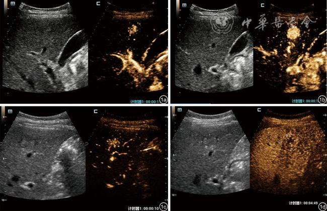
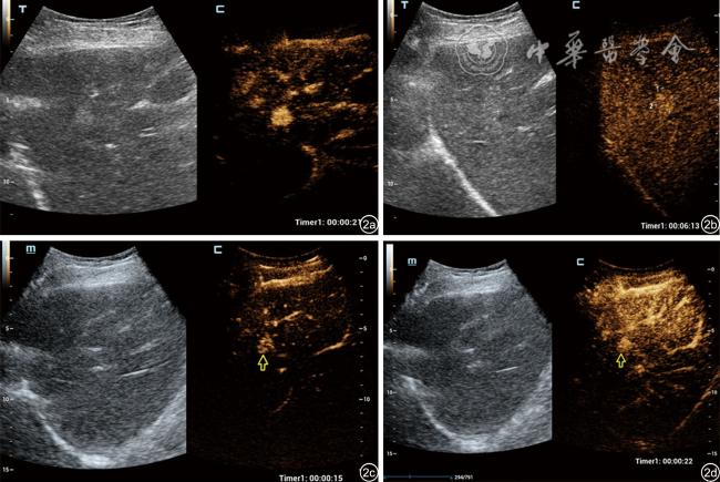
H-CEUS通过提高实时帧频,可提供FLL在动脉期增强方向和血管架构等方面的丰富诊断信息,有利于提高慢性肝病FLL良恶性鉴别诊断的能力。
任新平 , 郑丽丽 , 冯梅晶 , 肖俊飞 , 林艳艳 , 詹维伟 . 高帧频超声造影对慢性肝病患者≤3 cm肝局灶性病变定性诊断的应用价值[J]. 中华医学超声杂志(电子版), 2023 , 20(11) : 1119 -1124 . DOI: 10.3877/cma.j.issn.1672-6448.2023.11.003
The purpose of this study was to assess the value of high-frame-rate contrast-enhanced ultrasound (H-CEUS) in the diagnosis of small (≤3 cm) focal liver lesions in patients with chronic liver disease.
From January 2018 to April 2023, 153 patients with chronic liver disease (176 lesions, ≤3 cm) who underwent CEUS and H-CEUS examinations at Ruijin Hospital and its Wuxi Branch were retrospectively analyzed. CEUS and H-CEUS images were interpreted independently by two doctors and image features were statistically analyzed between CEUS and H-CEUS. The χ2 test was used to compare the differences in the characteristics of the two contrast-enhanced models (start enhancement time, enhancement degree, uniformity, fill-in direction, and vascular morphology) between the benign and malignant groups. The diagnostic performance of the two modalities was assessed by calculating sensitivity, specificity, accuracy, etc.
The start time, intensity, and homogeneity of enhancement did not differ significantly between CEUS and H-CEUS (P>0.05). Fill-in direction and vascular morphology differed significantly between CEUS and H-CEUS (χ2=6.480 and 58.284, P=0.011 and P<0.001, respectively) in the malignant group. H-CEUS had a higher sensitivity (93.68% vs 80.00%) and accuracy (92.61% vs 83.52%) than CEUS in the differential diagnosis of small focal liver lesions (χ2=7.784 and 6.921, P=0.005 and 0.009, respectively), but there was no statistical difference in specificity between the two modalities (P>0.05).
By increasing the frame rate of real-time images, H-CEUS can dynamically and accurately display fill-in patterns and vascular morphology in the arterial phase, thereby improving the differential diagnosis of small focal liver lesions.
表1 81个良性肝局灶性病变不同造影模式下声像图表现比较(个) |
| 造影模式 | 开始增强时间 | 增强程度 | 增强均匀性 | 增强方向 | 血管构型 | ||||||||
|---|---|---|---|---|---|---|---|---|---|---|---|---|---|
| 早期 | 非早期 | 高 | 非高 | 均匀 | 非均匀 | 向心 | 离心 | 其他 | 轮辐样 | 结节样 | 不规则 | 其他 | |
| 常规CEUS | 68 | 13 | 66 | 15 | 56 | 25 | 37 | 15 | 29 | 11 | 35 | 19 | 16 |
| H-CEUS | 68 | 13 | 66 | 15 | 53 | 28 | 38 | 21 | 22 | 21 | 36 | 22 | 2 |
| χ2值 | 0.000 | 0.000 | 0.251 | 1.974 | 14.247 | ||||||||
| P值 | 1.000 | 1.000 | 0.617 | 0.373 | 0.003 | ||||||||
注:CEUS为超声造影,H-CEUS为高帧频超声造影 |
表2 95个恶性肝局灶性病变不同超声造影模式下声像图表现比较(个) |
| 造影模式 | 开始增强时间 | 增强程度 | 增强均匀性 | 增强方向 | 血管构型 | ||||||||
|---|---|---|---|---|---|---|---|---|---|---|---|---|---|
| 早期 | 非早期 | 高 | 非高 | 均匀 | 非均匀 | 向心 | 离心 | 其他 | 轮辐样 | 结节样 | 不规则 | 其他 | |
| 常规CEUS | 93 | 2 | 91 | 4 | 69 | 26 | 28 | 0 | 67 | 0 | 0 | 59 | 36 |
| H-CEUS | 93 | 2 | 92 | 3 | 59 | 36 | 79 | 1 | 15 | 0 | 0 | 75 | 20 |
| χ2值 | 0.000 | 0.148 | 2.382 | 58.284 | 6.480 | ||||||||
| P值 | 1.000 | 0.701 | 0.123 | <0.001 | 0.011 | ||||||||
注:CEUS为超声造影,H-CEUS为高帧频超声造影 |
表3 不同超声造影模式在肝局灶性病变良恶性鉴别诊断中的诊断效能比较[(%)个/个] |
| 造影模式 | 敏感度 | 特异度 | 阳性预测值 | 阴性预测值 | 准确性 |
|---|---|---|---|---|---|
| 常规CEUS | 80.00(76/95) | 87.65(71/81) | 88.37(76/86) | 78.89(71/90) | 83.52(147/176) |
| H-CEUS | 93.68(89/95) | 91.36(74/81) | 92.70(89/96) | 92.50(74/80) | 92.61(163/176) |
| χ2值 | 7.784 | 0.591 | 1.007 | 6.255 | 6.921 |
| P值 | 0.005 | 0.442 | 0.316 | 0.012 | 0.009 |
注:CEUS为超声造影,H-CEUS为高帧频超声造影 |
| 1 |
中华人民共和国国家卫生健康委员会医政医管局. 原发性肝癌诊疗指南(2022年版) [J]. 中华消化外科杂志, 2022, 21(2): 143-168.
|
| 2 |
|
| 3 |
|
| 4 |
|
| 5 |
|
| 6 |
中国医师协会超声医师分会. 中国超声造影临床应用指南 [M]. 北京: 人民卫生出版社, 2017: 85-93.
|
| 7 |
|
| 8 |
丁红. 肝脏超声造影临床应用指南(2012)解读 [J/CD]. 中华医学超声杂志(电子版), 2014, 11(2): 99-101.
|
| 9 |
中华医学会肝病学分会肝癌学组. 肝细胞癌癌前病变的诊断和治疗多学科专家共识(2020版) [J]. 临床肝胆病杂志, 2020, 36(3): 514-518.
|
| 10 |
|
| 11 |
任新平, 林艳艳, 郑丽丽, 等. 超声造影在脂肪肝背景下肝局灶性病变诊断中的应用价值[J/OL]. 中华医学超声杂志(电子版), 2020, 17(9): 834-840.
|
| 12 |
费翔, 罗渝昆, 李楠, 等. 高帧频超声造影在肝富血供占位性病变 动脉期中的成像优势与临床价值 [J/OL]. 中华医学超声杂志(电子版), 2020, 17(9): 827-833.
|
| 13 |
戴全, 戴海鹏, 刘冬梅, 等. 实时超声造影鉴别诊断脂肪肝背景下肝内低回声病变良恶性的临床价值 [J]. 实用肿瘤学志, 2014(5): 415-419.
|
| 14 |
张丽丽, 顾金花, 齐云峰, 等. 不同肝背景下原发性肝癌超声造影灌注特征的定量分析 [J]. 吉林大学学报(医学版), 2016, 42(1): 164-167.
|
/
| 〈 |
|
〉 |