
2023 , Vol. 20 >Issue 11: 1181 - 1185
DOI: https://doi.org/10.3877/cma.j.issn.1672-6448.2023.11.012
超声造影评价肺腺癌与肺鳞癌血流灌注特征的价值研究
Copy editor: 吴春凤
收稿日期: 2022-10-20
网络出版日期: 2024-01-15
基金资助
山西省科技厅重点研发计划项目(201703D421029)
版权
Value of contrast-enhanced ultrasonography in evaluating blood perfusion characteristics of pulmonary adenocarcinoma and squamous cell carcinoma
Received date: 2022-10-20
Online published: 2024-01-15
Copyright
对比分析肺腺癌与肺鳞癌超声造影增强特征,探讨超声造影评价肺腺癌与肺鳞癌血流灌注特征差异的应用价值。
回顾性选取2017年10月至2021年12月于山西医科大学第二医院经病理确诊的54例(54个病灶)肺腺癌与46例(46个病灶)肺鳞癌患者,采用χ2检验对比分析二者超声造影增强特征的差异,包括增强时相(肺动脉增强、支气管动脉增强)、增强模式(基底树枝型增强、杂乱血管型增强)及达峰模式(均匀型增强、不均匀型增强)。
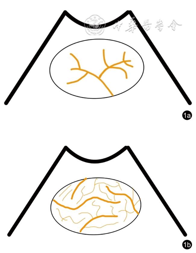
肺腺癌与肺鳞癌超声造影增强特征呈现一定差异,增强时相表现为肺动脉增强的病灶肺腺癌(70.4%,38/54)多于肺鳞癌(45.7%,21/46);增强模式表现为基底树枝型增强的病灶肺腺癌(20.4%,11/54)多于肺鳞癌(4.3%,2/46);达峰模式表现为不均匀型增强的病灶肺鳞癌(67.4%,31/46)多于肺腺癌(16.7%,9/54),差异均具有统计学意义(χ2=6.274、5.638、26.630,P=0.012、=0.018、<0.001)。
肺腺癌与肺鳞癌的超声造影增强特征存在一定差异,超声造影可评估二者血流灌注差异,对其临床治疗方案的选择及鉴别诊断具有潜在的临床应用价值。
赵鑫 , 郝磊 , 朱丽静 , 土继政 , 王博娟 , 张凯 , 王兴华 . 超声造影评价肺腺癌与肺鳞癌血流灌注特征的价值研究[J]. 中华医学超声杂志(电子版), 2023 , 20(11) : 1181 -1185 . DOI: 10.3877/cma.j.issn.1672-6448.2023.11.012
To compare and analyze the enhancement characteristics of pulmonary adenocarcinoma (ADC) and squamous cell carcinoma (SCC) on contrast-enhanced ultrasonography (CEUS), and to investigate the value of CEUS in evaluating the differences in blood perfusion characteristics of ADC and SCC.
A total of 54 patients with pulmonary ADC and 46 patients with SCC confirmed by pathology at the Second Hospital of Shanxi Medical University from October 2017 to December 2021 were retrospectively selected. The chi-square test was used to compare the CEUS characteristics between pulmonary ADC and SCC, including the enhancement phase (early pulmonary-arterial [PA] pattern of enhancement, delayed bronchial-arterial [BA] pattern of enhancement), enhancement pattern (basal dendritic enhancement, chaotic vascular enhancement), and peak pattern of CEUS (homogeneous enhancement, inhomogeneous enhancement).
There were some differences in the enhancement characteristics between ADC and SCC, with more ADC (70.4%, 38/54) than SCC (45.7%, 21/46) cases showing PA pattern of enhancement in the enhancement phase; more ADC (20.4%, 11/54) than SCC (4.3%, 2/46) cases showing basal dendritic enhancement in the enhancement pattern; and more SCC (67.4%, 31/46) than ADC (16.7%, 9/54) cases showing inhomogeneous enhancement in the peak pattern; the differences were statistically significant χ2=6.274, 5.638, and 26.630; P=0.012, =0.018, and <0.001, respectively).
There are some differences in enhancement characteristics of CEUS between ADC and SCC. CEUS can evaluate the differences in blood perfusion characteristics between them, and has potential clinical value in the selection of treatment plan and differential diagnosis of ADC and SCC.
 表示,肺腺癌组与肺鳞癌组组间差异比较采用独立样本t检验;性别、超声造影增强特征为计数资料,以例(%)表示,肺腺癌组与肺鳞癌组组间差异比较采用χ2检验,不同病灶最大径组内肺腺癌与肺鳞癌超声造影达峰模式的差异比较采用Fisher精确概率法。以P<0.05为差异具有统计学意义。
表1 肺腺癌与肺鳞癌患者基本临床特征比较 |
| 组别 | 例数 | 年龄(岁, ) ) | 病灶最大径(cm, ) ) | 男/女(例) |
|---|---|---|---|---|
| 肺腺癌组 | 54 | 64.3±9.9 | 5.31±2.57 | 36/18 |
| 肺鳞癌组 | 46 | 71.2±9.6 | 6.88±2.59 | 45/1 |
| 统计值 | t=-3.542 | t=-3.032 | χ2=15.671 | |
| P值 | 0.001 | 0.003 | <0.001 |
表2 肺腺癌与肺鳞癌超声造影增强特征比较[例(%)] |
| 组别 | 例数 | 增强时相 | 增强模式 | 达峰模式 | |||
|---|---|---|---|---|---|---|---|
| 肺动脉增强 | 支气管动脉增强 | 基底树枝型 | 杂乱血管型 | 不均匀型 | 均匀型 | ||
| 肺腺癌组 | 54 | 38(70.4) | 16(29.6) | 11(20.4) | 43(79.6) | 9(16.7) | 45(83.3) |
| 肺鳞癌组 | 46 | 21(45.7) | 25(54.3) | 2(4.3) | 44(95.7) | 31(67.4) | 15(32.6) |
| χ2值 | 6.274 | 5.638 | 26.630 | ||||
| P值 | 0.012 | 0.018 | <0.001 | ||||
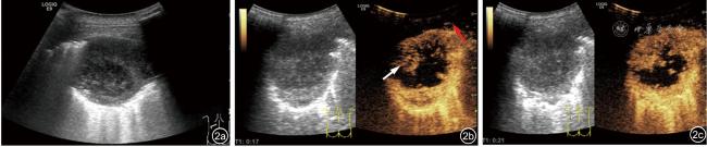
图2 78岁男性肺鳞癌患者超声图像。图a:普通二维超声表现。图b:超声造影17 s时病灶与胸壁同时增强,为支气管动脉增强,该病灶呈杂乱血管型增强,白色箭头为病灶增强,红色箭头为胸壁增强。图c:21 s时病灶达峰模式为不均匀型增强 |
表3 不同病灶最大径组内肺腺癌与肺鳞癌超声造影达峰模式比较[个(%)] |
| 组别 | 例数 | 不均匀型 | 均匀型 | P值 |
|---|---|---|---|---|
| A组 | - | |||
| 肺腺癌 | 11 | 0 | 11(100) | |
| 肺鳞癌 | 1 | 0 | 1(100) | |
| B组 | 0.013 | |||
| 肺腺癌 | 14 | 1(7.1) | 13(92.9) | |
| 肺鳞癌 | 13 | 7(53.8) | 6(46.2) | |
| C组 | 0.060 | |||
| 肺腺癌 | 18 | 6(33.3) | 12(66.7) | |
| 肺鳞癌 | 12 | 9(75.0) | 3(25.0) | |
| D组 | 0.007 | |||
| 肺腺癌 | 11 | 2(18.2) | 9(81.8) | |
| 肺鳞癌 | 20 | 15(75.0) | 5(25.0) |
注:A组病灶最大径≤3 cm,B组病灶最大径>3 cm且≤5 cm,C组病灶最大径>5 cm且≤7 cm,D组病灶最大径>7 cm;-表示无相应统计值 |
| 1 |
|
| 2 |
|
| 3 |
|
| 4 |
郭西源, 郝磊, 朱丽静, 等. 超声造影鉴别诊断周围型肺局灶性病变的临床价值 [J]. 临床超声医学杂志, 2020, 22(11): 866-868.
|
| 5 |
朱丽静, 王兴华. 超声造影定量参数对肺部良恶性病变鉴别诊断的研究 [J]. 中国临床医学影像杂志, 2020, 31(1): 34-37.
|
| 6 |
郭西源, 朱丽静, 土继政, 等. 超声造影与增强CT对周围型肺局灶性病变诊断价值的对比研究 [J]. 中国临床医学影像杂志, 2021, 32(11): 799-802.
|
| 7 |
|
| 8 |
|
| 9 |
董伟华, 肖湘生, 李惠民, 等. 支气管动脉和肺动脉多层螺旋CT血管造影对肺癌血供的研究 [J]. 中华放射学杂志, 2003, 37(7): 612-614.
|
| 10 |
|
| 11 |
|
| 12 |
|
| 13 |
|
| 14 |
|
| 15 |
李相生, 张挽时, 熊明辉, 等. 周围型肺鳞癌与周围型肺腺癌的螺旋CT动态增强对照 [J]. 中华肿瘤杂志, 2006, 28(1): 70-73.
|
| 16 |
|
| 17 |
陈亚男, 陈武飞, 滑炎卿. 肺结节倍增时间的CT研究进展 [J]. 中华解剖与临床杂志, 2017, 22(6): 522-527.
|
| 18 |
中国临床肿瘤学会血管靶向治疗专家委员会, 中国临床肿瘤学会非小细胞肺癌专家委员会, 中国临床肿瘤学会非小细胞肺癌抗血管生成药物治疗专家组. 晚期非小细胞肺癌抗血管生成药物治疗中国专家共识(2020版) [J]. 中华肿瘤杂志, 2020, 42(12): 1063-1077.
|
/
| 〈 |
|
〉 |