
2024 , Vol. 21 >Issue 03: 281 - 287
DOI: https://doi.org/10.3877/cma.j.issn.1672-6448.2024.03.006
Miller-Dieker综合征胎儿产前超声、磁共振影像学特征及遗传学分析
Copy editor: 吴春凤
收稿日期: 2023-08-16
网络出版日期: 2024-06-05
基金资助
南京市卫生科技发展专项资金(YKK20077)
南京鼓楼医院临床研究专项资金(2021-LCYJ-PY-11)
版权
Prenatal ultrasound and MRI characteristics and genetic analysis of Miller-Dieker syndrome
Received date: 2023-08-16
Online published: 2024-06-05
Copyright
探讨Miller-Dieker综合征胎儿的产前超声、磁共振影像学特征及其与遗传学结果的相关性。
回顾性分析2019年11月至2022年9月因“胎儿发育异常”转诊至南京大学医学院附属鼓楼医院妇产医学中心就诊的经产前诊断的4例Miller-Dieker综合征胎儿的资料,对其超声筛查、神经学超声检查、磁共振成像检查、遗传学和病理结果进行总结分析。
4例Miller-Dieker综合征胎儿中,(1)产前超声筛查发现宫内生长受限3例,心脏畸形2例,侧脑室扩张、室管膜下囊肿、小脑下蚓部缺失、头围小和羊水增多各1例;(2)其中2例接受了神经学超声检查,进一步发现无脑回畸形2例、胼胝体部分缺失和颅后窝Blake's囊肿各1例,排除小脑下蚓部缺失1例;(3)3例接受了头颅磁共振成像检查,发现无脑回畸形2例、胼胝体部分缺失和显示不清各1例;(4)4例孕妇均终止妊娠,尸检结果显示3例无脑回畸形、2例心脏畸形和3例面部轻度异常;(5)染色体微阵列检测显示4例胎儿均存在17p13.3区域不同片段缺失,累及基因包括PAFAH1B1、YWHAE和CRK;3例PAFAH1B1基因缺失胎儿均出现无脑回畸形,3例宫内生长受限胎儿均出现YWHAE基因缺失。
Miller-Dieker综合征具有特征性的产前超声征象,当产前超声发现无脑回畸形和宫内生长受限时应考虑本病的存在,部分病例有特殊面部征象,这些征象主要与17p13.3区域缺失的PAFAH1B1、YWHAE和CRK基因有关;神经学超声检查的加入,明显提高了Miller-Dieker综合征胎儿颅脑复杂畸形和无脑回畸形的检出率。
关键词: Miller-Dieker综合征; 超声; 神经学超声检查; 磁共振; 产前诊断
徐燕 , 茹彤 , 郑明明 , 顾燕 , 朱湘玉 , 严陈晨 , 陈玲 , 戴晨燕 . Miller-Dieker综合征胎儿产前超声、磁共振影像学特征及遗传学分析[J]. 中华医学超声杂志(电子版), 2024 , 21(03) : 281 -287 . DOI: 10.3877/cma.j.issn.1672-6448.2024.03.006
To explore the prenatal ultrasound and magnetic resonance imaging (MRI) features of Miller-Dieker syndrome and their correlation with genetic results.
A retrospective analysis was conducted to review the clinical data of 4 cases of pregnant women with Miller-Dieker syndrome fetuses who were referred to the Obstetrics and Gynecology Medical Center of Nanjing Drum Tower Hospital Affiliated to Nanjing University Medical College from November 2019 to September 2022 due to "fetal developmental abnormalities". The ultrasound screening, neurosonography, MRI examination, genetics, and pathological results were summarized and analyzed.
Prenatal ultrasound screening revealed 3 cases of intrauterine growth restriction, 2 cases of cardiac malformation, 1 case of ventriculomegaly, 1 case of subependymal cyst, 1 case of cerebellar vermis deficiency, 1 case of small head circumference, and 1 case of increased amniotic fluid. Neurosonography was further performed in two fetuses, which revealed 2 cases of lissencephaly, 1 case of partial agenesis of the corpus callosum, and 1 case of Blake's cyst in the posterior fossa, and excluded crebellar vermis deficiency in one fetus. Three fetuses underwent fetal MRI examination, which revealed 2 cases of lissencephaly, 1 case of partial agenesis, and 1 case of unclear display of the corpus callosum. All four pregnant women terminated their pregnancies, and fetal autopsy results showed 3 cases of lissencephaly, 2 cases of cardiac malformations, and 3 cases of mild facial abnormalities. Chromosome microarray detection showed different deletions in the 17p13.3 region in the 4 fetuses, involving the PAFAH1B1, YWHAE, and CRK genes. Three fetuses with PAFAH1B1 gene deletion all showed lissencephaly, and three cases with intrauterine growth restriction all showed YWHAE gene deletion.
Miller-Dieker syndrome has characteristic prenatal ultrasound features. When lissencephaly and intrauterine growth restriction are found by prenatal ultrasound, the presence of this disease should be considered. Some cases have special facial signs. All of these features are mainly related to the deletion of PAFAH1B1, YWHAE, and CRK genes in the 17p13.3 region. The addition of neurosonography may significantly improve the detection rate of complex cranial malformations and lissencephaly in Miller-Dieker syndrome fetuses.
表1 4例Miller-Dieker综合征胎儿CMA检测结果 |
| 病例 | CMA结果 | 片段大小(Mb) | OMIM基因数 | 涉及主要基因 |
|---|---|---|---|---|
| 1 | 17p13.3(525_2274390)×1 | 2.27 | 36 | YWHAE、CRK |
| 10q26.12q26.3(121748084_135426386)×3 | 13.67 | 53 | ||
| 2 | 17p13.3(1824913_2996959)×1 | 1.17 | 15 | PAFAH1B1 |
| 3 | 17p13.3p13.2(150733_6378093)×1 | 6.23 | 100 | YWHAE、PAFAH1B1 |
| 13q34(111070794_114342258)×3 | 3.27 | 21 | ||
| 4 | 17p13.3(150733_3204854)×1 | 3.054 | 43 | YWHAE、PAFAH1B1 |
注:CMA为染色体微阵列分析,OMIM为在线人类孟德尔遗传数据库 |
表2 4例Miller-Dieker综合征胎儿的产前超声筛查、神经学超声检查、MRI检查及随访结果 |
| 病例 | 孕妇年龄(岁) | 孕周(周) | 超声筛查 | 神经学超声检查 | MRI检查 | 随访 |
|---|---|---|---|---|---|---|
| 1 | 25 | 22 | 宫内生长受限,左心发育不良综合征(左心偏小、主动脉瓣及升主动脉狭窄) | - | - | 尸检结果:显示体质量偏轻(446 g),面容轻度异常(双眼线下斜、鼻梁塌陷、宽大、短鼻、鼻孔略上翻、双耳低位、上唇肥厚),左心室发育不良(左心室约为右心室1/3大小),主动脉狭窄(主动脉周径0.6 cm,肺动脉周径1.2 cm),主动脉弓离断 |
| 2 | 26 | 36 | 宫内生长受限,双侧脑室后角增宽(左侧11.1 mm,右侧12.0 mm),左侧室管膜下囊肿 | - | 胎儿脑实质、脑沟回、部分脑室形态及信号异常,考虑无脑回畸形,右侧脑室后角中度增宽(14.63 mm),左侧脑室后角轻度增宽(11.52 mm),双顶径小于孕周 | 尸检结果:胎儿相当于34周,面容轻度异常(眼距增宽,双耳低置,小下颌),无脑回畸形,脑室轻度扩张 |
| 3 | 28 | 24 | 小脑下蚓部缺失、室间隔缺损 | 透明隔腔偏窄(1.3 mm)、颅后窝Blake's囊肿(9.4 mm×6.1 mm×5.1 mm)、蛛网膜下腔增宽、无脑回畸形(外侧裂浅显,顶枕沟、距状沟及扣带沟未显示) | 双侧脑室后角轻度增宽(6.25 mm/8.00 mm),胼胝体显示欠清晰,脑沟回显示不满意 | 尸检结果:室间隔膜部缺损、无脑回畸形 |
| 4 | 31 | 30 | 宫内生长受限、头围小、羊水增多(羊水指数233 mm) | 小头畸形、胼胝体部分缺失(胼胝体短)、无脑回畸形(外侧裂及顶枕沟浅显,距状沟及扣带沟未显示、大脑表面光滑,脑沟回不明显)、蛛网膜下腔增宽 | 小头畸形、胼胝体部分缺失、无脑回畸形 | 尸检结果:各发育参数小于孕周,提示存在宫内生长受限,脑沟及脑回结构不明显,提示无脑回畸形,胼胝体部分缺失。外眦间距显著增宽,下牙床中部稍突起 |
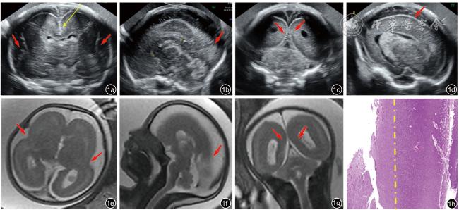
图1 病例4,孕30周胎儿,图a~d为神经学超声检查切面,图a经丘脑冠状切面:扣带沟未显示(黄箭头),双侧外侧裂发育迟缓(红箭头),蛛网膜下腔增宽;图b正中矢状切面:胼胝体部分缺失(胼胝体长26 mm),顶枕沟未显示(箭头),蛛网膜下腔增宽;图c经小脑冠状切面:距状沟未显示(箭头),图d旁矢状切面示右侧大脑皮质表面光滑,未见明显脑沟;图e~g为颅脑MRI检查切面,图e横轴位:双侧外侧裂浅显(箭头),蛛网膜下腔增宽;图f正中矢状位:胼胝体显示欠清晰,顶枕沟未显示(箭头);图g冠状位:距状沟未显示(箭头);图h为镜下无脑回畸形继发征象(HE×10):黄线为额叶皮层灰白质分界线,右侧是灰质区,相对增宽,左侧是白质区,相对变窄 |
| 1 |
|
| 2 |
|
| 3 |
|
| 4 |
|
| 5 |
章锦曼, 杨必成, 冯燕, 等. Miller-Dieker综合征新发染色体突变胎儿二例产前诊断 [J]. 中华妇产科杂志, 2017, 52(10): 700-702.
|
| 6 |
解丽梅. 产前超声发展及展望 [J]. 中国临床医学影像杂志, 2019, 30(1): 1-3.
|
| 7 |
|
| 8 |
|
| 9 |
Sonographic examination of the fetal central nervous system: guidelines for performing the 'basic examination' and the 'fetal neurosonogram' [J]. Ultrasound Obstet Gynecol, 2007, 29(1): 109-116.
|
| 10 |
|
| 11 |
|
| 12 |
|
| 13 |
|
| 14 |
|
| 15 |
|
| 16 |
|
| 17 |
|
| 18 |
|
| 19 |
陈芷萱, 文华轩, 钟晓红, 等. 正常胎儿脑表面沟回在三维反转水晶仿真成像上的发育变化规律[J/OL]. 中华医学超声杂志(电子版), 2022, 19(11): 1165-1172.
|
| 20 |
|
| 21 |
|
| 22 |
|
| 23 |
|
| 24 |
|
| 25 |
|
| 26 |
|
/
| 〈 |
|
〉 |