
2024 , Vol. 21 >Issue 12: 1124 - 1131
DOI: https://doi.org/10.3877/cma.j.issn.1672-6448.2024.12.005
新型微纳秒脉冲电场消融仪器的研发及动物实验研究
Copy editor: 汪荣
收稿日期: 2024-09-23
网络出版日期: 2025-01-23
基金资助
国家自然科学基金项目(82027803,81971623,82171937 和82202151)浙江省“尖兵”“领雁”科技计划项目(2024C03092)浙江省自然科学基金项目(Y24H180007)
版权
Development of novel microsecond-nanosecond pulsed electric field ablation equipment and its animal experimental study
Received date: 2024-09-23
Online published: 2025-01-23
Copyright
目的
探讨微纳秒脉冲电场(μs-nsPEFs)消融新技术的安全性和有效性,主要包含消融范围、肌肉收缩程度及对重要脉管结构的保护作用等。
方法
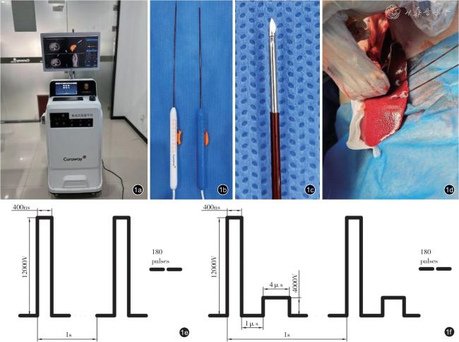
本研究利用3D 电场仿真软件建立三维模型,再进行超声引导下开腹消融手术。选用4 头雌性白猪,将白猪随机分为μs-nsPEFs 和纳秒脉冲电场(nsPEFs)两组(每组各2 头),比较两组术前仿真模拟电场分布、术中量化肌肉收缩程度、术后造影面积和组织病理学表现等,探索μs-nsPEFs 消融猪肝脏组织的安全性和有效性。计量资料组间比较采用Student’s t 检验。
结果
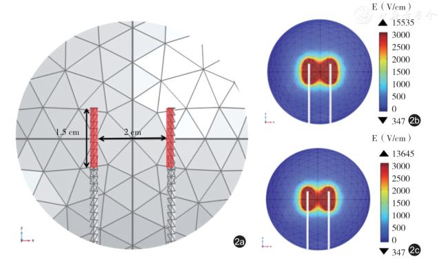
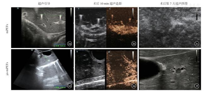
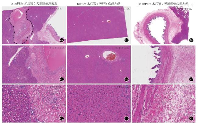
使用COMSOL 软件绘制的3D 仿真电场分布图显示,μs-nsPEFs的最大消融面积为(6.06±0.02)cm2;nsPEFs 的最大消融面积为(5.00±0.03)cm2(P<0.01)。术后10 min 的超声造影(CEUS)结果显示,μs-nsPEFs 组消融面积为(4.70±1.62)cm2,nsPEFs 组为(4.33±1.55)cm2(P>0.05)。术后第7 天超声探查μs-nsPEFs 组存在低回声区域,而nsPEFs 组未发现明显的低回声区域。术后第7 天的HE 染色切片评估提示μs-nsPEFs 消融区边界明确,面积为(0.15±0.08)cm2,而nsPEFs 组未提示消融灶。胆囊壁HE 染色切片提示μs-nsPEFs 术后第7 天消融病灶周围血流通畅,胆囊整体结构保持完整,肝肾功能指标未出现异常。
结论
新型μs-nsPEFs 技术不仅能够有效控制术中肌肉收缩和保护脉管结构,还能使肝脏消融效果持久有效,具有良好的临床应用前景。
李琚 , 陈强 , 张洵 , 谢丽婷 , 蒋天安 . 新型微纳秒脉冲电场消融仪器的研发及动物实验研究[J]. 中华医学超声杂志(电子版), 2024 , 21(12) : 1124 -1131 . DOI: 10.3877/cma.j.issn.1672-6448.2024.12.005
Objective
To investigate the safety and efficacy of the new microsecond-nanosecond pulsed electric field (μs-nsPEF) ablation technology, focusing on ablation range, degree of muscle contraction,and protection of vital vascular structures.
Methods
A three-dimensional (3D) model was established using 3D electric field simulation software, followed by an ultrasound-guided open-abdomen ablation procedure.Four pigs were randomly assigned to μs-nsPEF (n=2) and nanosecond pulsed electric field (nsPEF) groups(n=2).Safety and efficacy of μs-nsPEFs in porcine liver tissue were evaluated by comparing preoperative simulated electric field distributions, intraoperative muscle contraction measurements, postoperative ablation area, and histopathological findings between the two groups.Student's t-test was used for comparisons between groups.
Results
3D electric field distribution maps created with COMSOL software showed a maximum ablation area of 6.06±0.02 cm2 for the μs-nsPEF group and 5.00±0.03 cm2 for the nsPEF group (P<0.01).Contrast-enhanced ultrasound (CEUS) imaging 10 minutes post-ablation indicated an ablation area of 4.70±1.62 cm2 for the μs-nsPEF group and 4.33±1.55 cm2 for the nsPEF group (P>0.05).Ultrasound examination on postoperative day 7 revealed a hypoechoic area in the μs-nsPEF group, whereas no such area was observed in the nsPEF group.H&E staining on postoperative day 7 showed a well-defined ablation boundary in the μs-nsPEF group with an area of 0.15±0.08 cm2, while no ablation lesion was observed in the nsPEF group.H&E staining of the gallbladder wall indicated that on postoperative day 7, blood flow around the μs-nsPEF ablation site remained unobstructed, the gallbladder structure was intact, and liver and kidney function indicators showed no abnormalities.
Conclusion
The novel μs-nsPEF technique demonstrates sustained and effective liver ablation while ensuring muscle contraction and vascular protection during surgery, indicating promising clinical applications.
 ±s 表示,组间比较采用Student’s t 检验,P<0.05 为差异具有统计学意义。CEUS 造影图像和3D 仿真电场分布图像通过Image J 软件进行勾画和分析消融面积。
表1 μs-nsPEFs 消融前后P01 号白猪血清电解质及肝肾生化指标分析 |
| 电解质及生化指标 | 术前2h | 术后2h | 术后7d |
|---|---|---|---|
| 天门冬氨酸氨基转移酶(U/L) | 36.00 | 88.00 | 29.20 |
| 丙氨酸氨基转移酶(U/L) | 77.60 | 99.30 | 68.20 |
| 白蛋白(g/L) | 39.16 | 37.97 | 35.28 |
| 总蛋白(g/L) | 60.11 | 67.63 | 64.97 |
| 球蛋白(g/L) | 29.82 | 29.66 | 29.69 |
| 碱性磷酸酶(U/L) | 77.70 | 78.20 | 58.90 |
| 胆固醇(mmol/L) | 1.69 | 1.42 | 1.77 |
| 肌酐(μmol/L) | 106.10 | 110.50 | 106.40 |
| 谷氨酰转肽酶(U/L) | 35.10 | 41.00 | 36.70 |
| 葡萄糖(mmol/L) | 5.99 | 4.77 | 5.13 |
| 总胆红素(μmol/L) | 1.22 | 1.30 | 1.20 |
| 甘油三酯(mmol/L) | 0.25 | 0.60 | 0.22 |
| 尿素(mmol/L) | 3.37 | 8.23 | 3.46 |
| 钙(mmol/L) | 2.33 | 2.37 | 2.29 |
| 无机磷(μmol/L) | 1.99 | 1.98 | 1.96 |
| 钾(mmol/L) | 4.53 | 4.33 | 4.49 |
| 钠(mmol/L) | 144.50 | 141.70 | 141.20 |
| 氯(mmol/L) | 98.20 | 97.80 | 98.90 |
| 1 |
Sung H, Ferlay J, Siegel RL, et al.Global cancer statistics 2020:GLOBOCAN estimates of incidence and mortality worldwide for 36 cancers in 185 countries[J].CA Cancer J Clin, 2021, 71(3): 209-249.
|
| 2 |
Rumgay H, Ferlay J, de Martel C, et al.Global, regional and national burden of primary liver cancer by subtype[J].Eur J Cancer, 2022, 161:108-118.
|
| 3 |
Kondo Y, Shiina S, Tateishi R, et al.Intrahepatic bile duct dilatation after percutaneous radiofrequency ablation for hepatocellular carcinoma: impact on patient’s prognosis[J].Liver Int,2011, 31(2):197-205.
|
| 4 |
Sparchez Z, Mocan T, Radu P, et al.Prognostic factors after percutaneous radiofrequency ablation in the treatment of hepatocellular carcinoma.Impact of incomplete ablation on recurrence and overall survival rates[J].J Gastrointestin Liver Dis, 2018, 27(4): 399-407.
|
| 5 |
Xu M, Xie LT, Xiao YY, et al.Chinese clinical practice guidelines for ultrasound-guided irreversible electroporation of liver cancer (version 2022) [J].Hepatobiliary Pancreat Dis Int, 2022, 21(5): 462-471.
|
| 6 |
Geboers B, Scheffer HJ, Graybill PM,et al.High-voltage electrical pulses in oncology: irreversible electroporation, electrochemotherapy,gene electrotransfer, electrofusion, and electroimmunotherapy[J].Radiology, 2020, 295(2): 254-272.
|
| 7 |
Golberg A, Bruinsma B, Uygun B, et al. Tissue heterogeneity in structure and conductivity contribute to cell survival during irreversible electroporation ablation by “electric field sinks” [J].Sci Rep, 2015, 5:8485.
|
| 8 |
Rubinsky B, Onik G, Mikus P.Irreversible electroporation: a new ablation modality—clinical implications[J].Technol Cancer Res Treat,2007, 6(1): 37-48.
|
| 9 |
Martin RC, Schwartz E, Adams J, et al.Intra - operative anesthesia management in patients undergoing surgical irreversible electroporation of the pancreas, liver, kidney, and retroperitoneal tumors[J].Anesth Pain Med, 2015, 5(3): e22786.
|
| 10 |
Qasrawi R, Silve L, Burdío F, et al.Anatomically realistic simulations of liver ablation by irreversible electroporation: impact of blood vessels on ablation volumes and undertreatment[J].Technol Cancer Res Treat, 2017, 16(6): 783-792.
|
| 11 |
Xu M, Xu D, Dong G, et al.The safety and efficacy of nanosecond pulsed electric field in patients with hepatocellular carcinoma: a prospective phase 1 clinical study protocol[J].Front Oncol, 2022, 12:869316.
|
| 12 |
Wang Y, Ma R, Huang Z, et al.Investigation of lethal thresholds of nanosecond pulsed electric field in rabbit VX2 hepatic tumors through finite element analysis and verification with a single-needle bipolar electrode: A prospective strategy employing three-dimensional comparisons[J].Comput Biol Med, 2024, 168: 107824.
|
| 13 |
Pethig R.Dielectric properties of body tissues[J].Clin Phys Physiol Meas, 1987, 8(Suppl A): 5-12.
|
| 14 |
Piliopoulos S, Reppas L, Filippiadis D, et al.Irreversible electroporation for the management of pancreatic cancer: Current data and future directions[J].World J Gastroenterol, 2023, 29(2): 223-231.
|
| 15 |
Zeng J, Liu G, Li ZH, et al.The safety and efficacy of irreversible electroporation for large hepatocellular carcinoma[J].Technol Cancer Res Treat, 2017, 16(1): 120-124.
|
| 16 |
Qian J, Liu J, Hong L, et al.Upregulation of PDGF mediates robust liver regeneration after nanosecond pulsed electric field ablation by promoting the HGF/c-Met pathway[J].Biomed Res Int, 2020, 2020:3635787.
|
| 17 |
Qian J, Chen T, Wu Q, et al.Blocking exposed PD-L1 elicited by nanosecond pulsed electric field reverses dysfunction of CD8+ T cells in liver cancer.Cancer Lett[J].2020, 495: 1-11.
|
| 18 |
Golberg A, Bruinsma BG, Uygun BE, et al.Tissue heterogeneity in structure and conductivity contribute to cell survival during irreversible electroporation ablation by “electric field sinks”[J].Sci Rep, 2015, 5: 8485.
|
| 19 |
Distelmaier M, Barabasch A, Heil P, et al.Midterm safety and efficacy of irreversible electroporation of malignant liver tumors located close to major portal or hepatic veins[J].Radiology, 2017, 285(3): 1023-1031.
|
| 20 |
Arena CB, Sano MB, Rossmeisl JH Jr, et al.High-frequency irreversible electroporation (H-FIRE) for non-thermal ablation without muscle contraction[J].Biomed Eng Online, 2011, 10: 102.
|
| 21 |
董守龙, 姚陈果, 储贻道, 等.不可逆电穿孔对兔组织阻抗的影响[J].高电压技术, 2015, 41(4): 1402-1408.
|
| 22 |
Dunki-Jacobs EM, Philips P, Martin RC 2nd.Evaluation of thermal injury to liver, pancreas and kidney during irreversible electroporation in an in vivo experimental model[J].Br J Surg, 2014, 101(9): 1113-1121.
|
| 23 |
Rogers WR, Merritt JH, Comeaux JA, et al.Strength-duration curve for an electrically excitable tissue extended down to near 1 nanosecond[J].IEEE Trans Plasma Science, 2004, 32(4): 1587-1599.
|
| 24 |
Long G, Shires PK, Plescia D, et al.Targeted tissue ablation with nanosecond pulses[J].IEEE Trans Biomed Eng, 2011, 58(8).
|
| 25 |
Sugimoto K, Abe M, Yoshimasu Y, et al.Irreversible electroporation of hepatocellular carcinoma: the role of ultrasonography[J].Ultrasonography, 2020, 39(3): 229-237.
|
| 26 |
Kim KH, An JS, Park YJ, et al.Tissue ablation using irreversible electrolytic electroporation with reduced voltage[J].Electronics, 2023,12(13): 2916.
|
/
| 〈 |
|
〉 |