
2024 , Vol. 21 >Issue 11: 1057 - 1067
DOI: https://doi.org/10.3877/cma.j.issn.1672-6448.2024.11.008
成年健康人群膈肌超声正常值参考范围及影响因素
Copy editor: 吴春凤
收稿日期: 2024-09-19
网络出版日期: 2025-01-24
基金资助
河北省医学科学研究计划项目(20230347,20211264)
版权
Reference ranges for normal values of diaphragmatic ultrasound indices and influencing factors in healthy adults
Received date: 2024-09-19
Online published: 2025-01-24
Copyright
目的
构建成年健康人群膈肌超声指标正常值参考范围并分析相关影响因素。
方法
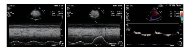
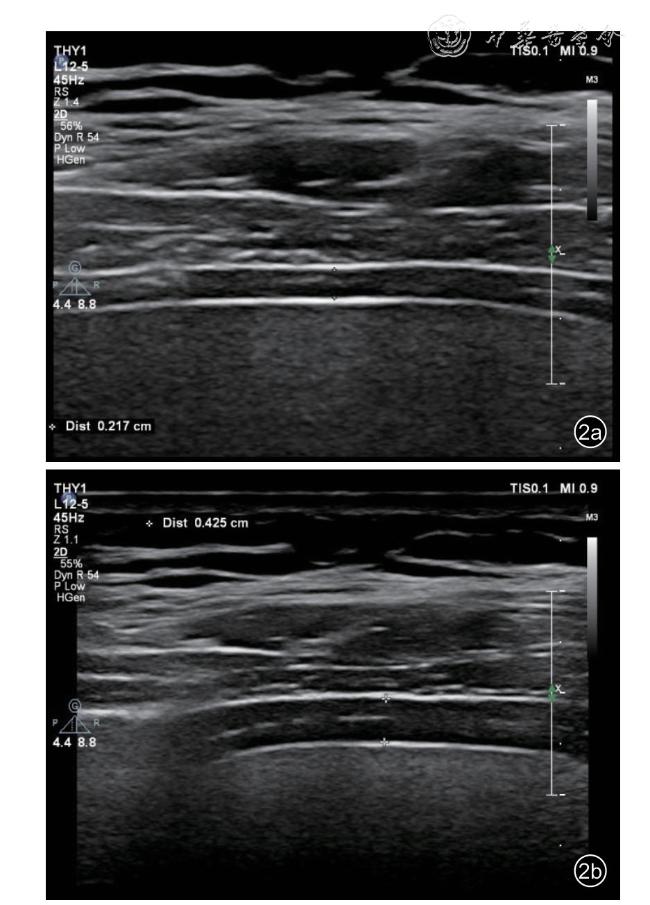
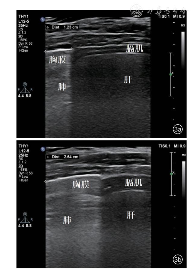
本研究为前瞻性、观察性研究,选择2023年12月至2024年9月河北省人民医院门诊或体检的健康受试者、本院职工及家属共1035人,使用超声获取膈肌移动度(DD)、深吸气膈肌移动度(DDdi)、膈肌收缩期峰值速度(DPSV)、膈肌舒张期峰值速度(DPDV)、呼气末膈肌厚度(DD-ee)、吸气末膈肌厚度(DD-ei)、深吸气末膈肌厚度(DD-edi)、膈肌增厚率(DTF)、深吸气膈肌增厚率(DTF-di)、胸膜滑动位移(PSD),并计算DD储备值、DT储备值和DTF储备值。根据年龄分层分为18~39岁组、40~59岁组、60~79岁组、80~99岁组,计算各组间整体及不同性别各超声指标,以计量资料各指标区间的第5%、第95%位数值作为正常值参考范围的下限和上限。采用Pearson法或Spearman法进行年龄、性别、体质量指数(BMI)、呼吸频率与上述膈肌超声参数的相关性分析,采用组内相关系数(ICC)评价观察者内及观察者间重复性。
结果
(1)不同年龄段之间,80~99岁组(21人)DD、DD-di、DD储备值、DT-ei、DT-edi、DT储备值、DTF、DTF-di、DTF储备值均低于18~39岁组(344人)、40~59岁组(403人)及60~79岁组(267人),PSD低于18~39岁组及40~59岁组。60~79岁组DD-di、PSD低于18~39岁组及40~59岁组,DD储备值、DTedi、DT储备值、DTF-di、DTF储备值均低于18~39岁组,DPDV高于40~59岁组。40~59岁组DD-di、DD储备值、DTF-di、DTF储备值均低于18~39岁组(P均<0.05)。(2)各年龄段不同性别之间,总例数组内,男性DD-di、DD储备值、DT-ee、DT-ei、DT-edi、DT储备值均高于女性。18~39岁组内,男性DD-di、DD储备值、DT-ee、DT-ei、DT-edi均高于女性,DTF、DTF-di均低于女性。40~59岁组内,男性DD-di、DD储备值、DT-ee、DT-ei、DT-edi、DT储备值均高于女性。80~99岁组内,男性DD-di、DT-ee、DT-ei、DT-edi、DT储备值均高于女性(P均<0.05)。(3)性别与DT-ee呈中度负相关性(r =-0.407),与DD储备值、DT-ei、DT-edi呈弱负相关性(r =-0.208、-0.378、-0.283,P均<0.05)。BMI与DT-ee、DT-ei呈弱正相关性(r =0.337、0.287,P均<0.001)。呼吸频率与DD、DT-edi、DT储备值、DTF-di呈弱负相关性(r =-0.384、-0.266、-0.272、-0.205,P均<0.05)。(3)PSD与DD-di在总例数组(r =0.425)、18~39岁组(r =0.447)、40~59岁组(r =0.415)、60~79岁组(r =0.379)、80~99岁组(r =0.530)内均为中度正相关性。
结论
膈肌超声指标正常值参考范围的建立具有重要意义,有助于为膈肌结构和功能评估提供参考依据。
赵浩天 , 王晓娜 , 刘奕 , 李丽 , 刘凯 , 姚光耀 , 薛红元 , 赵鹤龄 . 成年健康人群膈肌超声正常值参考范围及影响因素[J]. 中华医学超声杂志(电子版), 2024 , 21(11) : 1057 -1067 . DOI: 10.3877/cma.j.issn.1672-6448.2024.11.008
Objective
To establish the reference ranges for normal values of diaphragmatic ultrasound indices in healthy adult populations and analyze the related influencing factors.
Methods
This is a prospective, observational study in which 1035 healthy subjects, staff and family members of Hebei General Hospital from December 2023 to September 2024 were enrolled. Ultrasound was used to obtain diaphragm displacement (DD), deep inspiratory DD (DD-di), peak systolic velocity of the diaphragm (DPSV), peak diastolic velocity of the diaphragm (DPDV), end expiratory diaphragm thickness (DT-ee), end inspiratory DT (DT-ei), end deep inspiratory DT (DT-edi), diaphragm thickening fraction (DTF), deep inspiratory DTF(DTF-di), and pleural sliding displacement (PSD). The DD reserve value and DT reserve value were also calculated. Subjects were stratified by age into groups of 18-39 years, 40-59 years, 60-79 years, and 80-99 years. The ultrasound indices were compared among the groups and between genders within each group.The 5th and 95th percentiles of each index in the quantitative data were used as the lower and upper limits of the reference ranges. Pearson’s or Spearman’s method was used to analyze the correlation between age, sex,body mass index(BMI), respiratory frequency and the above phrenic ultrasound parameters, and intraclass correlation coefficient (ICC) was used to assess the intra- and inter-observer reproducibility.
Results
Among the different age groups, the group of 80-99 years had lower DD, DD-di, DD reserve value, DT-ei, DT-edi,DT reserve value, DTF, DTF-di, and DTF reserve value compared to the 18-39 years, 40-59 years, and 60-79 years groups, and lower PSD compared to the 18-39 years and 40-59 years groups. The 60-79 years group had lower DD-di and PSD compared to the 18-39 years and 40-59 years groups, and the 60-79 years group had lower DD reserve value, DT-edi, DT reserve value, DTF-di, and DTF reserve value compared to the 18-39 years group, and higher DPDV compared to the 40-59 years group. The 40-59 years group had lower DD-di, DD reserve value, DTF-di, and DTF reserve value compared to the 18-39 years group (P<0.05 for all). Between genders within each age group, males had higher DD-di, DD reserve value, DT-ee, DT-ei, DTedi, and DT reserve value than females in the overall cohort. Within the 18-39 years group, males had higher DD-di, DD reserve value, DT-ee, DT-ei, and DT-edi, but lower DTF and DTF-di compared to females. Within the 40-59 years group, males had higher DD-di, DD reserve value, DT-ee, DT-ei, DT-edi, and DT reserve value than females. Within the 80-99 years group, males had higher DD-di, DT-ee, DT-ei, DT-edi, and DT reserve value than females (P<0.05 for all). Gender showed a moderate negative correlation with DT-ee(r =-0.407) and a weak negative correlation with DD reserve value, DT-ei, and DT-edi (r =-0.208, -0.378,and -0.283, respectively, P<0.05 for all). BMI showed a weak positive correlation with DT-ee and DT-ei(r =0.337 and 0.287, respectively, P<0.001 for both). Respiratory rate showed a weak negative correlation with DD, DT-edi, DT reserve value, and DTF-di (r =-0.384, -0.266, -0.272, and -0.205, respectively,P<0.05 for all). PSD showed a moderate positive correlation with DD-di in the overall cohort (r =0.425)and in the age groups of 18-39 years (r =0.447), 40-59 years (r =0.415), 60-79 years (r =0.379), and 80-99 years (r =0.530).
Conclusion
The establishment of reference ranges for diaphragmatic ultrasound indices is of great significance and helps to provide a reference basis for the assessment of diaphragmatic structure and function.
 ±s表示,2组间比较采用t检验,多组间比较采用ANOVA单因素方差分析法,组间两两比较采用LSD-t检验;不符合正态分布的计量资料以M(QR)表示,2组间比较采用秩和检验,多组间比较采用Kruskal-Wallis H检验,组间两两比较采用Nemenyi检验;计数资料以例数(%)表示,采用χ2检验比较组间差异,以计量资料各指标区间的第5%、第95%位数值作为正常值参考范围的下限和上限。采用Pearson法或Spearman法进行年龄、性别、BMI、呼吸频率与膈肌超声参数的相关性分析,相关系数r≥0.8为高度相关,0.6≤r<0.8为强相关,0.4≤r<0.6为中等相关,0.2≤r<0.4为弱相关,r<0.2为无相关性。观察者重复性检验采用组内相关系数(intraclass correlation coefficient,ICC)评价,ICC>0.9为优秀,0.75≤ICC<0.9为良好,0.4≤ICC<0.75为中等,ICC<0.4为差。P<0.05为差异有统计学意义。
表1 所有受试者整体及年龄分层膈肌运动相关超声参数正常值 |
| 组别 | 人数 | 膈肌移动度指标 | 膈肌峰速度指标 | 胸膜滑动指标 | |||
|---|---|---|---|---|---|---|---|
| DD(mm,±s) | DD-di(mm,±s) | DD储备值(mm,±s) | DPSV(cm/s,±s) | DPDV(cm/s,±s) | PSD[mm,M(QR)] | ||
| 所有受试者 | 1035 | 18.00±5.05 | 56.23±12.07 | 38.23±10.78 | 2.41±0.45 | 2.17±0.43 | 17.30(12.70,22.80) |
| 男性 | 454 | 18.14±5.17 | 59.00±11.51 | 40.86±10.37 | 2.42±0.43 | 2.18±0.42 | 17.85(12.50,24.20) |
| 女性 | 581 | 17.90±4.94 | 54.07±12.07* | 36.17±10.66* | 2.39±0.46 | 2.17±0.44 | 17.10(12.90,21.65) |
| 18~39岁组 | 344 | 18.08±4.97 | 58.29±11.67 | 40.20±10.63 | 2.43±0.43 | 2.18±0.43 | 17.85(14.10,23.58) |
| 男性 | 178 | 17.97±4.88 | 60.49±11.38 | 42.52±10.21 | 2.43±0.41 | 2.16±0.39 | 18.35(14.05,24.33) |
| 女性 | 166 | 18.20±5.07 | 55.92±11.55* | 37.72±10.54* | 2.43±0.46 | 2.21±0.47 | 17.55(14.08,22.98) |
| 40~59岁组 | 403 | 18.39±5.29 | 56.31±12.51a | 37.92±11.01a | 2.39±0.47 | 2.13±0.42 | 17.50(13.30,23.00) |
| 男性 | 153 | 18.78±5.78 | 59.19±11.76 | 40.41±10.44 | 2.40±0.49 | 2.16±0.45 | 17.10(12.90,25.70) |
| 女性 | 250 | 18.15±5.00 | 54.55±12.65* | 36.39±11.10* | 2.37±0.45 | 2.11±0.40 | 17.55(13.58,21.85) |
| 60~79岁组 | 267 | 17.69±4.72 | 54.46±11.17ab | 36.77±10.25a | 2.42±0.43 | 2.23±0.44b | 16.60(11.40,21.40)ab |
| 男性 | 116 | 17.77±4.78 | 57.01±10.99 | 39.24±10.26 | 2.45±0.39 | 2.27±0.43 | 17.50(10.70,23.18) |
| 女性 | 151 | 17.62±4.69 | 52.50±10.94* | 34.87±9.87* | 2.39±0.46 | 2.21±0.45 | 16.10(12.00,20.30) |
| 80~99岁组 | 21 | 13.34±2.74abc | 43.60±10.49abc | 30.26±8.55abc | 2.23±0.43 | 2.12±0.47 | 12.10(9.35,20.25)ab |
| 男性 | 7 | 14.90±2.54 | 49.99±11.28 | 35.09±9.70 | 2.28±0.42 | 2.08±0.48 | 12.10(9.80,26.00) |
| 女性 | 14 | 12.56±2.56 | 40.40±8.81* | 27.84±7.09 | 2.21±0.44 | 2.15±0.48 | 12.00(8.60,17.65) |
| 统计值 | F=7.273 | F=13.377 | F=9.644 | F=1.853 | F=3.272 | H=20.981 | |
| P值 | <0.001 | <0.001 | <0.001 | 0.136 | 0.021 | <0.001 | |
表2 所有受试者整体及年龄分层膈肌厚度相关超声参数正常值 |
| 组别 | 人数 | 膈肌厚度指标 | 膈肌厚度变化指标 | |||||
|---|---|---|---|---|---|---|---|---|
| DT-ee(mm,±s) | DT-ei(mm,±s) | DT-edi(mm,±s) | DT储备值[mm,M(QR)] | DTF(%,±s) | DTF-di[%,M(QR)] | DTF储备值[%,M(QR)] | ||
| 所有受试者 | 1035 | 1.74±0.37 | 2.24±0.49 | 3.88±0.98 | 1.45(1.05,2.02) | 29.32±11.53 | 114.29(89.71,152.38) | 84.21(61.90,124.68) |
| 男性 | 454 | 1.91±0.39 | 2.45±0.50 | 4.17±0.97 | 1.56(1.11,2.12) | 28.59±10.60 | 111.59(88.27,150.00) | 83.62(61.38,122.74) |
| 女性 | 581 | 1.61±0.30* | 2.08±0.43* | 3.64±0.93* | 1.38(1.00,1.99)* | 29.89±12.18 | 116.67(90.62,154.73) | 85.37(62.05,126.43) |
| 18~39岁组 | 344 | 1.71±0.38 | 2.22±0.50 | 3.99±0.99 | 1.58(1.11,2.20) | 29.92±12.08 | 122.19(95.02,167.35) | 95.09(67.27,137.50) |
| 男性 | 178 | 1.89±0.38 | 2.42±0.48 | 4.24±0.94 | 1.70(1.22,2.22) | 28.68±10.82 | 118.58(91.91,155.25) | 89.47(65.52,131.25) |
| 女性 | 166 | 1.53±0.29* | 2.00±0.42* | 3.71±0.97* | 1.44(1.03,2.14) | 31.25±13.20* | 125.00(99.84,178.43)* | 100.77(69.23,141.35) |
| 40~59岁组 | 403 | 1.76±0.38 | 2.27±0.51 | 3.88±1.00 | 1.43(1.07,1.99) | 29.13±11.85 | 112.23(88.24,147.37)a | 81.25(61.90,119.28)a |
| 男性 | 153 | 1.97±0.39 | 2.51±0.52 | 4.24±1.01 | 1.57(1.11,2.15) | 27.67±10.52 | 109.64(87.13,141.92) | 78.95(60.24,114.38) |
| 女性 | 250 | 1.63±0.31* | 2.12±0.45* | 3.66±0.93* | 1.39(1.00,1.90)* | 30.02±12.52 | 116.24(89.98,149.27) | 82.31(62.47,122.57) |
| 60~79岁组 | 267 | 1.75±0.35 | 2.26±0.46 | 3.81±0.92a | 1.40(1.00,1.89)a | 29.40±10.44 | 108.22(87.59,147.62)a | 78.57(57.76,115.87)a |
| 男性 | 116 | 1.86±0.39 | 2.41±0.52 | 4.01±0.95 | 1.42(1.04,1.96) | 29.98±10.51 | 108.03(88.25,147.09) | 77.40(57.49,107.20) |
| 女性 | 151 | 1.66±0.28* | 2.14±0.37* | 3.65±0.86* | 1.38(0.99,1.80) | 28.95±10.39 | 108.70(86.67,149.43) | 78.82(57.76,116.67) |
| 80~99岁组 | 21 | 1.60±0.36 | 1.96±0.44abc | 2.83±0.76abc | 0.77(0.54,1.08)abc | 22.01±5.80abc | 68.42(58.68,88.38)abc | 44.27(37.36,67.42)abc |
| 男性 | 7 | 1.94±0.35 | 2.39±0.42 | 3.57±0.55 | 1.16(0.78,1.60) | 23.10±2.63 | 67.42(64.09,105.00) | 44.27(43.09,80.00) |
| 女性 | 14 | 1.44±0.22* | 1.74±0.27* | 2.46±0.55* | 0.68(0.47,0.80)* | 21.47±6.90 | 70.38(52.11,80.17) | 45.68(31.04,59.89) |
| 统计值 | F=1.861 | F=3.004 | F=10.044 | H=37.300 | F=3.187 | H=42.561 | H=38.707 | |
| P值 | 0.134 | 0.030 | <0.001 | <0.001 | 0.023 | <0.001 | <0.001 | |
表3 不同年龄段及性别分层的膈肌运动超声正常值参考范围(第5%~第95%位数值) |
| 组别 | 人数 | 膈肌移动度指标 | 膈肌峰速度指标 | 胸膜滑动指标 | |||
|---|---|---|---|---|---|---|---|
| DD(mm) | DD-di(mm) | DD储备值(mm) | DPSV(cm/s) | DPDV(cm/s) | PSD(mm) | ||
| 所有受试者 | 1035 | 10.98~27.64 | 37.38~77.52 | 21.58~57.68 | 1.80~3.19 | 1.55~3.00 | 8.20~31.12 |
| 男性 | 454 | 10.48~28.20 | 40.48~78.35 | 24.98~59.78 | 1.80~3.19 | 1.55~3.00 | 8.08~32.38 |
| 女性 | 581 | 11.20~27.50 | 34.43~75.37 | 19.40~56.10 | 1.75~3.19 | 1.55~3.00 | 8.30~30.08 |
| 18~39岁组 | 344 | 11.63~27.73 | 39.40~78.20 | 24.20~60.25 | 1.85~3.19 | 1.55~3.00 | 9.23~32.45 |
| 男性 | 178 | 11.52~26.67 | 41.07~80.83 | 27.08~61.80 | 1.85~3.15 | 1.55~2.95 | 9.20~33.70 |
| 女性 | 166 | 11.70~27.87 | 37.71~76.03 | 21.41~56.53 | 1.85~3.20 | 1.55~3.00 | 9.44~31.23 |
| 40~59岁组 | 403 | 11.12~29.30 | 36.54~77.88 | 20.02~57.38 | 1.75~3.20 | 1.51~3.00 | 8.02~30.56 |
| 男性 | 153 | 9.67~30.21 | 40.00~79.08 | 23.54~58.79 | 1.76~3.41 | 1.58~3.00 | 7.39~32.16 |
| 女性 | 250 | 11.80~27.55 | 34.58~77.80 | 18.40~57.35 | 1.75~3.15 | 1.50~2.91 | 8.41~29.61 |
| 60~79岁组 | 267 | 10.78~26.96 | 36.64~73.76 | 21.94~55.72 | 1.79~3.17 | 1.58~3.00 | 7.34~29.82 |
| 男性 | 116 | 10.70~26.92 | 40.49~77.19 | 24.80~58.86 | 1.91~3.12 | 1.59~3.02 | 6.69~31.49 |
| 女性 | 151 | 10.82~27.70 | 35.12~70.18 | 19.90~51.68 | 1.75~3.21 | 1.57~3.00 | 7.76~29.04 |
| 80~99岁组 | 21 | 8.87~19.74 | 30.71~64.08 | 18.21~48.59 | 1.66~3.26 | 1.51~2.99 | 6.94~29.51 |
| 男性 | 7 | 12.50~19.92 | 37.60~64.09 | 24.60~48.90 | 1.90~3.00 | 1.50~2.84 | 7.60~29.74 |
| 女性 | 14 | 8.80~16.46 | 30.60~56.76 | 17.90~41.08 | 1.65~3.27 | 1.58~2.99 | 6.90~25.13 |
表4 不同年龄段及性别分层的膈肌厚度超声正常值参考范围(第5%~第95%位数值) |
| 组别 | 人数 | 膈肌厚度指标 | 膈肌厚度变化指标 | |||||
|---|---|---|---|---|---|---|---|---|
| DT-ee(mm) | DT-ei(mm) | DT-edi(mm) | DT储备值(mm) | DTF(%) | DTF-di(%) | DTF储备值(%) | ||
| 所有受试者 | 1035 | 1.20~2.43 | 1.51~3.17 | 2.50~5.80 | 0.60~3.37 | 14.39~52.94 | 59.05~236.79 | 32.62~204.42 |
| 男性 | 454 | 1.39~2.60 | 1.71~3.39 | 2.86~6.19 | 0.60~3.52 | 14.27~50.00 | 53.91~222.53 | 27.62~193.81 |
| 女性 | 581 | 1.14~2.10 | 1.46~2.80 | 2.30~5.47 | 0.60~3.23 | 14.96~53.80 | 64.33~241.69 | 37.50~214.17 |
| 18~39岁组 | 344 | 1.12~2.48 | 1.49~3.18 | 2.56~5.90 | 0.68~3.45 | 14.40~53.72 | 66.19~257.00 | 37.68~218.29 |
| 男性 | 178 | 1.39~2.60 | 1.74~3.27 | 2.91~6.19 | 0.69~3.44 | 14.36~50.13 | 60.24~222.29 | 31.73~185.45 |
| 女性 | 166 | 1.10~2.00 | 1.40~2.70 | 2.43~5.68 | 0.67~3.62 | 14.50~54.73 | 74.08~277.85 | 42.91~253.65 |
| 40~59岁组 | 403 | 1.20~2.45 | 1.51~3.22 | 2.47~5.90 | 0.60~3.37 | 14.09~53.25 | 55.57~232.60 | 33.40~202.63 |
| 男性 | 153 | 1.40~2.59 | 1.78~3.47 | 2.80~6.26 | 0.56~3.71 | 13.55~48.69 | 51.15~220.27 | 24.71~194.20 |
| 女性 | 250 | 1.19~2.10 | 1.50~2.90 | 2.30~5.43 | 0.64~3.15 | 14.35~55.87 | 65.90~239.15 | 38.12~209.03 |
| 60~79岁组 | 267 | 1.25~2.37 | 1.60~3.06 | 2.59~5.54 | 0.56~3.23 | 15.03~48.29 | 58.20~227.13 | 28.55~200.97 |
| 男性 | 116 | 1.37~2.70 | 1.65~3.50 | 2.77~5.97 | 0.55~3.42 | 14.85~55.83 | 47.22~231.50 | 26.17~205.31 |
| 女性 | 151 | 1.21~2.20 | 1.53~2.80 | 2.42~5.43 | 0.55~3.15 | 15.05~46.67 | 61.38~224.50 | 36.18~200.64 |
| 80~99岁组 | 21 | 1.15~2.57 | 1.36~3.15 | 1.67~4.35 | 0.25~2.00 | 14.98~40.87 | 32.08~149.90 | 16.67~127.70 |
| 男性 | 7 | 1.54~2.60 | 1.87~3.19 | 2.97~4.37 | 0.77~1.91 | 19.81~27.33 | 58.45~144.46 | 38.65~122.89 |
| 女性 | 14 | 1.15~1.89 | 1.35~2.19 | 1.63~3.89 | 0.25~1.96 | 14.96~41.59 | 31.01~147.59 | 15.82~125.06 |
表5 膈肌超声参数与个体因素相关性分析 |
| 因素 | DD | DD-di | DD储备值 | DPSV | DPDV | DT-ee | DT-ei | DT-edi | DT储备值 | DTF | DTF-di | DTF储备值 | PSD | |
|---|---|---|---|---|---|---|---|---|---|---|---|---|---|---|
| 年龄 | r值 | -0.074 | -0.167 | -0.153 | -0.034 | 0.022 | 0.048 | 0.027 | -0.110 | -0.151 | -0.042 | -0.166 | -0.167 | -0.162 |
| P值 | 0.017 | <0.001 | <0.001 | 0.279 | 0.480 | 0.125 | 0.382 | <0.001 | <0.001 | 0.179 | <0.001 | <0.001 | <0.001 | |
| 性别 | r值 | -0.025 | -0.198 | -0.208 | -0.041 | -0.026 | -0.407 | -0.378 | -0.283 | -0.107 | 0.045 | 0.053 | 0.040 | -0.059 |
| P值 | 0.431 | <0.001 | <0.001 | 0.190 | 0.405 | <0.001 | <0.001 | <0.001 | 0.001 | 0.146 | 0.091 | 0.196 | 0.059 | |
| BMI | r值 | -0.004 | 0.050 | 0.064 | -0.030 | -0.041 | 0.337 | 0.287 | 0.196 | 0.060 | -0.125 | -0.081 | -0.063 | 0.046 |
| P值 | 0.895 | 0.105 | 0.038 | 0.337 | 0.192 | <0.001 | <0.001 | <0.001 | 0.054 | <0.001 | 0.009 | 0.042 | 0.139 | |
| 呼吸频率 | r值 | -0.384 | -0.122 | 0.046 | 0.110 | 0.101 | -0.081 | -0.056 | -0.266 | -0.272 | 0.062 | -0.205 | -0.231 | -0.065 |
| P值 | <0.001 | <0.001 | 0.137 | <0.001 | 0.001 | 0.009 | 0.074 | <0.001 | <0.001 | 0.046 | <0.001 | <0.001 | 0.035 | |
表6 膈肌超声参数观察者内及观察者间重复性检验的组内相关系数值 |
| 指标 | DD | DD-di | DPSV | DPDV | DT-ee | DT-ei | DT-edi | PSD |
|---|---|---|---|---|---|---|---|---|
| 观察者内 | 0.995(0.978~0.999) | 0.961(0.857~0.990) | 0.814(0.444~0.949) | 0.692(0.158~0.913) | 0.998(0.991~0.999) | 0.990(0.962~0.997) | 0.961(0.857~0.990) | 0.853(0.504~0.962) |
| 观察者间 | 0.975(0.904~0.994) | 0.905(0.674~0.975) | 0.640(0.059~0.897) | 0.644(0.104~0.896) | 0.994(0.976~0.999) | 0.991(0.966~0.998) | 0.953(0.763~0.989) | 0.779(0.340~0.940) |
表7 膈肌超声测量方法及超声提示意义 |
| 膈肌超声指标 | 测量位点 | 测量方法 | 超声提示意义 | |
|---|---|---|---|---|
| 低于阈值 | 高于阈值 | |||
| DD | 右侧锁骨中线和腋前线之间与肋弓下缘交界,探头倾斜指向头侧,获取膈肌顶部切面 | M模式,观察平静呼吸时相膈肌移动距离 | 膈肌收缩功能减低 | 膈肌主动收缩增强(提示存在潜在吸气努力) |
| DD-di | 同DD | M模式,嘱受试者做最大深吸气,观察膈肌移动距离 | 膈肌最大收缩功能减低 | — |
| DD储备值 | 同DD | DD-di与DD差值 | 膈肌储备收缩功能减低 | — |
| DPSV | 同DD | TDI模式,将取样框置于膈顶位置,获取膈肌运动速度频谱,测量收缩期峰值速度 | — | 膈肌存在潜在吸气努力 |
| DPDV | 同DD | TDI模式,将取样框置于膈顶位置,获取膈肌运动速度频谱,测量舒张期峰值速度 | — | 膈肌存在潜在呼气努力 |
| PSD | 右侧腋中线肺与肝交界点 | 测量肺肝交界点于呼气末期和吸气末期移动距离,二者差值即PSD | 提示肺下界移动度降低 | — |
| DT-ee | 右侧腋中线8~10肋间隙,经肝获取膈肌切面 | 测量呼气末期胸膜与腹膜之间肌肉厚度 | 膈肌变薄 | 膈肌增厚 |
| DT-ei | 同DT-ee | 测量吸气末期胸膜与腹膜之间肌肉厚度 | 膈肌变薄 | 膈肌增厚 |
| DT-edi | 同DT-ee | 嘱受试者做最大深吸气,测量胸膜与腹膜之间肌肉厚度 | 膈肌变薄 | 膈肌增厚 |
| DT储备值 | 同DT-ee | DT-edi与DT-ei差值 | 膈肌储备厚度减低 | — |
| DTF | 同DT-ee | 计算公式(DT-ei-DT-ee)/DT-ee | 膈肌收缩功能减低 | 膈肌主动收缩增强(提示存在潜在吸气努力) |
| DTF-di | 同DT-ee | 计算公式(DT-edi-DT-ee)/DT-ee | 膈肌最大收缩功能减低 | — |
| DTF储备值 | 同DT-ee | DTF-di与DTF差值 | 膈肌储备收缩功能减低 | — |
| 1 |
曹文悦, 申锷, 刘奇志. 膈肌超声在呼吸支持中的研究进展 [J/OL].中华医学超声杂志(电子版), 2023, 20(5): 557-560.
|
| 2 |
Mu H, Zhang Q. The application of diaphragm ultrasound in chronic obstructive pulmonary disease: a narrative review [J]. COPD, 2024,21(1): 2331202.
|
| 3 |
张成, 黄怀, 沈丹彤, 等. 膈肌超声对脑卒中后机械通气患者脱机的评估研究 [J/CD]. 中华医学超声杂志(电子版), 2019, 16(11):832-837.
|
| 4 |
赵敏, 倪卫星, 郑永科, 等. 床旁多脏器联合超声在重症患者机械通气脱机风险评估中的应用价值 [J/CD]. 中华医学超声杂志(电子版), 2019, 16(2): 95-101.
|
| 5 |
Luo L, Li Y, Wang L, et al. Ultrasound evaluation of cardiac and diaphragmatic function at different positions during a spontaneous breathing trial predicting extubation outcomes: a retrospective cohort study [J]. BMC Med Imaging, 2024, 24(1): 217.
|
| 6 |
Boussuges A, Gole Y, Blanc P. Diaphragmatic motion studied by m-mode ultrasonography: methods, reproducibility, and normal values[J]. Chest, 2009, 135(2): 391-400.
|
| 7 |
Boussuges A, Finance J, Chaumet G, et al. Diaphragmatic motion recorded by M-mode ultrasonography: limits of normality [J]. ERJ Open Res, 2021, 7(1): 00714-2020.
|
| 8 |
Haaksma ME, Smit JM, Boussuges A, et al. EXpert consensus On Diaphragm Ultrasonography in the critically ill (EXODUS): a Delphi consensus statement on the measurement of diaphragm ultrasoundderived parameters in a critical care setting [J]. Crit Care, 2022, 26(1):99.
|
| 9 |
Bruni A, Garofalo E, Pasin L, et al. Diaphragmatic dysfunction after elective cardiac surgery: a prospective observational study [J]. J Cardiothorac Vasc Anesth, 2020, 34(12): 3336-3344.
|
| 10 |
张璇, 马宇童, 苗玉倩, 等. 超声对Duchenne肌营养不良儿童膈肌功能的评价 [J/OL]. 中华医学超声杂志(电子版), 2023, 20(10):1068-1073.
|
| 11 |
高子瑞, 李硕. 超声测量新生儿机械通气中膈肌的变化规律 [J]. 中国超声医学杂志, 2024, 40(4): 430-433.
|
| 12 |
Skala Kavanagh H, Aksentijevic J, Grubisic F, et al. Thoracic manifestations and respiratory function alterations in axial spondyloarthritis and newest possibilities of ultrasound to detect changes in diaphragm-a narrative review [J]. J Thorac Dis, 2024,16(5): 3441-3449.
|
| 13 |
Boussuges A, Rives S, Finance J, et al. Assessment of diaphragmatic function by ultrasonography: current approach and perspectives [J].World J Clin Cases, 2020, 8(12): 2408-2424.
|
| 14 |
Wang L, Muhetaer Y, Zhu L, et al. Is it reasonable to predict weaning by measuring diaphragm activity under ultrasound especially its reduction of excursion? [J]. Crit Care, 2023, 27(1): 309.
|
| 15 |
Corbellini C, Boussuges A, Villafañe JH, et al. Diaphragmatic mobility loss in subjects with moderate to bery severe COPD may improve after in-patient pulmonary rehabilitation [J]. Respir Care, 2018, 63(10):1271-1280.
|
| 16 |
南淑良, 姚筱, 刘博, 等. 慢性阻塞性肺疾病患者膈肌移动度分析[J]. 实用心脑肺血管病杂志, 2023, 31(12): 91-94.
|
| 17 |
An TJ, Yoo YJ, Lim JU, et al. Diaphragm ultrasound is an imaging biomarker that distinguishes exacerbation status from stable chronic obstructive pulmonary disease [J]. Int J Chron Obstruct Pulmon Dis,2022, 17: 3-12.
|
| 18 |
Boon AJ, Ba CJH, Ghahfarokhi LS, et al. Two-dimensional ultrasound imaging of the diaphragm: quantitative values in normal subjects [J].Muscle Nerve, 2013, 47(6): 884-889.
|
| 19 |
Carrillo-Esper R, Pérez-Calatayud AA, Arch-Tirado E, et al.Standardization of sonographic diaphragmthickness evaluations in healthy volunteers [J]. Respir Care, 2016, 61(7): 920-924.
|
| 20 |
Yamada T, Minami T, Yoshino S, et al. Relationship between diaphragm thickness, thickening fraction, dome excursion, and respiratory pressures in healthy subjects: an ultrasound study [J]. Lung,2024, 202(2): 171-178.
|
| 21 |
Orde SR, Boon AJ, Firth DG, et al. Diaphragm assessment by two dimensional speckle tracking imaging in normal subjects [J]. BMC Anesthesiol, 2016, 16(1): 43.
|
| 22 |
Hellyer NJ, Andreas NM, Bernstetter AS, et al. Comparison of diaphragm thickness measurements among postures via ultrasound imaging [J]. PM R. 2017, 9(1): 21-25.
|
| 23 |
Soilemezi E, Savvidou S, Sotiriou P, et al. Tissue Doppler imaging of the diaphragm in healthy subjects and critically ill patients [J]. Am J Respir Crit Care Med, 2020, 202(7): 1005-1012.
|
| 24 |
赵浩天, 刘奕, 孙丽, 等. 组织多普勒法监测膈肌运动峰速度对机械通气老年患者膈肌功能评价 [J]. 中国超声医学杂志, 2021, 37(9):1006-1009.
|
| 25 |
Jonkman AH, Wennen M, Sklar MC, et al. Tissue Doppler imaging of the diaphragm: a novel approach but too early for clinical implementation? [J]. Am J Respir Crit Care Med, 2020, 202(12):1741-1742.
|
| 26 |
McCool FD, Tzelepis GE. Tissue Doppler imaging of the diaphragm:a new kid on the block? [J]. Am J Respir Crit Care Med, 2020, 202(7):921-922.
|
| 27 |
赵浩天, 刘元琳, 刘奕, 等. 胸膜超声表现的临床意义 [J]. 中国超声医学杂志, 2023, 39(1): 12-16.
|
/
| 〈 |
|
〉 |