
2025 , Vol. 22 >Issue 02: 139 - 145
DOI: https://doi.org/10.3877/cma.j.issn.1672-6448.2025.02.007
缩窄性心包炎超声心动图漏诊分析
Copy editor: 汪荣
网络出版日期: 2025-04-01
基金资助
四川省自然科学基金面上项目(2024NSFSC0646)成都市第三人民医院院内科研项目(CSYYN-01-2023-066)
版权
Causes of missed diagnosis of constrictive pericarditis by echocardiography
Online published: 2025-04-01
Copyright
目的
分析超声心动图漏诊的缩窄性心包炎患者的临床及影像学特征。
方法
回顾性纳入2020 年1 月至2024 年12 月于成都市第三人民医院就诊的缩窄性心包炎患者共92 例。所有患者均最终经手术及术后病理证实,诊断明确。所有患者均行超声心动图及CT 检查,其中82 例首次超声心动图检查确诊缩窄性心包炎(超声诊断组),10 例首次检查漏诊(超声漏诊组)。对比分析2 组的临床及影像学特征。
结果
与超声诊断组比较,漏诊组患者病程较短[9.00(11.00)个月 vs 2.50(7.90)个月,P<0.05]。超声诊断组中有更高比例的患者合并肝静脉扩张(31.7%,26/82)、腹腔积液(54.9%,45/82)及胸腔积液(73.2%,60/82),2 组比较差异有统计学意义(P均<0.05)。与超声诊断组比较,漏诊组心脏形态改变较不明显。超声诊断组与漏诊组的左心房前后径[41.00(8.30)mm vs 37.50(4.30)mm,P<0.05]、左心室舒张末期内径[40.50(4.00)mm vs 45.50(4.30)mm,P<0.05]、右心房横径[41.00(7.00)mm vs 36.50(9.00)mm,P<0.05]、右房室角[(80.71±30.55)°vs(106.50±35.87)°,P<0.05]、心尖球形指数[69.00(29.00) vs 84.00(25.00),P<0.05]及左心室质量指数 [(69.79±15.91)g/m2vs(84.51±13.53)g/m2,P<0.05]比较,差异均有统计学意义。2 组患者心包均增厚,但漏诊组不合并心包钙化。超声诊断组与漏诊组行心包剥脱术后围术期死亡率[(3.7%,3/82)vs (0,0/10),P>0.05]及不良事件发生率差异无统计学意义。
结论
缩窄性心包炎的超声心动图表现具有一定特征性,但临床实践中存在漏诊。漏诊病例多为病程早期患者,这部分患者虽有心包增厚,但无心包钙化,同时心脏形态改变不明显,临床症状相对较轻。结合患者病史特征进行多模态影像学评估比单一依赖超声心动图更有利于做出正确诊断。
谭焜月 , 郭静 , 赵正凯 , 蔡秋艺 , 王淑珍 , 高小强 , 熊峰 . 缩窄性心包炎超声心动图漏诊分析[J]. 中华医学超声杂志(电子版), 2025 , 22(02) : 139 -145 . DOI: 10.3877/cma.j.issn.1672-6448.2025.02.007
Objective
To analyze the causes of missed diagnosis of constrictive pericarditis by echocardiography, and investigate the safety of pericardiectomy in patients with atypical constrictive pericarditis.
Methods
A total of 92 patients with constrictive pericarditis who visited the Third People's Hospital of Chengdu from January 2020 to December 2024 were retrospectively included.All patients underwent echocardiography and CT examination.Based on the first echocardiographic examination, the patients were divided into an accurate diagnosis group(n=82)and a missed diagnosis group(n=10),and the clinical and imaging characteristics of the two groups were compared and analyzed.
Results
Compared to the accurate diagnosis group, the missed diagnosis group had a shorter disease course [9.00(11.00)months vs 2.50(7.90)months, P<0.05] and milder liver function impairment.The accurate diagnosis group exhibited higher proportions of hepatic vein dilation, ascites, and pleural effusion (all P<0.05).The missed diagnosis group demonstrated milder cardiac morphological changes, including the changes of left atrial size [41.00(8.30)mm vs 37.50(4.30)mm, P<0.05], left ventricular size [40.50(4.00)mm vs 45.50(4.30)mm, P<0.05], right atrial size [41.00(7.00)mm vs 36.50(9.00)mm,P<0.05], right atrioventricular angle [(80.71±30.55)° vs(106.50±35.87)°, P<0.05], apical sphericity index [69.00(29.00) vs 84.00(25.00), P<0.05], and left ventricular mass index [(69.79±15.91)g/m2 vs(84.51±13.53)g/m2, P<0.05].Both groups had thickened pericardium, but the missed diagnosis group did not have calcifled pericardium.Postoperative mortality [3.7% (3/82) vs 0 (0/10)] and adverse event rates showed no signiflcant difference between the two groups (P>0.05).
Conclusion
Echocardiography exhibits characteristic flndings for constrictive pericarditis, but misdiagnosis occurs in clinical practice.Missed cases are often early-stage patients with pericardial thickening but no calciflcation, subtle cardiac morphological changes, and milder symptoms.Multimodal imaging evaluation combined with clinical history can improve diagnostic accuracy compared with reliance on echocardiography alone.
Key words: Constrictive pericarditis; Echocardiogram; Missed diagnosis
 ±s 表示,组间比较采用独立样本t 检验;非正态分布的计量资料以M(QR)表示,组间比较采用 Mann-Whitney U 检验。计数资料以例(%)表示,组间比较采用χ2 检验。以P<0.05 为差异有统计学意义。
表1 缩窄性心包炎超声诊断组与超声漏诊组临床资料比较[M(QR)] |
| 临床资料 | 超声诊断组(n=82) | 超声漏诊组(n=10) | 统计值 | P 值 |
|---|---|---|---|---|
| 年龄(岁) | 49.00(33.30) | 48.50(15.30) | Z=-0.78 | 0.437 |
| 男性[ 例(%)] | 56(68.3) | 6(60.0) | χ 2=0.28 | 0.597 |
| BMI(kg/m2,±s) | 22.78±3.77 | 21.90±1.89 | t=0.73 | 0.470 |
| 病程(月) | 9.00(11.00) | 2.50(7.90) | Z=-2.50 | 0.012 |
| 白细胞(109/L) | 5.33(2.40) | 5.96(2.11) | Z=-0.92 | 0.357 |
| 中性粒细胞(109/L) | 3.75(2.22) | 3.78(2.20) | Z=-0.18 | 0.856 |
| 淋巴细胞(109/L) | 0.87(0.64) | 1.21(0.88) | Z=-2.18 | 0.030 |
| C 反应蛋白(mg/L) | 7.36(19.57) | 7.26(63.05) | Z=-0.12 | 0.908 |
| 谷丙转氨酶(U/L) | 16.60(12.80) | 13.40(11.20) | Z=-0.88 | 0.376 |
| 谷草转氨酶(U/L) | 27.30(13.50) | 22.35(9.00) | Z=-1.71 | 0.087 |
| 总胆红素(μmol/L) | 18.24(16.59) | 12.07(7.89) | Z=-2.10 | 0.036 |
| 直接胆红素(μmol/L) | 6.89(6.96) | 2.90(2.80) | Z=-2.58 | 0.010 |
| 间接胆红素(μmol/L) | 10.88(8.53) | 8.42(5.60) | Z=-1.47 | 0.142 |
| 乳酸脱氢酶(U/L) | 187.60(53.80) | 171.70(39.40) | Z=-2.16 | 0.031 |
| 胆碱酯酶(U/L,±s) | 5138.71±1979.13 | 6868.22±2074.17 | t=-2.47 | 0.015 |
| 甘油三酯(mmol/L) | 0.86(0.40) | 0.96(0.38) | Z=-0.92 | 0.357 |
| 总胆固醇(mmol/L) | 3.74±1.00 | 4.04±1.15 | t=-0.74 | 0.462 |
| 肝静脉扩张[ 例(%)] | 26(31.7) | 0 | χ 2=4.42 | 0.036 |
| 肝淤血[ 例(%)] | 18(22.0) | 0 | χ 2=2.73 | 0.099 |
| 腹腔积液[ 例(%)] | 45(54.9) | 2(20.0) | χ 2=4.34 | 0.037 |
| 胸腔积液[ 例(%)] | 60(73.2) | 4(40.0) | χ 2=4.63 | 0.031 |
注:BMI 为体质量指数 |
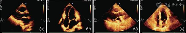
图1 缩窄性心包炎超声诊断与超声漏诊图像。图a、b 分别为超声心动图诊断的缩窄性心包炎左心室长轴及心尖四腔二维图像;图c、d 分别为超声心动图漏诊的缩窄性心包炎左心室长轴及心尖四腔二维图像 |
表2 缩窄性心包炎超声诊断组与超声漏诊组的超声心动图结果比较[M(QR)] |
| 超声心动图参数 | 超声诊断组(n=82) | 超声漏诊组(n=10) | 统计值 | P 值 |
|---|---|---|---|---|
| 左心房前后径(mm) | 41.00(8.30) | 37.50(4.30) | Z=-2.10 | 0.036 |
| 左心室舒张末期内径(mm) | 40.50(4.00) | 45.50(4.30) | Z=-3.03 | 0.002 |
| 右心房横径(mm) | 41.00(7.00) | 36.50(9.00) | Z=-2.43 | 0.015 |
| 右心室前后径(mm) | 20.00(2.30) | 21.00(3.30) | Z=-0.68 | 0.498 |
| 左心室质量指数(g/m2,±s) | 69.79±15.91 | 84.51±13.53 | t=-2.99 | 0.004 |
| LVEF(%) | 58.00(9.00) | 59.50(5.50) | Z=-0.11 | 0.915 |
| e'(cm/s, ±s) | 11.84±4.33 | 8.80±2.97 | t=2.16 | 0.034 |
| 下腔静脉宽度(mm) | 24.00(3.50) | 20.50(7.80) | Z=-2.52 | 0.012 |
| 室间隔“弹跳征”[ 例(%)] | 67(81.7) | 3(30.0) | χ 2=13.10 | < 0.001 |
| 二尖瓣前向E-E 变化率>25%[ 例(%)] | 49(59.8) | 2(20.0) | χ 2=5.70 | 0.017 |
注:LVEF 为左心室射血分数;e’为二尖瓣间隔瓣环舒张早期速度 |
表3 缩窄性心包炎超声诊断组与超声漏诊组的CT 结果比较[M(QR)] |
| CT 参数 | 超声诊断组(n=82) | 超声漏诊组(n=10) | 统计值 | P 值 |
|---|---|---|---|---|
| 左房室角(°,±s) | 95.72±23.66 | 104.70±18.48 | t=-1.16 | 0.251 |
| 右房室角(°,±s) | 80.71±30.55 | 106.50±35.87 | t=-2.47 | 0.015 |
| 心尖球形指数 | 69.00(29.00) | 84.00(25.00) | Z=-2.51 | 0.012 |
| 最厚心包厚度(mm) | 12.00(8.00) | 14.50(8.30) | Z=-0.70 | 0.482 |
| 左心室周围心包(mm) | 8.00(7.00) | 10.50(9.00) | Z=-1.22 | 0.222 |
| 右心室周围心包(mm) | 8.00(6.50) | 9.50(7.80) | Z=-0.52 | 0.602 |
| 心尖部心包(mm) | 3.00(3.00) | 2.50(4.00) | Z=-0.43 | 0.669 |
| 前室尖沟心包(mm) | 5.00(5.00) | 5.00(4.30) | Z=-0.95 | 0.341 |
| 后室尖沟心包(mm) | 5.00(4.50) | 5.50(8.50) | Z=-0.77 | 0.444 |
| 左心房心包(mm) | 3.00(3.00) | 4.50(4.80) | Z=-1.06 | 0.289 |
| 右心房心包(mm) | 6.00(6.00) | 8.50(10.30) | Z=-1.91 | 0.056 |
| 上腔静脉心包(mm) | 2.00(2.00) | 2.00(2.00) | Z=-1.30 | 0.192 |
| 下腔静脉心包(mm) | 4.00(3.50) | 5.00(5.30) | Z=-0.97 | 0.332 |
| 右心室流出道心包(mm) | 5.00(3.00) | 4.00(4.50) | Z=-1.52 | 0.129 |
| 心包积液[ 例(%)] | 36(43.9) | 6(60.0) | χ 2=0.93 | 0.335 |
| 心包钙化[ 例(%)] | 33(40.2) | 0 | χ 2=6.28 | 0.012 |
| 钙化心包嵌入心肌[ 例(%)] | 5(6.1) | 0 | χ 2=0.65 | 0.422 |
表4 缩窄性心包炎超声诊断组与超声漏诊组的围术期不良事件比较[例(%)] |
| 围术期不良事件 | 超声诊断组(n=82) | 超声漏诊组(n=10) | 统计值 | P 值 |
|---|---|---|---|---|
| 死亡 | 3(3.7) | 0 | χ 2=0.38 | 0.539 |
| 术后低心排血量综合征 | 12(14.6) | 0 | χ 2=1.68 | 0.195 |
| 术中输血 | 3(3.7) | 1(1.3) | χ 2=0.86 | 0.353 |
| 术后输血 | 17(20.7) | 1(1.3) | χ 2=0.65 | 0.419 |
| 术后低蛋白 | 34(41.5) | 6(60.0) | χ 2=1.25 | 0.264 |
| 术后电解质紊乱 | 32(39.0) | 3(3.8) | χ 2=0.31 | 0.579 |
| 术后腹腔感染 | 1(1.2) | 0 | χ 2=0.12 | 0.725 |
| 休克 | 4(4.9) | 0 | χ 2=0.51 | 0.475 |
| 胃肠功能紊乱 | 60(73.2) | 8(80.0) | χ 2=0.22 | 0.642 |
| 切口愈合不良 | 2(2.4) | 0 | χ 2=0.25 | 0.618 |
| 消化道出血 | 1(1.2) | 0 | χ 2=0.12 | 0.725 |
| 急性肾损伤 | 3(3.7) | 0 | X 2=0.38 | 0.539 |
| 肝损伤 | 2(2.4) | 0 | χ 2=0.25 | 0.618 |
| 住院时间(d,±s) | 24.66±9.40 | 22.30±15.49 | t=0.69 | 0.491 |
| ICU 住院时间(d,±s) | 3.13±2.59 | 1.80±1.03 | t=1.60 | 0.113 |
| 术后住院时间(d,±s) | 11.51±7.03 | 10.40±6.88 | t=0.47 | 0.639 |
| 1 |
Welch TD.Constrictive pericarditis:diagnosis, management and clinical outcomes[J].Heart, 2018, 104(9):725-731.
|
| 2 |
夏雪, 周建中.缩窄性心包炎诊断技术的研究进展[J].心血管病学进展, 2024, 45(7):608-611, 621.
|
| 3 |
郑嘉荣, 邓剑玲, 邢超, 等.超声心动图对冠心病心衰患者左室心尖形态与功能的分析[J].中国超声医学杂志, 2020, 36(4):336-338.
|
| 4 |
曹安强, 罗勇, 袁武, 等.心包剥脱术治疗缩窄性心包炎患者的近期疗效分析:单中心结果[J].岭南心血管病杂志, 2020, 26(4):424-428.
|
| 5 |
Schwefer M, Aschenbach R, Heidemann J, et al.Constrictive pericarditis, still a diagnostic challenge:comprehensive review of clinical management[J].Eur J Cardiothorac Surg, 2009, 36(3):502-510.
|
| 6 |
Adler Y, Charron P.The 2015 ESC Guidelines on the diagnosis and management of pericardial diseases[J].Eur Heart J, 2015, 36(42):2873-2874.
|
| 7 |
Oh JK, Hatle LK, Seward JB, et al.Diagnostic role of Doppler echocardiography in constrictive pericarditis[J].J Am Coll Cardiol,1994, 23(1):154-162.
|
| 8 |
Welch TD, Ling LH, Espinosa RE, et al.Echocardiographic diagnosis of constrictive pericarditis:Mayo Clinic criteria[J].Circ Cardiovasc Imaging, 2014, 7(3):526-534.
|
| 9 |
Li J, Li R, Cheng G, et al.A case series of constrictive pericarditis and suggested echocardiographic diagnostic criteria[J].J Int Med Res,2022, 50(11):3000605221134468.
|
| 10 |
李国英, 徐明, 刘玲玲, 等.缩窄性心包炎超声特征的logistic 回归分析[J].放射学实践, 2023, 38(8):1065-1069.
|
| 11 |
Chao CJ, Jeong J, Arsanjani R, et al.Echocardiography-based deep learning model to differentiate constrictive pericarditis and restrictive cardiomyopathy[J].JACC Cardiovasc Imaging, 2024, 17(4):349-360.
|
| 12 |
陈结, 彭光高, 刘向阳, 等.缩窄性心包炎的三种影像诊断比较分析[J].医学临床研究, 2006,23(5):737-739.
|
| 13 |
Lin J, Li M, Huang Y, et al.Evaluation of pericardial thickening and adhesion using high-frequency ultrasound[J].J Am Soc Echocardiogr,2023, 36(8):841-848.
|
| 14 |
张连仲, 赵冰, 张慧君.缩窄性心包炎心脏几何形态的二维超声心动图特征[J].中国临床医学影像杂志, 2000, 11(2):99-100.
|
| 15 |
Tumkosit M, Martin CG, Bayram E, et al.Left ventricular spherical remodeling and apical myocardial relaxation:cardiovascular MR imaging measurement of myocardial segments[J].Radiology,2007,244(2):411-418.
|
| 16 |
李汉美, 佟明汇, 王巍, 等.慢性缩窄性心包炎行心包剥脱术的预后及危险因素:单中心二十年经验[J].中国体外循环杂志, 2018,16(3):160-164.
|
| 17 |
张耀中, 张雅娉, 郭建中, 等.左室质量与慢性缩窄性心包炎手术效果的关系研究[J].中国心血管杂志, 2022, 27(6):548-551.
|
/
| 〈 |
|
〉 |