
2025 , Vol. 22 >Issue 10: 982 - 987
DOI: https://doi.org/10.3877/cma.j.issn.1672-6448.2025.10.012
急性肾静脉闭塞肾脏不同区域杨氏模量差异性的实验研究
通信作者:
王兴华,Email:wangxhus@163.comCopy editor: 汪荣
收稿日期: 2025-04-25
网络出版日期: 2025-12-24
基金资助
山西省基础研究计划资助项目(202403021221318)
版权
Differences in Young's modulus across renal regions following acute renal vein occlusion: an experimental study
Corresponding author:
Wang Xinghua, Email: wangxhus@163.comReceived date: 2025-04-25
Online published: 2025-12-24
Copyright
分析急性肾静脉闭塞后肾脏不同区域的杨氏模量差异性并探讨诊断急性肾静脉闭塞的肾脏最佳区域。
取20只新西兰大白兔为研究对象,结扎左肾静脉后2 h测量并比较肾脏不同侧别(健侧组、患侧组)、不同解剖部位(上极、中部、下极)及不同组织结构(皮质、髓质、肾窦)的杨氏模量。采用点二列相关系数分析肾脏不同区域的杨氏模量与急性肾静脉闭塞状态的相关性,采用组内相关系数(ICC)评价杨氏模量测量的可重复性。
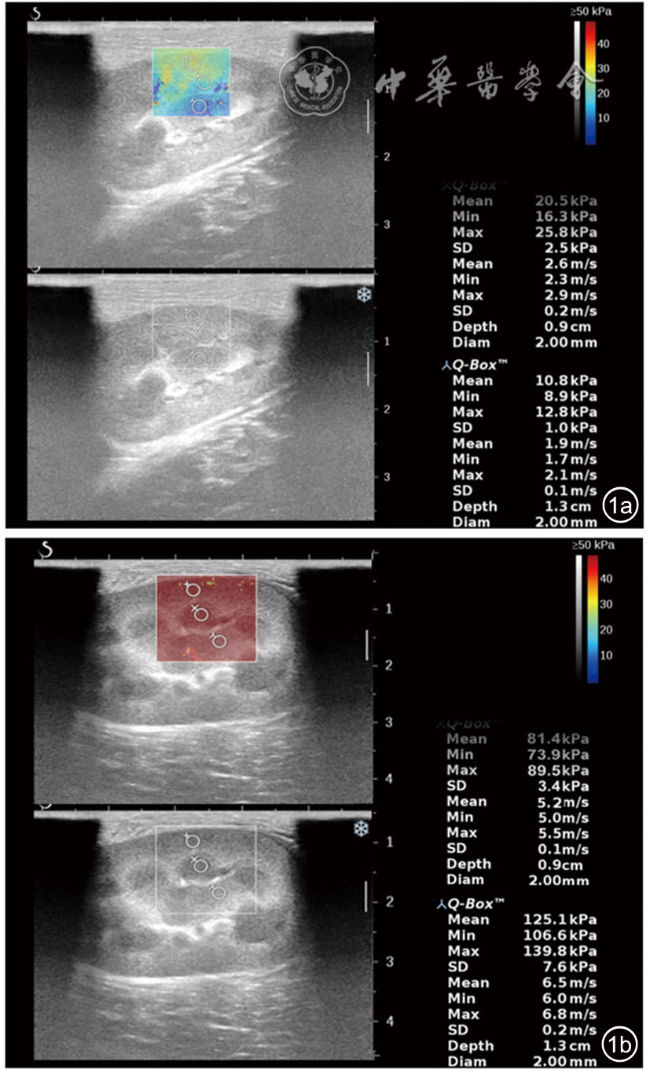
在不同解剖部位,健侧组上极与中部、中部与下极间杨氏模量差异有统计学意义(P<0.05)。患侧组上极、中部及下极间杨氏模量差异均无统计学意义(P>0.05)。在不同组织结构,健侧组皮质与肾窦、髓质与肾窦间杨氏模量差异有统计学意义(P<0.05)。患侧组皮质、髓质及肾窦间杨氏模量差异均有统计学意义(P<0.05)。多因素方差分析显示,三向交互效应(急性肾静脉闭塞状态×解剖部位×组织结构)差异无统计学意义(F=1.575,P=0.190)。患侧组相同解剖部位及相同组织结构的杨氏模量均高于健侧组,差异均有统计学意义(P<0.05)。其中,中部皮质的效应量最突出(Cohen's d=2.770),其与急性肾静脉闭塞的相关性最强(r=0.867),测量可重复性最优(ICC=0.987)。
正常肾脏的弹性具有区域异质性,病理状态下肾脏的弹性有重塑的可能,急性肾静脉闭塞使肾脏硬度明显增加,中部皮质可作为诊断急性肾静脉闭塞的肾脏最佳区域。
张涛 , 徐梓祎 , 徐景竹 , 王兴华 . 急性肾静脉闭塞肾脏不同区域杨氏模量差异性的实验研究[J]. 中华医学超声杂志(电子版), 2025 , 22(10) : 982 -987 . DOI: 10.3877/cma.j.issn.1672-6448.2025.10.012
To analyze regional heterogeneity in Young's modulus of the kidney following acute renal vein occlusion and identify optimal regions for diagnosis.
A total of 20 New Zealand White rabbits were used as the study subjects. At 2 hours after ligation of the left renal vein, Young's modulus was measured and compared across different sides (healthy side vs affected side), anatomical locations (upper pole vs mid portion vs lower pole), and organizational structures (cortex vs medulla vs renal sinus) of the kidney. The point-biserial correlation coefficient was employed to analyze the correlation between Young's modulus in various renal regions and the status of acute renal vein occlusion. The intraclass correlation coefficient (ICC) was used to assess the reproducibility of the Young's modulus measurements.
Regarding anatomical locations, in the healthy side group, the differences in Young's modulus were statistically significant between the upper pole and mid portion, and between the mid portion and lower pole (P<0.05). In the affected side group, no statistically significant differences in Young's modulus were observed among the upper pole, mid portion, and lower pole (P>0.05). Regarding organizational structures, in the healthy side group, the differences in Young's modulus were statistically significant between the cortex and renal sinus, and between the medulla and renal sinus (P<0.05). In the affected side group, the differences in Young's modulus among the cortex, medulla, and renal sinus were all statistically significant (P<0.05). Three-way ANOVA revealed that the three-way interaction effect (acute renal vein occlusion status × anatomical location × organizational structure) was not statistically significant (F=1.575, P=0.190). The Young's modulus values in the affected side group were consistently higher than those in the healthy side group for identical anatomical locations and organizational structures, and all these differences were statistically significant (P<0.05). Among these comparisons, the mid-portion cortex demonstrated the most prominent effect size (Cohen's d=2.770), eexhibited the strongest correlation with acute renal vein occlusion (r=0.867), and showed the optimal measurement reproducibility (ICC=0.987).
Renal elasticity in normal kidneys shows regional heterogeneity and undergoes remodeling under pathological conditions. Acute renal vein occlusion causes a significant increase in stiffness, identifying the mid-portion cortex as the optimal diagnostic region.

表1 急性肾静脉闭塞后肾脏不同区域杨氏模量测量的ICC |
| 肾脏组织结构 | 肾脏解剖部位 | ||
|---|---|---|---|
| 上极 | 中部 | 下极 | |
| 皮质 | 0.957 | 0.987 | 0.981 |
| 髓质 | 0.965 | 0.958 | 0.953 |
| 肾窦 | 0.942 | 0.969 | 0.921 |
注:ICC为组内相关系数 |
表2 肾脏不同解剖部位杨氏模量比较(kPa, |
| 组别 | 例数 | 上极 | 中部 | 下极 | F值 | P值 |
|---|---|---|---|---|---|---|
| 健侧组 | 20 | 20.21±5.79 | 16.45±4.62 | 18.76±4.44 | 8.665 | <0.001 |
| 患侧组 | 20 | 55.95±22.02 | 55.86±22.70 | 56.71±21.32 | 0.027 | 0.973 |
| t值 | 12.839 | 14.005 | 16.617 | |||
| P值 | <0.001 | <0.001 | <0.001 |
注:与上极比较,aP<0.001;与中部比较,bP=0.032 |
表3 肾脏不同组织结构杨氏模量比较(kPa, |
| 组别 | 例数 | 皮质 | 髓质 | 肾窦 | F值 | P值 |
|---|---|---|---|---|---|---|
| 健侧组 | 20 | 19.15±6.80 | 19.21±4.29 | 17.04±3.76 | 3.465 | 0.033 |
| 患侧组 | 20 | 66.14±18.53 | 57.03±17.63 | 45.34±24.09 | 15.849 | <0.001 |
| t值 | 19.516 | 16.878 | 11.091 | |||
| P值 | <0.001 | <0.001 | <0.001 |
注:健侧组中,与皮质比较,aP=0.026;与髓质比较,bP=0.043。患侧组中,与皮质比较,aP(髓质)=0.007,aP(肾窦)<0.001;与髓质比较,bP<0.001 |
表4 急性肾静脉闭塞后肾脏不同区域杨氏模量比较(kPa, |
| 组别 | 例数 | 皮质 | 髓质 | 肾窦 | F值 | P值 |
| 健侧组(上极) | 20 | 23.19±6.94 | 20.71±4.18 | 16.71±3.99 | 7.859 | <0.001 |
| 患侧组(上极) | 20 | 69.54±18.09 | 58.87±18.07 | 39.44±19.01 | 13.776 | <0.001 |
| t值 | 9.873 | 9.160 | 5.408 | |||
| P值 | <0.001 | <0.001 | <0.001 | |||
| Cohen's d | 2.208 | 2.048 | 1.209 | |||
| 组别 | 例数 | 皮质 | 髓质 | 肾窦 | F值 | P值 |
| 健侧组(中部) | 20 | 15.77±6.34 | 17.21±4.00 | 16.36±2.97 | 0.486 | 0.617 |
| 患侧组(中部) | 20 | 66.57±19.02 | 54.87±18.05 | 41.35±18.00 | 9.449 | <0.001 |
| t值 | 12.388 | 8.953 | 6.267 | |||
| P值 | <0.001 | <0.001 | <0.001 | |||
| Cohen's d | 2.770 | 2.002 | 1.402 | |||
| 组别 | 例数 | 皮质 | 髓质 | 肾窦 | F值 | P值 |
| 健侧组(下极) | 20 | 18.48±5.06 | 19.72±4.12 | 18.07±4.16 | 0.737 | 0.483 |
| 患侧组(下极) | 20 | 61.73±17.44 | 54.74±12.45 | 46.49±16.66 | 4.740 | 0.012 |
| t值 | 11.905 | 11.487 | 7.497 | |||
| P值 | <0.001 | <0.001 | <0.001 | |||
| Cohen's d | 2.662 | 2.569 | 1.676 |
注:*P值为经过Holm-Bonferroni校正后的值 |
表5 肾脏不同区域杨氏模量与急性肾静脉闭塞状态的点二列相关系数(r值,n=20) |
| 肾脏组织结构 | 肾脏解剖部位 | ||
|---|---|---|---|
| 上极 | 中部 | 下极 | |
| 皮质 | 0.856 | 0.867 | 0.855 |
| 髓质 | 0.820 | 0.818 | 0.861 |
| 肾窦 | 0.639 | 0.696 | 0.759 |
张涛, 徐梓祎, 徐景竹, 等. 急性肾静脉闭塞肾脏不同区域杨氏模量差异性的实验研究[J/OL]. 中华医学超声杂志(电子版), 2025, 22(10): 982-987.
| 1 |
|
| 2 |
|
| 3 |
|
| 4 |
|
| 5 |
|
| 6 |
|
| 7 |
|
| 8 |
|
| 9 |
李娜, 刘晓娜, 郑海宁, 等. 肾脏中部及下极皮质杨氏模量值差异的动物实验研究[J/CD]. 中华医学超声杂志(电子版), 2018, 15(4): 309-312.
|
| 10 |
|
| 11 |
|
| 12 |
|
| 13 |
邹震宇, 刘泽政, 陈玲, 等. 二维剪切波弹性成像评估高血压肾病小鼠肾纤维化的实验研究[J]. 临床超声医学杂志, 2023, 25(12): 945-950.
|
| 14 |
|
| 15 |
|
| 16 |
|
| 17 |
|
| 18 |
|
| 19 |
郑金龙, 韩萍, 柳曦, 等. 下腔静脉病变的螺旋CT诊断[J]. 临床放射学杂志, 2006, 2006(9): 838-842.
|
| 20 |
黄志芳, 吕仁华, 丁红, 等. 以剪切波速度评估慢性肾病分期的最佳肾脏靶区[J]. 中国医学影像技术, 2024, 40(11): 1745-1748.
|
| 21 |
|
| 22 |
李凤, 张蔚蓝, 黄伟俊, 等. 超声及超声造影定量分析在移植肾术后肾功能延迟恢复评价中的应用[J]. 海南医学, 2022, 33(4):494-497.
|
/
| 〈 |
|
〉 |
